


Introduction
Brown-Séquard syndrome (BSS) was first described in 1849 by Charles-Édouard Brown-Séquard, who documented the constellation of findings in a sea captain stabbed in the neck [1]. A hemisection or hemicompression of the cord underlies the syndrome, and because the affected pathways cross the midline at different points, the deficit splits down the middle of the body: Weakness, proprioceptive loss, and diminished vibration sense on the side of the lesion, paired with lost pain and temperature sensation on the opposite side [2]. Reported causes include traumatic injury, tumor, and hemorrhage; cervical disc herniation (CDH) is a comparatively rare etiology [3]. Spinal cord injury following manual manipulation of the cervical spine is itself uncommon [4], and in a review of all previously published cases, the manipulation was invariably performed by a second party, typically a chiropractor or other manual therapist. We report a case of BSS caused by CDH that developed after a man manipulated his own cervical spine; to our knowledge, this is the first such case in the literature.
Case Report
A previously healthy 38-year-old male, working as a software engineer, presented to the emergency department with sudden right-sided weakness immediately after “cracking” his own neck. He denied any recent trauma. Five years earlier, he had experienced an episode of cervical pain treated with 2 weeks of medication and physiotherapy, after which he was left with only mild, intermittent discomfort recurring roughly every 3–4 months. The chronology of his history, presentation, and treatment is summarized as a CARE-guideline timeline in Table 1.
CARE guideline timeline of history, presentation, investigations, and treatment
| Time | Event |
|---|---|
| 5 years before injury | Episode of cervical pain treated with 2 weeks of medication and physiotherapy; left with mild, intermittent discomfort recurring every 3–4 months |
| 5 years before injury to day of injury | Habitual self-manipulation of the cervical spine several times daily to relieve recurrent discomfort, without prior complication |
| Day 0, 0 h (symptom onset) | Self-manipulation of the neck followed immediately by shooting neck pain and right-sided weakness |
| Day 0, shortly after onset | Presentation to emergency department; right hemiparesis and dissociated sensory loss on examination; dexamethasone started |
| Day 0, hours after onset | MRI cervical spine: Large C4-C5 and C5-C6 disc herniations with severe cord compression and T2 signal change |
| Day 0, +16 h | Emergent C4-C6 ACDF |
| POD 1 | Post-operative MRI: resolution of canal stenosis and cord compression; new focal T2 signal (evolving contusion) |
| POD 2 | Discharged to acute inpatient rehabilitation facility; 14-day dexamethasone taper begun; AM-PAC 32/48 on admission |
| POD 9 | Discharged from inpatient rehabilitation; AM-PAC 48/48, independent in ADLs and mobility without an assistive device; outpatient therapy arranged |
| 2 months | Motor strength fully recovered; mild residual sensory deficit in the right upper limb |
ADLs: Activities of daily living, AM-PAC: Activity measure for post-acute care, MRI: Magnetic resonance imaging, POD: Post-operative day
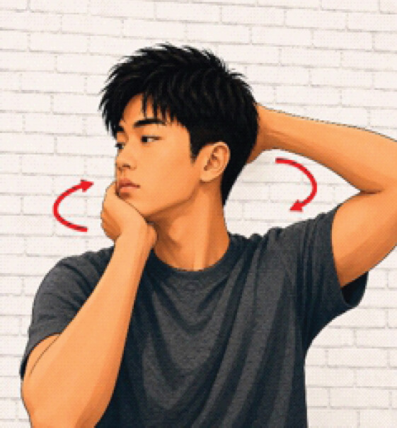
To relieve this recurrent discomfort, he had developed a habit of manipulating his own cervical spine: Grasping his chin with one hand and placing the other on top of his head (Fig. 1), then forcefully bending his neck laterally while rotating his chin toward the opposite shoulder until he produced an audible crack. He performed the maneuver in both directions (left bend with right rotation and right bend with left rotation) several times a day for several years, without prior complication.
On the day of injury, immediately after the maneuver, he felt a shooting pain radiate down his neck and instantly developed right-sided weakness, prompting presentation to the emergency department. Examination revealed right hemiparesis, weakest distally with 0/5 wrist extension and grip strength, whereas strength on the left remained fully intact; muscle-by-muscle grades are given in Table 2. Sensory examination showed decreased pinprick sensation on the left side of the body from the C5 level downward, and decreased vibratory sense on the right side from the C4 level downward, the classic dissociated sensory pattern of BSS.
Serial neurological and functional findings from presentation to 2-month follow-up
| Parameter | Presentation | Rehab admission (POD 2) | Rehab discharge (POD 9) | 2-month follow-up |
|---|---|---|---|---|
| Neurological level (NLI) | C5 | C5 | C5 | C5 |
| AIS grade | D | D | D | D |
| Right upper-limb strength (MRC) | 3/5 shoulder abduction | 5/5 shoulder abduction | 5/5 all groups | 5/5 all groups |
| 2/5 elbow flexion | 5/5 elbow flexion | except grip 4/5 | Documented sum*: 30/30 | |
| 2/5 elbow extension | 5/5 elbow extension | Documented sum*: 29/30 | ||
| 1/5 wrist flexion | 4/5 wrist flexion | |||
| 0/5 wrist extension | 4/5 wrist extension | |||
| 0/5 grip | 3/5 grip | |||
| Documented sum*: 8/30 | Documented sum*: 26/30 | |||
| Right lower-limb strength (MRC) | 3/5 hip flexion | 5/5 hip flexion | 5/5 all groups | 5/5 all groups |
| 3/5 knee extension | 5/5 knee extension | Documented sum*: 25/25 | Documented sum*: 25/25 | |
| 3/5 knee flexion | 5/5 knee flexion | |||
| 2/5 ankle dorsiflexion | 3/5 ankle dorsiflexion | |||
| 2/5 ankle plantar flexion | 3/5 ankle plantar flexion | |||
| Documented sum*: 13/25 | Documented sum*: 21/25 | |||
| Left upper-limb strength | Intact, 5/5 all groups† | Intact† | Intact† | Intact† |
| Documented sum*: 30/30 | Documented sum*: 30/30 | Documented sum*: 30/30 | Documented sum*: 30/30 | |
| Left lower-limb strength | Intact, 5/5 all groups† | Intact† | Intact† | Intact† |
| Documented sum*: 25/25 | Documented sum*: 25/25 | Documented sum*: 25/25 | Documented sum*: 25/25 | |
| Pinprick sensation | Decreased on left | Unchanged | Unchanged | Not documented |
| from C5 | ||||
| Vibration sensation | Decreased on right | Unchanged | Unchanged | Not specifically |
| from C4 | documented | |||
| Sacral sparing | Present‡ | Present‡ | Present‡ | Present‡ |
| Functional status (AM-PAC) | Acute right | 32/48 | 48/48 | Functionally |
| hemiparesis | independent in ADLs | recovered | ||
| and mobility | ||||
| Assistive device | Not applicable | Not documented | None required | Not documented |
ADLs: Activities of daily living, AIS: ASIA impairment scale, AM-PAC: Activity measure for post-acute care, MRC: Medical research council, NLI: neurological level of injury, POD: Post-operative day.
*
Documented sum reflects the muscle groups actually recorded in the case notes (six upper-limb and five lower-limb groups per side), not the standardized five-muscle ISNCSCI upper- and lower-extremity motor score panel.
†
Left-sided strength was documented as fully intact at presentation and assumed stable thereafter; it was not re-tested muscle-by-muscle at every subsequent visit.
‡
Voluntary anal contraction and/or deep anal pressure were intact at all time points, consistent with AIS grade D throughout the patient’s course.
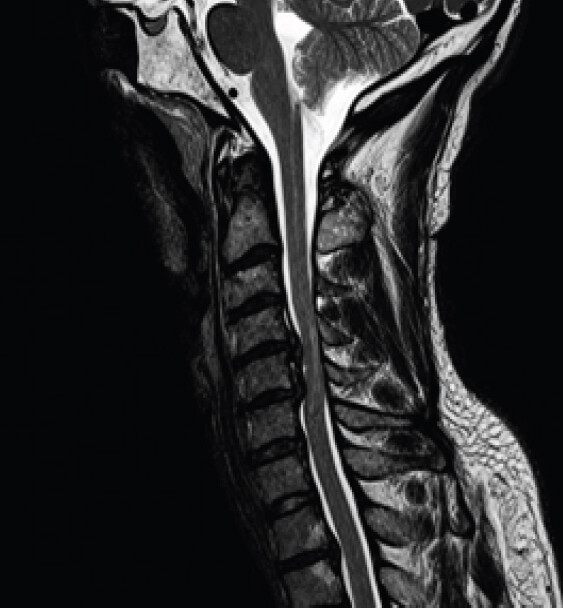
Magnetic resonance imaging (MRI) of the cervical spine showed large disc herniations at C4-C5 and C5-C6 with severe cord compression and T2 signal change, superimposed on chronic degenerative changes (facet arthropathy and endplate remodeling) more advanced than expected for his age (Fig. 2). He was started on dexamethasone and taken emergently for a C4-C6 anterior cervical discectomy and fusion 16 h after symptom onset. Intraoperatively, the C4-C5 disc had ruptured through the annulus and posterior longitudinal ligament (PLL), with large free fragments lying between the PLL and the thecal sac; a similar but smaller herniation was found at C5-C6. The PLL behind the C5 vertebral body was notably thickened, contributing mild additional canal stenosis.
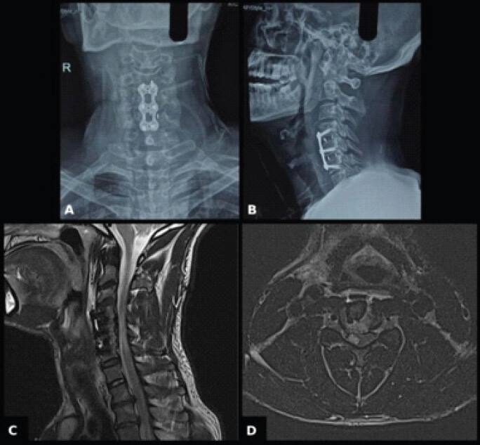
Post-operative day (POD)-1 MRI confirmed resolution of the canal stenosis and cord compression, with new focal T2 signal in the right hemicord at C4-C5 consistent with evolving contusion (Fig. 3). His strength improved rapidly, and he was discharged to an acute inpatient rehabilitation facility on POD 2 with a planned 14-day dexamethasone taper.
Rehabilitation and outcome
An interdisciplinary inpatient spinal cord injury rehabilitation team, comprising physiatry, physical therapy, occupational therapy, and nursing, managed his admission. Physical therapy focused on gait re-education and lower-limb strengthening; occupational therapy addressed upper-limb and grip retraining and activities-of-daily-living practice; and nursing provided bowel, bladder, and skin surveillance appropriate to his ASIA impairment scale (AIS) D status. Functional gains were tracked with the Activity Measure for Post-Acute Care (AM-PAC) (“6-Clicks”), which improved from 32/48 on admission (19 for activities of daily living, 13 for mobility) to the maximum of 48/48 by POD 9, when he was independent in all activities and mobility without an assistive device; muscle-group strength over the same period is detailed in Table 2. His sensory deficit had not yet improved at discharge. He was then discharged home with a structured outpatient physiotherapy program, and at 2-month follow-up his motor strength had fully normalized, though he continued to notice mildly reduced light-touch sensation in the right arm.
Discussion
The severity and mix of deficits in BSS depend on the level and extent of the lesion, which is why atypical presentations are grouped under the term “Brown-Séquard-plus syndrome” rather than treated as exceptions to the rule [5]. The pattern follows from where each pathway sits and crosses within the cord: The corticospinal tract has already crossed in the medulla, so injury produces weakness below the lesion on the same side, whereas the still-uncrossed dorsal columns produce a matching ipsilateral loss of proprioception and vibration. The spinothalamic tract is the outlier, crossing within a few segments of entering the cord, so a hemicord lesion instead strips pain and temperature sensation from the opposite side, typically beginning two to three levels below the injury [2].
Spinal cord injury after cervical manipulation is rare (a systematic review identified only 32 cases of all causes [4]), and we identified 11 previously published cases of BSS specifically following manual cervical manipulation (Table 3). Only four of these were attributable to CDH, the remainder resulting from spinal epidural hematoma or vascular anomaly causing cord contusion. The four CDH cases had a mean age of 49 years, 75% were female, and in every instance the manipulation had been performed by another individual, presumably a licensed practitioner [6, 7, 8, 9, 10, 11, 12, 13, 14, 15]. An updated PubMed search covering 2022 through 2026 using the terms “Brown-Séquard syndrome,” “cervical disc herniation,” and “manual manipulation” did not identify any further cases of manipulation-induced CDH causing BSS, reinforcing how rare this specific combination remains.
Reported cases of Brown-Séquard syndrome following manual manipulation of the cervical spine, including the case described in this report
| N | Reference | Prior symptoms | Trauma history | Age | Sex | Level | Motor at presentation (MMT) | Tx | Motor at recovery (MMT) | Recovery (weeks) | Dx |
|---|---|---|---|---|---|---|---|---|---|---|---|
| 1 | Our patient | MNP | No | 38 | M | C4-C5, C5- C6 | 0/5 | ACDF | 05-May | ~8 | CDH |
| 2 | Hsieh et al. [6] | MNP | NR | 61 | F | C3-C4 | 03-May | ACDF | 05-May | 12 | CDH |
| 3 | Finelli et al. [7] | MNP w/ Rad | No | 46 | F | C4-C6 | NR | ACDF | Incomplete | 16 | CDH |
| 4 | Malone et al. [8] | MNP | No | 35 | F | C5-C6 | NR | ACDF | 05-May | 12 | CDH |
| 5 | Malone et al. [8] | MNP | No | 55 | M | C4-C5 | NR | ACDF | Incomplete | NR | CDH |
| 6 | Tsou et al. [9] | MNP | Major | 83 | M | C1-C2 | 0/5 | ORIF | 03-May | 8 | Other |
| 7 | Lipper et al. [10] | MNP | No | 58 | F | C4 | 03-May | Steroids only | Incomplete | 15 | Other |
| 8 | Segal et al. [11] | MNP | Minor | 33 | F | C4-C6 | 03-May | Lami | Incomplete | 0.5 | SEH |
| 9 | Tseng et al. [12] | MNP | NR | 67 | F | C3-C5 | 01-May | Lami | 05-May | 52 | SEH |
| 10 | Neetu et al. [13] | MNP | No | 55 | M | C1-C5 | 01-May | Steroids only | 04-May | 0.2 | SEH |
| 11 | Zupruk et al. [14] | None | No | 86 | M | C2-C7 | 0/5 | Lami | 05-May | 12 | SEH |
| 12 | Domenicucci et al. [15] | MNP | No | 52 | F | C3-T1 | NR | Lami | 05-May | 26 | SEH |
ACDF: Anterior cervical discectomy and fusion, CDH: Cervical disc herniation, Dx: Diagnosis, Lami: Laminectomy, MMT: Manual muscle testing, MNP: Mild neck pain, MNP w/Rad: Mild neck pain with radicular symptoms, NR: Not recorded, ORIF: Open reduction and internal fixation, SEH: Spinal epidural hematoma, Tx: Treatment, M: Male, F: Female.
That search did, however, identify five recent reports of BSS caused by CDH through other mechanisms, most notably intradural disc migration, a recently characterized entity in which extruded disc material breaches the dura and enters the subarachnoid space [16, 17, 18, 19, 20]. Jamaleddine et al. and Guo et al. each confirmed the intradural rupture at surgery and recovered their patients well after anterior decompression with dural repair; both suggested that marked T2 hyperintensity in the disc, together with a split or “Y-shaped” ventral dura, can flag the diagnosis before the disc space is even opened [19, 20]. Xiang et al. and Yen et al. reported similar presentations managed with early anterior surgery [16, 17], and Groussis et al. described a C3-C4 extrusion treated with disc replacement [18]. None involved manipulation, but together with our patient, these cases show that even a degenerated but previously asymptomatic disc can decompensate catastrophically with relatively modest additional load.
Manual manipulation outside a formal clinical setting is not confined to Western chiropractic practice. In India, a forceful neck “crack,” colloquially gardan chatkana (गद चटकाना, literally “to snap the neck”), is commonly offered by barbers as a finishing flourish to a shave or head massage, and is also a widespread self-administered habit for relieving neck stiffness. Nair et al. reported a 57-year-old male in Mumbai whose barber twisted and wrenched his neck sideways as a finishing touch to a routine salon head massage, precipitating acute paraparesis in a spine already carrying multilevel degenerative disease and ultimately requiring anterior decompression for a large C4-C5 extrusion [21]. Although his sensory findings were bilateral rather than a discrete hemicord pattern, the case shows that informal or self-administered cervical manipulation is a real, probably under-reported exposure across South Asia, wherever a degenerated disc meets a forceful, untrained hand.
The other four previously reported CDH-BSS cases did not specify the time from symptom onset to surgery or the intensity of post-operative rehabilitation, but our case suggests that both may matter [22, 23]. Our patient underwent surgery within 16 h and completed a full interdisciplinary inpatient rehabilitation admission and, despite presenting with the most severe weakness among the reported CDH cases, achieved the fastest return to full strength (Table 3). His AM-PAC score rose 16 points over 7 days, from needing assistance with all activities and mobility to complete independence without an assistive device, consistent with evidence that AIS D patients with greater initial motor impairment benefit most from formal inpatient rather than home-based rehabilitation [24].
To our knowledge, this is the first reported spinal cord injury resulting from a patient manipulating his own cervical spine. The pronounced chronic degenerative changes and thickened PLL found intraoperatively, disproportionate for his age, raise the possibility that years of repeated forceful self-manipulation may have caused cumulative subclinical injury to his discs and facet joints, eventually culminating in frank rupture; alternatively, those same degenerative changes may have been the original source of the discomfort that led him to manipulate his neck in the first place. Either way, this case is a cautionary example for anyone who regularly self-manipulates the cervical spine, particularly in the presence of underlying degenerative change. This report is limited by its single-patient design and 2-month follow-up; longer-term sensory recovery and the risk of adjacent-level injury from continued neck manipulation remain unknown.
Conclusion
Self-administered cervical spine manipulation, though seemingly benign after years without incident, can precipitate acute BSS in a degenerated cervical spine. Sudden lateralizing weakness after neck manipulation, whether by a therapist, an untrained third party, or the patient, warrants a low threshold for urgent MRI; when cord compression is confirmed, early decompression combined with intensive rehabilitation can produce excellent recovery even from a severe initial deficit.
Clinical Message
A history of habitual, forceful self-manipulation of the neck should raise suspicion for underlying cervical disc disease, and lateralizing weakness after such a maneuver warrants urgent imaging: Early anterior decompression with intensive inpatient rehabilitation can convert a severe Brown-Séquard presentation into a near-complete recovery.
Conflict of Interest:
Nil
Source of Support:
Nil
Consent:
The authors confirm that informed consent was obtained from the patient for publication of this article
How to Cite this Article
Raj S, Kumar D, VB S, Jose A, Goni VG. Brown-Séquard Syndrome after Self-administered Manual Manipulation of the Cervical Spine: A Case Report. Journal of Orthopaedic Case Reports 2026 October;16(10): 97-102.
References
- Rengachary SS, Colen C, Guthikonda M. Charles-edouard brown-séquard: An eccentric genius. Neurosurgery 2008;62:954-64. [Google Scholar] | [PubMed]
- Shams S, Farrell CL, Arain A. Brown-Séquard Syndrome. StatPearls.. Treasure Island, FL:StatPearls Publishing 2026 Available from: https://www.ncbi.nlm.nih.gov/books/nbk538135 (Last accessed on 16 Aug 2026). [Google Scholar] | [PubMed]
- Zeng Y, Ren H, Wan J, Lu J, Zhong F, Deng S. Cervical disc herniation causing Brown-Sequard syndrome: Case report and review of literature (CARE-compliant). Medicine (Baltimore) 2018;97:e12377. [Google Scholar] | [PubMed]
- Kranenburg HA, Schmitt MA, Puentedura EJ, Luijckx GJ, Van Der Schans CP. Adverse events associated with the use of cervical spine manipulation or mobilization and patient characteristics: A systematic review. Musculoskelet Sci Pract 2017;28:32-8. [Google Scholar] | [PubMed]
- Roth EJ, Park T, Pang T, Yarkony GM, Lee MY. Traumatic cervical brown-sequard and brown-sequard-plus syndromes: The spectrum of presentations and outcomes. Paraplegia 1991;29:582-9. [Google Scholar] | [PubMed]
- Hsieh JH, Wu CT, Lee ST. Cervical intradural disc herniation after spinal manipulation therapy in a patient with ossification of posterior longitudinal ligament: A case report and review of the literature. Spine (Phila Pa 1976) 2010;35:E149-51. [Google Scholar] | [PubMed]
- Finelli PF, Leopold N, Tarras S. Brown-Sequard syndrome and herniated cervical disc. Spine (Phila Pa 1976) 1992;17:598-600. [Google Scholar] | [PubMed]
- Malone DG, Baldwin NG, Tomecek FJ, Boxell CM, Gaede SE, Covington CG . Complications of cervical spine manipulation therapy: 5-Year retrospective study in a single-group practice. Neurosurg Focus 2002;13 ecp1. [Google Scholar] | [PubMed]
- Tsou A, Juan YH, Chen TY, Lin SK. Thrombolysis for atlantoaxial dislocation mimicking acute ischemic stroke. Am J Emerg Med 2019;37:1216.e3-5. [Google Scholar] | [PubMed]
- Lipper MH, Goldstein JH, Do HM. Brown-séquard syndrome of the cervical spinal cord after chiropractic manipulation. AJNR Am J Neuroradiol 1998;19:1349-52. [Google Scholar] | [PubMed]
- Segal DH, Lidov MW, Camins MB. Cervical epidural hematoma after chiropractic manipulation in a healthy young woman: Case report. Neurosurgery 1996;39:1043-5. [Google Scholar] | [PubMed]
- Tseng SH, Chen Y, Lin SM, Wang CH. Cervical epidural hematoma after spinal manipulation therapy: Case report. J Trauma 2002;52:585-6. [Google Scholar] | [PubMed]
- Neetu R, Chandra MS, Rashmi M. Cervical spinal epidural hematoma with acute brown-séquard presentation. Neurol India 2006;54:107-8. [Google Scholar] | [PubMed]
- Zupruk GM, Mehta Z. Brown-Séquard syndrome associated with posttraumatic cervical epidural hematoma: Case report and review of the literature. Neurosurgery 1989;25:278-80. [Google Scholar] | [PubMed]
- Domenicucci M, Ramieri A, Salvati M, Brogna C, Raco A. Cervicothoracic epidural hematoma after chiropractic spinal manipulation therapy. Case report and review of the literature. J Neurosurg Spine 2007;7:571-4. [Google Scholar] | [PubMed]
- Xiang Z, Cen B, Zhang Y, Shang H. Acute non-traumatic cervical disc herniation producing brown-séquard Syndrome: A case report. Asian J Surg 2023;46:3318-9. [Google Scholar] | [PubMed]
- Yen TC, Worley JR, St Clair D, Crim JR, Moore DK. Intradural cervical herniated nucleus pulposus presentation and management: A case report. AME Case Rep 2023;7:28. [Google Scholar] | [PubMed]
- Groussis M, Issa R, Taheri MR, Pourmand A. C3-C4 cervical disc herniation producing Brown-Séquard syndrome: A case report and review of the literature. Am J Emerg Med 2024;80:229.e5-7. [Google Scholar] | [PubMed]
- Jamaleddine Y, Ali M, Dib EY, Assi C, Moucharafieh R, Badra M. Acute Brown-Sequard syndrome from an intradural C5-C6 disc herniation: Case report and literature review. Int J Surg Case Rep 2025;135:111905. [Google Scholar] | [PubMed]
- Guo G, Shi X, Qiu Y, Gao D, Fang P, Ma H . Cervical intradural disc herniation: A new diagnostic clue based on MRI T2 hyperintensity and intraoperative increased water content in the intervertebral disc-a case report and mechanistic insights. Front Surg 2025;12:1653174. [Google Scholar] | [PubMed]
- Nair VV, Kundnani V, Anand M, Shetty A, Jain M, Chodavadiya SS. Acute paraparesis in a patient after high velocity manipulation of cervical spine and its management. J Orthop Case Rep 2022;12:50-3. [Google Scholar] | [PubMed]
- Badhiwala JH, Wilson JR, Witiw CD, Harrop JS, Vaccaro AR, Aarabi B . The influence of timing of surgical decompression for acute spinal cord injury: A pooled analysis of individual patient data. Lancet Neurol 2021;20:117-26. [Google Scholar] | [PubMed]
- Fehlings MG, Vaccaro A, Wilson JR, Singh A, Cadotte DW, Harrop J . Early versus delayed decompression for traumatic cervical spinal cord injury: Results of the Surgical Timing in Acute Spinal Cord Injury Study (STASCIS). PLoS One 2012;7:e32037. [Google Scholar] | [PubMed]
- Richard-Denis A, Dionne A, Mputu PM, Mac-Thiong JM. Do all patients with functional motor-incomplete (AIS-D) traumatic spinal cord injury need specialized inpatient functional rehabilitation? A prospective observational cohort study proposing clinical criteria for home-based rehabilitation after acute care. J Spinal Cord Med 2024;47:753-64. [Google Scholar] | [PubMed]
© 2026 Journal of Orthopaedic Case Reports - Published by Indian Orthopaedic Research Group
About the Authors
-

Sharath Raj
-

Deepak Kumar
-

Shrinivas VB
-

Ajay Jose
-

Vijay G Goni
Related Articles in Journal of Orthopaedic Case Reports
 September 1, 2026 Blunt Traumatic Posterior Cord Syndrome Following a Hangman Fracture: A Case Report
 September 1, 2026 Three-Grade Neurological Recovery (ASIA Impairment Scale A to D) Following Surgical Stabilization of Traumatic D11–D12 Meyerding Grade IV Spondylolisthesis: A Case Report
 September 1, 2026 Efficacy and Safety of Fixed Dose Combination of Inosine Monophosphate, Agmatine Sulphate, and L-Carnosine (CGXNW) as Add-on Therapy for the Management of Spinal Cord Injury – A Randomized Controlled Clinical Trial
 April 1, 2026 Anterior Cervical Discectomy and Fusion with Polyetheretherketone Cage or Anterior Cervical Plate: A Comparative Evaluation of Short-term Outcomes