


Introduction
Anterior cruciate ligament (ACL) tears are among the most common ligamentous injuries in active individuals and frequently necessitate surgical reconstruction to restore knee stability and function. Traditional autograft options such as bone-patellar tendon-bone (BPTB) and hamstring tendons have well-documented advantages, but each is associated with specific donor-site complications including anterior knee pain, patellar fractures, and hamstring weakness or sensory disturbances [1].
Peroneus longus tendon (PLT) autograft has emerged as a promising alternative due to its adequate length, favorable graft diameter, and high tensile strength, and is increasingly utilized not only in ACL reconstruction but also in procedures such as medial patellofemoral ligament reconstruction and deltoid ligament repair. Despite encouraging biomechanical and clinical data, the potential for donor ankle morbidity – particularly altered eversion strength or lateral ankle instability – remains a major barrier to its widespread acceptance [2].
Because the peroneus longus and peroneus brevis tendons act synergistically, it has been proposed that peroneus brevis can compensate for the harvested PLT, thereby preserving ankle function [3]. Several clinical series have reported minimal or no donor-site morbidity after PLT harvest, whereas a few have raised concerns about transient strength deficits or mild instability [4,5]. This study aims to prospectively quantify donor-site ankle morbidity following PLT harvest for ACL reconstruction using the Kitaoka Ankle-Hindfoot Scale, a validated, joint-specific functional score.
Materials and Methods
Study design and setting
This was a prospective observational cohort study conducted in the department of orthopedics at a tertiary care teaching hospital. Forty-five consecutive patients undergoing arthroscopic single-bundle ACL reconstruction with autologous PLT graft were enrolled.
Inclusion criteria included patients with age between 18 and 45 years, isolated ACL tear diagnosed clinically and confirmed radiologically, no pre-existing ankle or foot pathology or deformity on the donor side and patients’ willingness and ability to comply with the scheduled follow-up visits for at least 6 months. Exclusion criteria include any existing ankle or foot pathology or deformity on the side of graft harvest, professional athletes or elite sportspersons, multi-ligamentous knee injuries, and associated fractures around the knee or ankle.
Pre-operative assessment
All patients underwent detailed clinical and radiological evaluation of the knee to confirm isolated ACL insufficiency and of the donor ankle to document baseline status. Pre-operative Kitaoka Ankle-Hindfoot Scores were recorded for the donor ankle in all patients.
Surgical technique
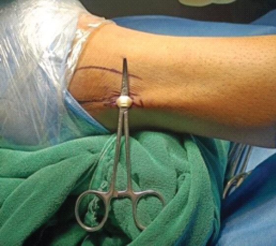
All patients fulfilled the inclusion criteria before surgery. A full-thickness PLT autograft was harvested from the ipsilateral limb. A longitudinal skin incision of approximately 1.5–2 cm was made 2–3 cm proximal and 1 cm posterior to the posterior prominence of the lateral malleolus to optimize exposure while minimizing soft-tissue trauma and risk to neurovascular structures (Fig. 1).
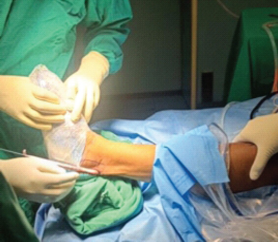
The sural nerve was carefully identified and protected throughout the dissection. The PLT was isolated, sharply transected distally, and harvested proximally using a closed tendon stripper (Fig. 2). The graft was then prepared on the back table and pre-tensioned. Depending on the native tendon diameter, it was folded into a triple-strand configuration, aiming for a final composite graft diameter of at least 8 mm. Graft dimensions were checked to ensure compatibility with the planned femoral and tibial tunnels.
Standard arthroscopic single-bundle ACL reconstruction was performed using established tunnel placement and fixation techniques. Graft fixation was achieved with appropriate devices according to surgeon preference; intra-articular tension and range of motion were verified at the end of the procedure.
Post-operative protocol and rehabilitation
A standardized post-operative protocol was followed for all patients. Immediate post-operative care included limb elevation, cryotherapy, and analgesia as needed. Early quadriceps activation and knee range-of-motion exercises were initiated as per standard ACL rehabilitation protocols, with protected weight-bearing using crutches initially and progression to full weight-bearing based on pain tolerance and knee stability.
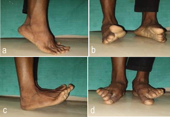
Specific ankle rehabilitation included active and passive range-of-motion exercises for plantarflexion, dorsiflexion, inversion, and eversion as pain allowed. Strengthening exercises for ankle evertors and inverters were gradually introduced. Final ankle movements and strength were evaluated at follow-up visits, with representative movements at 6 months, as shown in Fig. 3.
Outcome measures
Donor-site morbidity was assessed using the Kitaoka Ankle-Hindfoot Scale, which allocates up to 40 points for pain, 50 for function, and 10 for alignment, for a total score of 100. Scores were categorized as: Excellent: ≥90, Good: 75–89, Fair: 60–74, and Poor: <60.
The scale was administered preoperatively and at 14 days, 4 weeks (30 days), 12 weeks (90 days), and 6 months (180 days) postoperatively for the donor ankle.
Statistical analysis
Data were analyzed using the Statistical Package for the Social Sciences version 22.0. Continuous variables were expressed as mean ± standard deviation. Paired t-tests were used to compare pre-operative and post-operative Kitaoka scores for pain, function, alignment, and total score at final follow-up. Repeated-measures analysis and paired comparisons were performed between early post-operative time points (14 days, 30 days, and 90 days) and 180 days. P < 0.05 was considered statistically significant.
Results
Demographic profile
The mean age of the study population was 29.95 years. The mean interval between injury and surgical intervention was 4.4 months. Males constituted approximately 75.6% of the cohort. All patients had normal donor ankle function preoperatively, with a mean Kitaoka Ankle-Hindfoot Score of 100.
Temporal variation in total Kitaoka score
There was a transient decline in donor ankle function in the early post-operative period, followed by progressive improvement (Table 1). The mean total Kitaoka score decreased from 100 preoperatively to 71.42 ± 6.92 at 14 days, indicating early post-operative morbidity.
Variation in Kitaoka ankle hindfoot scale
| Total | Max | Min | Mean | Standard deviation |
|---|---|---|---|---|
| Pre-operative | 100 | 100 | 100 | 0 |
| 14 Days | 78 | 61 | 71.42 | 6.92 |
| 30 Days | 97 | 85 | 92.95 | 5.19 |
| 90 Days | 100 | 88 | 97.86 | 4.63 |
| 180 Days | 100 | 88 | 99.11 | 3.81 |
Comparison of ankle hindfoot scale during follow-up
| Scale | Follow-up period | Mean | Standard deviation | t-test | P-value |
|---|---|---|---|---|---|
| Pain | Pre-operative | 40 | 0 | 1.131 | 0.160 |
| 180 Days | 38.88 | 3.17 | |||
| Function | Pre-operative | 50 | 0 | 1.89 | 0.065 |
| 180 Days | 49.7 | 0.63 | |||
| Alignment | Pre-operative | 10 | 0 | — | — |
| 180 Days | 10 | 0 | |||
| Total | Pre-operative | 100 | 0 | 1.902 | 0.064 |
| 180 Days | 99.11 | 3.81 |
Subsequently, scores improved steadily to 92.95 ± 5.19 at 30 days and 97.86 ± 4.63 at 90 days, reaching 99.11 ± 3.81 at 180 days (Graph 1). The differences in total scores between 14 days and 180 days, and between 30 days and 180 days, were highly significant (P < 0.0001 for both comparisons), confirming substantial recovery of donor ankle function over time (Table 3).

Variation in total ankle hindfoot scale during follow-up
| Follow-up period | Mean ankle hindfoot score | Standard deviation | Paired t-test | P-value |
|---|---|---|---|---|
| Pre-operative | 100 | 0 | 1.902 | 0.064 |
| 180 Days | 99.11 | 3.81 | ||
| 14 Days | 71.42 | 6.92 | 29.933 | 0.0001 |
| 180 Days | 99.11 | 3.81 | ||
| 30 Days | 92.95 | 5.19 | 9.035 | 0.0001 |
| 180 Days | 99.11 | 3.81 | ||
| 90 Days | 97.86 | 4.63 | 2.527 | 0.15 |
| 180 Days | 99.11 | 3.81 |
Pain, function, and alignment components
Component-wise analysis of the Kitaoka scale is summarized in Table 2.
Pain: The mean pain score decreased marginally from 40 preoperatively to 38.88 ± 3.17 at 180 days (P = 0.160), a difference that was not statistically significant. Function: The mean function score changed from 50 preoperatively to 49.74 ± 0.63 at 180 days (P = 0.065), again without statistical significance. Alignment: Alignment remained unchanged at 10 preoperatively and at 6 months for all patients.
The overall mean total score decreased slightly from 100 to 99.11 ± 3.81 at 180 days (P = 0.064), indicating that donor ankle function was effectively preserved without clinically meaningful long-term compromise.
Paired comparisons over time
Paired analysis of total Kitaoka scores revealed: Pre-operative versus 180 days: 100 versus 99.11 ± 3.81 (P = 0.064; not significant), 14 days versus 180 days: 71.42 ± 6.92 versus 99.11 ± 3.81 (P < 0.0001; significant), 30 days versus 180 days: 92.95 ± 5.19 versus 99.11 ± 3.81 (P < 0.0001; significant), and 90 days versus 180 days: 97.86 ± 4.63 versus 99.11 ± 3.81 (P = 0.15; not significant).
Categorical outcomes
Kitaoka outcome categories at each follow-up are presented in Table 4. At 14 days, 57.8% of patients had good scores and 42.2% had fair scores, reflecting early morbidity but no poor outcomes. 30 days, 71.1% had excellent and 28.9% good outcomes. At 90 days, 82.2% of patients achieved excellent scores and 17.8% remained good. At the final 6-month follow-up, 42 patients (93.3%) had excellent and 3 (6.7%) had good donor ankle function, with no fair or poor results.
Kitaoka ankle hindfoot score outcome on follow-up
| Outcome (100) | 14 Days | 30 Days | 90 Days | 180 Days |
|---|---|---|---|---|
| Excellent (90–100) | 0 | 32 | 37 | 42 |
| Good (75–89) | 26 | 13 | 8 | 3 |
| Fair (60–74) | 19 | 0 | 0 | 0 |
| Poor (<60) | 0 | 0 | 0 | 0 |
Clinical examination and complications
On clinical examination at final follow-up, patients demonstrated excellent ankle range of motion and strength in plantarflexion, dorsiflexion, inversion, and eversion (Fig. 3). No patient developed ankle instability, significant gait abnormality, or functional limitation affecting daily activities or return to work. There were no neurovascular complications related to graft harvest.
One superficial wound infection (approximately 2% of cases) at the harvest site was noted, which responded to local care and antibiotics without residual sequelae.
Discussion
Graft selection in ACL reconstruction remains an area of active debate, with several autograft and allograft options available. Commonly utilized grafts include BPTB, hamstring tendon, quadriceps tendon, PLT, synthetic grafts, and allografts. Each graft option offers distinct biomechanical properties and donor-site profiles that influence surgical choice and post-operative outcomes.
Historically, the BPTB graft has been regarded as the gold standard due to its excellent biomechanical strength and bone-to-bone healing characteristics. However, its use is frequently associated with donor-site morbidity, including anterior knee pain, kneeling discomfort, patellar tendon shortening, patellar fracture, chondromalacia patellae, patellar tendon rupture, and persistent quadriceps weakness. These complications may significantly affect patient satisfaction and functional recovery. Hamstring tendon grafts, another widely used option, are generally associated with lower rates of anterior knee pain; however, they are not without drawbacks. Harvest of the semitendinosus and gracilis tendons may result in medial thigh muscle atrophy, hamstring weakness, and sensory disturbances due to injury to the infrapatellar branch of the saphenous nerve. Such complications may manifest as long-term hypoesthesia or paresthesia and may negatively influence patient-reported outcomes.
In response to these limitations, the PLT has emerged as a promising alternative autograft for ACL reconstruction. The PLT provides adequate graft diameter and tensile strength while potentially minimizing donor-site morbidity around the knee. In the present study, the PLT was selected as the graft of choice in an effort to avoid the complications associated with more traditional graft sources while maintaining favorable functional outcomes.
Donor-site functional assessment in our cohort was performed using the Kitaoka Ankle–Hindfoot Score, a validated scoring system that evaluates pain, functional activity, and alignment of the ankle joint. At 6 months postoperatively, the changes observed in pain (from 40 ± 0.0 to 38.88 ± 3.17), function (from 50 ± 0.0 to 49.74 ± 0.63), and alignment (no change) were minimal and statistically insignificant. The overall mean score decreased slightly from 100.0 ± 0.0 to 99.11 ± 3.81 (P = 0.064), indicating that donor ankle function remained largely preserved following PLT harvest.
These findings are consistent with previously published literature demonstrating minimal functional compromise of the donor ankle after PLT harvesting. Hackla et al. reported a small, statistically insignificant reduction in American Orthopedic Foot and Ankle Society (AOFAS) scores from 100.0 to 96.0 at 6 months postoperatively (P = 0.06) [6]. Similarly, Sharma et al. observed a mean post-operative AOFAS score of 94.5 ± 1.5, while Cao et al. reported a mean score of 96.3 (range 84–100) with no significant difference compared with the contralateral ankle (P > 0.05) [7,8]. Kumar et al., in a study involving 100 patients, reported a mean score of 97.87 ± 3.21, further supporting the functional safety of PLT autograft harvesting [9].
Additional studies have corroborated these favorable outcomes. Rhatomy et al. reported a high mean AOFAS score of 97.3 ± 4.2 following PLT graft harvest [10]. Angthong et al., in a biomechanical and clinical evaluation, observed no statistically significant difference between pre-operative (97.7 ± 1.1) and post-operative scores (95.4 ± 12) at a mean follow-up of 13 months (P = 0.09) [5].
Longitudinal studies have also demonstrated sustained preservation of ankle function. Sahu et al. reported progressive improvement in AOFAS scores between 6 weeks, 3 months, and 6 months postoperatively (P = 0.001) [11]. Bi et al. demonstrated durable ankle function over a 3-year follow-up period, with mean AOFAS scores of 98.4 preoperatively, 91.6 at 1 year, 96.2 at 2 years, and 96.8 at 3 years [12]. Similarly, Kerimoglu et al. noted only minor complaints in 2 of 29 patients following PLT harvest, without any long-term functional deficits[13]. Khajotia et al. further confirmed preservation of ankle function in nearly all cases, supported by objective testing of foot evertor strength[14].
Other studies have reported comparable results. Duan En Trang et al. documented no significant donor-site complaints or neurological deficits following PLT harvest [15]. In a comparative analysis of 130 patients, Keyhani et al. found no significant differences in ankle function between patients undergoing PLT harvest and those with anatomically intact ankles [16].
Gok et al., in a comparative evaluation, reported slightly lower AOFAS scores in the donor ankle (93.46 ± 3.8) compared with the contralateral ankle (97 ± 1.8) [17]. Similarly, the mean foot and ankle disability index (FADI) score at the donor site was 93.48 ± 4.6 compared with 98.19 ± 2.1 on the unaffected side. Although these differences were statistically significant, they did not translate into clinically meaningful impairment. Importantly, no donor-site pain, ankle weakness, neurological deficits, or limitations in return to sporting activity were observed.
Comparative graft studies further support the safety of PLT harvest. Saeed et al. reported a higher incidence of donor-site morbidity in patients undergoing hamstring tendon harvest compared with those in whom the PLT was used [18]. Agarwal et al. similarly demonstrated no significant differences in post-operative ankle function scores between the two cohorts [19]. In one of the largest reported series involving 439 patients, Hossain et al. documented excellent post-operative ankle function with mean AOFAS and FADI scores of 97.63 ± 3.20 and 98.46 ± 2.31, respectively, indicating minimal functional compromise following PLT harvest [20].
In our cohort, no patients demonstrated poor functional outcomes at the donor ankle. At 2 weeks postoperatively, 57.8% of patients had good function while 42.2% had fair outcomes. By 1 month, 71.1% achieved excellent scores. This proportion increased to 82.2% at 3 months and further improved to 93.3% by 6 months, with the remaining patients maintaining good functional status. These progressive improvements suggest that any early post-operative functional limitations are transient and resolve with rehabilitation.
Collectively, the findings from the present study and previously published literature support the use of the PLT as a reliable autograft option for ACL reconstruction. Donor-site morbidity appears minimal, and ankle function remains well preserved over time. Biomechanically, the preserved function may be explained by compensatory activity of the peroneus brevis muscle and the intrinsic adaptability of ankle biomechanics following tendon harvest. Consequently, PLT autograft represents a viable and safe alternative graft choice in ACL reconstruction, particularly in patients where avoidance of knee donor-site morbidity is desirable.
Conclusion
Harvesting the PLT for ACL reconstruction results in minimal and reversible donor-site morbidity, with donor ankle function returning to near normal levels by 6 months. PLT autograft is a safe and mechanically reliable graft choice with preservation of ankle biomechanics as reflected by sustained Kitaoka Ankle-Hindfoot scores.
Clinical Message
Surgeons can confidently consider the PLT as an ACL graft source, as donor ankle morbidity is transient, clinically insignificant, and largely resolved within 6 months, with preservation of ankle stability and function.
Conflict of Interest:
Nil
Source of Support:
Nil
Consent:
The authors confirm that informed consent was obtained from the patient for publication of this article
References
- Adam F, Pape D, Schiel K, Steimer O, Kohn D, Rupp S. Biomechanical properties of patellar and hamstring graft tibial fixation techniques in anterior cruciate ligament reconstruction: Experimental study with roentgen stereometric analysis. Am J Sports Med 2004;32:71-8. [Google Scholar] | [PubMed]
- Rhatomy S, Hartoko L, Setyawan R, Soekarno NR, Zainal Asikin AI, Pridianto D. Single bundle ACL reconstruction with peroneus longus tendon graft: 2-year follow-up. J Clin Orthop Trauma 2020;11 Suppl 3:S332-6. [Google Scholar] | [PubMed]
- Romanini E, D'Angelo F, De Masi S, Adriani E, Magaletti M, Lacorte E. Graft selection in arthroscopic anterior cruciate ligament reconstruction. J Orthop Traumatol 2010;11:211-9. [Google Scholar] | [PubMed]
- Rhatomy S, Asikin AI, Wardani AE, Rukmoyo T, Lumban-Gaol I, Budhiparama NC. peroneus longus autograft can be recommended as a superior graft to hamstring tendon in single-bundle ACL reconstruction. Knee Surg Sports Traumatol Arthrosc 2019;27:3552-9. [Google Scholar] | [PubMed]
- Angthong C, Chernchujit B, Apivatgaroon A, Chaijenkit K, Nualon P, Suchao-in K. The anterior cruciate ligament reconstruction with the peroneus longus tendon: A biomechanical and clinical evaluation of the donor ankle morbidity. J Med Assoc Thai 2015;98:555-60. [Google Scholar] | [PubMed]
- Hackla S, Sohi S, Bora M, Singh S. Ipsilateral peroneus longus tendon graft in failed ACL reconstruction: Our experience. Int J Orthop 2021;7:10-2. [Google Scholar] | [PubMed]
- Sharma D, Agarwal A, Shah K, Shah R, Shah H. Peroneus longus: Most promising autograft for arthroscopic ACL reconstruction [Internet. Indian J Orthop Surg 2019. [cited 2026 Aug 15];5(3):172-175. [Google Scholar] | [PubMed]
- Cao HB, Liang J, Xin JY. Treatment of anterior cruciate ligament injury with peroneus longus tendon. Zhonghua Yi Xue Za Zhi 2012;92:2460-2. [Google Scholar] | [PubMed]
- Kumar R, Singh B, Gautam A. Single bundle ACL reconstruction with peroneus longus tendon autograft: A short-term study. Eur J Mol Clin Med 2021;7:5460-6. [Google Scholar] | [PubMed]
- Quang VP. Anatomical and mechanical feature of peroneus longus tendon-applying ACL reconstruction. Ho Chi Minh Med Univ 2017. [Google Scholar] | [PubMed]
- Sahu MM, Pragassame AS, Mohanty P, Sahoo PK. Evaluation of donor ankle function after harvesting peroneus longus tendon graft for anterior cruciate ligament reconstruction: A prospective cohort study. J Clin Diagn Res 2021;15:YC13-7. [Google Scholar] | [PubMed]
- Bi M, Zhao C, Zhang Q, Cao L, Chen X, Kong M. All-inside anterior cruciate ligament reconstruction using an anterior half of the peroneus longus tendon autograft. Orthop J Sports Med 2021;9:2325967121991226. [Google Scholar] | [PubMed]
- Kerimoğlu S, Aynacı O, Saraçoğlu M, Aydin H, Turhan AU. Peroneus longus tendonu ile ön çapraz bağ rekonstrüksiyonu [Anterior cruciate ligament reconstruction with the peroneus longus tendon. Acta Orthop Traumatol Turc 2008;42:38-43. [Google Scholar] | [PubMed]
- Khajotia BL, Chauhan S, Sethia R, Chopra BL. Functional outcome of arthroscopic reconstruction of anterior cruciate ligament tear using peroneus longus tendon autograft. Int J Res Orthop 2018;4:898-903. [Google Scholar] | [PubMed]
- Trung DT, Le Manh S, Thanh LN, Dinh TC, Dinh TC. Preliminary result of arthroscopic anterior cruciate ligament reconstruction using anterior half of peroneus longus tendon autograft. Open Access Maced J Med Sci 2019;7:4351-6. [Google Scholar] | [PubMed]
- Keyhani S, Qoreishi M, Mousavi Ronaghi H, Soleymanha M. Peroneus longus tendon autograft versus hamstring tendon autograft in anterior cruciate ligament reconstruction: A comparative study with a mean follow-up of two years. Arch Bone Jt Surg 2022;10:695-701. [Google Scholar] | [PubMed]
- Gök B, Kanar M, Tutak Y. Peroneus longus vs hamstring tendon autografts in ACL reconstruction: A comparative study of 106 patients' outcomes. Med Sci Monit 2024;30:e945626. [Google Scholar] | [PubMed]
- Saeed UB, Ramzan A, Anwar M, Mehmood T, Tariq H, Yasin A. Earlier return to sports, reduced donor-site morbidity with doubled peroneus longus versus quadrupled hamstring tendon autograft in ACL reconstruction. JBJS Open Access 2023;8:e23.00051. [Google Scholar] | [PubMed]
- Agarwal A, Singh S, Singh A, Tewari P. Comparison of functional outcomes of an anterior cruciate ligament (ACL) reconstruction using a peroneus longus graft as an alternative to the hamstring tendon graft. Cureus 2023;15:e37273. [Google Scholar] | [PubMed]
- Hossain GM, Islam MS, Rahman Khan MM, Rafiqul Islam M, Rahman SM, Jahan MS. A prospective study of arthroscopic primary ACL reconstruction with ipsilateral peroneus longus tendon graft: Experience of 439 cases. Medicine (Baltimore) 2023;102:e32943. [Google Scholar] | [PubMed]
© 2026 Journal of Orthopaedic Case Reports - Published by Indian Orthopaedic Research Group
About the Authors
-

Dr. Pradeep Patel
-

Dr. Jeetendra Singh Lodhi
-

Dr. Vishnu Jayaprakasan
-

Dr. Salman Khan
-

Dr. H S Varma
Related Articles in Journal of Orthopaedic Case Reports
 November 1, 2024 Randomized Controlled Study Comparing Hamstring Graft and Peroneus Longus Tendon Graft in Arthroscopic ACL Reconstruction
 August 1, 2026 Comparative Outcomes of Hamstring Tendon versus Peroneus Longus Tendon Autograft in Arthroscopic Single-Bundle Anterior Cruciate Ligament Reconstruction: A Prospective Comparative Study
 July 1, 2026 Clinical and Donor-site Outcomes of Full-thickness Peroneus Longus Autograft in Arthroscopic Anterior Cruciate Ligament Reconstruction: A Prospective Indian Cohort Study
 May 1, 2026 Comparative Evaluation of Peroneus Longus Tendon and Hamstring Tendon Autografts in Primary Anterior Cruciate Ligament Reconstruction: A Randomized Controlled Trial