


Introduction
Fractures of acetabulum are high-energy trauma fractures with incidences more in the middle-aged men [1,2]. Acetabular fractures often result from high-impact falls or motor vehicle accidents that transmit force most commonly from an impact to the greater trochanter or the flexed knee. As industrialization and urbanization are progressing, with rapid increase in traffic, incidence of high-energy trauma is increasing as well. With efforts of hard-working surgeons and imaging assistance, management of these acetabular fractures had witnessed a sea of change [2]. Operative treatment for any anatomical disruption of acetabulum is complex due to the complex three-dimensional (3D) anatomy, limitations of the surgical approaches due to important structures surrounding the acetabulum making access difficult and extensive dissection of the large muscles around the hip [3,4,5].
Until the 1960s, the vast majority of patients with acetabular fractures were treated conservatively which involved bed rest, compression devices, extensions and/or slings, traction, and closed reduction techniques. Non-operative management of displaced acetabular fractures has been associated with significant complications including chronic pain and stiffness of hip due to arthritic changes, backache, impaired gait, pelvic obliquity, and neurological deficit, which leads to poor functional outcome.
In the early 1960s, the management of acetabular fractures was revolutionized by the work of Judet and Letournel [6]. They recognized that the principles applied to the treatment of displaced articular fractures (anatomical reduction, stable fixation, and early mobilization) should also be applied to the acetabulum. Their work led to the development of new surgical approaches and a classification system which has been tested in mean time and is currently used all over the world.
It has become clear over the years that these injuries should be managed in a multi-disciplinary fashion to minimize early mortality secondary to hemodynamic instability. In patients with multiple injuries who are in an unstable or in poor physiological states, damage control orthopedics is the current treatment of choice [7]. The widespread use of the Advanced Trauma Life Support (ATLS) protocol and the advances made in intensive care medicine contributed to a reduction in mortality rates [8]. A thorough knowledge of the anatomical structures contributing to pelvic stability and the source of the bleeding is essential for the assessment and treatment of these injuries [9].
Only 10% of the pelvic disruptions involve acetabulum. Most acetabular fractures, approximately one-third, involve the posterior wall. Most posterior wall fractures are comminuted and also associated with an impaction injury of the articular surface into the underlying cancellous bone [2,6]. The soft tissues are frequently detached from fragments at the time of injury or during the surgery [10]. Therefore, surgeons should make every effort to obtain a stable congruous hip joint with complete union of the fragments during the primary surgery as quality of reduction is the main determinant for risk of post-traumatic arthritis and goal should be a reduction within 1 mm [11,12,13,14,15].
With improvements in the methods of restoration of physiological states of polytrauma patients, it helped surgeons to experiment with newer approaches for the stable fixation of acetabular fractures. The advent of computed tomography (CT) and development of 3D reconstructions has allowed for profound advances in the understanding of complex acetabular fractures and has helped the surgeons to plan the management better [16]. In addition, modified, less invasive approaches have been developed. Newer generations of implants have been introduced as well as percutaneous stabilization techniques and image guided surgery for the reconstruction of acetabulum. Acetabular fractures are generally associated with other injuries of the pelvis, lower limbs and head which may influence treatment options, surgical approach, hospital stay, and overall clinical outcomes [17]. Patient age, fracture stability, the presence of co morbidities, and osteoporosis combined with surgeon experience also influence treatment options and outcome as well [18]. Due to the increase in interest in managing such patients surgically, there is a need to evaluate the functional outcome of the patients [5,9,15,19,20].
Although surgical fixation is the established treatment for displaced posterior wall acetabular fractures, functional outcomes remain variable because of differences in fracture complexity, associated injuries, timing of surgery, and quality of reduction. Prospective evidence evaluating the influence of these factors on post-operative recovery remains limited. Therefore, this study aimed to assess functional outcomes following open reduction and internal fixation (ORIF) using the modified Harris Hip Score (HHS) and to determine whether age, interval between injury and surgery, quality of reduction, associated posterior column fracture, and posterior hip dislocation influence the final clinical outcome in patients treated at a high-volume tertiary trauma center [12,13,14,21].
Materials and Methods
This single-center prospective observational case series was conducted in the Department of Orthopedics at a tertiary trauma referral center from July 2019 to August 2021. Eligible patients with posterior wall acetabular fractures, with or without associated posterior column fractures and posterior hip dislocation, were enrolled and followed prospectively after surgical treatment. No formal a priori sample-size calculation was performed because all eligible patients presenting during the predefined 2-year study period were considered for inclusion. The final study sample comprised 20 patients. Approval was obtained from the Institutional Ethics Committee on January 09, 2019 (IEC No. 804/15.0.19) before commencement of the study. Written informed consent was obtained from all participants.
The inclusion criteria consisted of the following:
-
Age group 18–70 years of either sex.
-
Fractures duration <21 days after hemodynamic stabilization.
-
Closed fracture of posterior wall acetabulum with or without associated posterior column fractures and with or without posterior dislocation of hip.
-
Patients who give informed consent and willing for follow-up.
The exclusion criteria consisted of the following:
-
Compound fractures of pelvis.
-
Patients <18 years of age and more than 70 year.
-
Associated co morbid conditions, history of suffering from myocardial infarction <1year, psychiatric illness, head injury.
-
Uncontrolled diabetes mellitus, hypertension, patients with clinically detectable focus of active infection
-
Pathological fracture.
-
Peri prosthetic fractures.
-
Associated major visceral injuries
-
Acetabular fractures that were more than 3 weeks old were excluded from our study.
All the cases were initially investigated with radiograph-anteroposterior and oblique Judet views of the pelvis.
In fractures with associated hip dislocation, the dislocation was reduced under genetic algorithms and dynamic stress fluoroscopic examination under anesthesia was done to determine hip joint stability followed by skeletal pin traction till surgical fixation.
After primary treatment and initial stabilization according to ATLS protocol, patients were admitted under the accident and trauma ward.
CT scan of pelvis with 3D reconstruction was done in all cases for better understanding of the fracture pattern and pre-operative planning. The fracture lines were marked out on a pelvic bone model which was helpful in surgery [15,19].
Radiological assessment from radiographs (AP and Judet views) and CT scan were used to
-
Analyze the fracture pattern using Letournel and Judet classification.
-
Evaluate the extent of fracture displacement and the degree of comminution.
Written informed consent was obtained from all the patients. Treatment modalities and complications were discussed with the patients. Anesthetic fitness was obtained.
All patients were treated with ORIF with reconstruction plate and screws.
Surgical technique
Kocher-Langenbeck approach [3] was used in all cases for posterior wall and posterior column fracture. Patient was placed in a floppy lateral position with the affected hip facing up and slightly tilting towards the floor. Extremity is maintained with extension of the hip and flexion at the knee at all times to avoid stretching of the sciatic nerve during operative procedure. The injured side was adequately scrubbed and painted with 10% povidone-iodine solution after which the limb was draped free [9,20,22,23].
Matta’s Quadrangular clamp of various sizes and Picador ball spike pusher were very important instruments in acetabular surgery. Various reduction clamps were available to facilitate reduction and holding. Schanz screws were used as joy sticks that aided in the fracture reduction in some cases.
Osteochondral free fragments and marginally impacted fragments were sequentially reduced (assuming there is sufficient attached cancellous bone to allow fragment healing), usually using the femoral head as a template. First, the marginal impaction was elevated; leaving a void underneath from the impacted cancellous bone. This void was filled with autogenous cancellous bone either from the greater trochanter or the posterior iliac crest. After reduction of the wall fragments, provisional fixation with Kirschner wires was performed [24].
In posterior wall fractures, reduction was fixed with lag screws whenever possible using 4 mm cancellous screws or 3.5 mm cortical screw with or without washer. 3.5 mm reconstruction plates were used as neutralization plates which provide a “tension buttress” effect when secured to the posterior wall. The plate was bent to approximate the shape, and then secured to the ischial tuberosity and to the ilium superior to the acetabulum. Joint penetration of the screw was carefully watched for. Passive flexion, extension, and rotation of the hip were done after fixation to ensure a smooth range of movement [23,24].
In posterior column fractures, the distal fragment was displaced medially and medially rotated. At times, reduction was simply achieved by lifting the distal fragment with bone hook. In some cases, Matta’s reduction clamps were used for maintaining reduction at the sciatic notch and the iliac wing or the Steinman pin at the iliac wing, if inserted previously. In other cases, a Farabeuf clamp with two cortical screws helped in dislodging the fracture and reduction of the fracture. Definitive fixation was followed with a reconstruction plate after provisional fixation. The quality of reduction of fragments was assessed on operating table with image intensifier. Posterior capsule of hip was repaired. Short external rotators were repaired. Then, soft-tissue closure was done in layers. Skin closure was made after securing a removable no-16 drain in situ. Sterile dressing was applied over the wound. Patient was shifted to recovery room and subsequently to the ward with proper care.
Postoperatively, blood was transfused when needed and parenteral antibiotics according to the standard institutional protocol were administered for 5 days. Drains were removed at 24–48 h. Deep vein thrombosis prophylaxis in the form of low-molecular-weight heparin was given for 6 weeks in selected patients (according to hematologist’s opinion) and graduated compression stockings were used in the presence of pedal edema. Indomethacin prophylaxis to prevent heterotrophic ossification was not usually given.
24–48 h postoperatively, anteroposterior and 45° oblique (Judet) radiographs were taken. The reduction of the fracture was evaluated by measuring the residual post-operative displacements on the three plain radiographs. For each of these radiographs, the maximum displacement seen at any to the normal radiographic lines of the acetabulum or the innominate bone was recorded in milli-meters, and the highest of the three values was used to grade the reduction according to one of three categories:
Post-operative fracture reduction was graded according to Matta’s radiographic criteria [2] as follows:
-
Anatomical: 0–1 mm residual displacement
-
Imperfect: 2–3 mm residual displacement
-
Poor: >3 mm residual displacement
Suture removal was done on post-operative day 10–12. Passive mobilization through intermittent, pain-free quadriceps, and hip- and knee-flexion exercises was started on post-operative day 2. Gradually, the patient was shifted to toe touch weight bearing after 6 weeks with single crutch/walker. Full weight-bearing was permitted following radiological union of the fracture.
Patients were followed up at 6 weeks, 12 weeks, 6 months, 12 months, and 18 months post-injury. Functional outcome was assessed at 12 months to 18 months follow-up. The functional outcome was assessed by modified HHS. Follow-up radiographs were also assessed for the union of fracture, failure of fixation, aseptic necrosis of femoral head, presence of heterotopic calcification, sclerosis, diminution of joint space, spur formation on the femoral head, congruence of the femoral head within the acetabulum, and other signs of degenerative changes. Patients were also assessed for the amount and cause of pain (if present), ROM of hip and knee, wound status, local signs of infection, and walking ability [14,25].
Data were analyzed using IBM Statistical Package for the Social Sciences (SPSS) Statistics, version 21.0. Continuous variables were summarized using the mean and range, while categorical variables were presented as frequencies and percentages. Functional outcomes assessed using the modified HHS were categorized as excellent, good, fair, or poor. Associations between functional outcome and age, interval between injury and surgery, quality of reduction, associated posterior column fracture, and posterior hip dislocation were assessed using Pearson’s Chi-square test. A two-sided P < 0.05 was considered statistically significant.
Results
Our study was done to evaluate the functional outcome of surgical management of properly indicated posterior wall acetabular fractures with or without posterior hip dislocation at a short-term follow-up. This prospective study was undertaken in a tertiary care hospital. Ethical clearance was obtained from the Institutional Review Board. The study was conducted between July 2019 and August 2021. During this period, 193 cases of pelvic injury were reported in our institute, out of which 54 cases had acetabular fractures. However, 20 cases of posterior wall acetabular fractures satisfying the inclusion criteria were included in our study.
The observations were made and the results were statistically analyzed using SPSS software (version 21.0). Significance of association of all the predictive factors with the clinical outcome was done by means of Pearson Chi-square test. P < 0.05 was considered to be statistically significant.
The basic demographic data are summarized in the Table 1.
Demographic information of patients
| S. No. | Age | Sex | Diagnosis | Concurrent injuries | Mode of injury | Associated hip dislocation | Side affected | Time delay for surgery | Surgical time | Final radiological score | Final Harris Hip Score |
|---|---|---|---|---|---|---|---|---|---|---|---|
| 1 | 32 | M | Fracture posterior wall acetabulum | Fracture lateral condyle right tibia | RTA | Posterior dislocation hip | Right | 3 days | 75 min | Anatomical | Excellent |
| 2 | 36 | M | Fracture posterior wall acetabulum | Nil | RTA | Posterior dislocation hip | Left | 3 days | 90 min | Anatomical | Good |
| 3 | 45 | M | Fracture posterior wall acetabulum | Nil | RTA | No dislocation | Right | 3 days | 75 min | Anatomical | Good |
| 4 | 48 | M | Fracture posterior wall acetabulum | Fracture right tibia and fibula | RTA | No dislocation | Right | 10 days | 90 min | Satisfactory | Fair |
| 5 | 43 | M | Fracture posterior wall acetabulum | Nil | Fall from height | No dislocation | Right | 10 days | 120 min | Anatomical | Good |
| 6 | 50 | M | Fracture posterior wall acetabulum | Fracture posterior columnar acetabulum | RTA | Posterior dislocation hip | Right | 17 days | 120 min | Poor | Poor |
| 7 | 25 | M | Fracture posterior wall acetabulum | Fracture right humerus | RTA | Posterior dislocation hip | Right | 7 days | 90 Min | Anatomical | Good |
| 8 | 60 | M | Fracture posterior wall acetabulum | Nil | RTA | Posterior dislocation hip | Left | 17 days | 90 Min | Anatomical | Poor |
| 9 | 45 | M | Fracture posterior wall acetabulum | Pelvic ring injury | RTA | Posterior dislocation hip | Right | 10 days | 120 Min | Anatomical | Good |
| 10 | 44 | M | Fracture posterior wall acetabulum | Nil | Fall from height | No dislocation | Right | 3 days | 90 Min | Anatomical | Excellent |
| 11 | 54 | M | Fracture posterior wall acetabulum | Nil | RTA | No dislocation | Left | 3 days | 75 Min | Anatomical | Excellent |
| 12 | 45 | M | Fracture posterior wall acetabulum | Nil | RTA | Posterior dislocation hip | Right | 5 days | 120 Min | Satisfactory | Good |
| 13 | 18 | F | Fracture posterior wall acetabulum | Head injury and Fracture posterior columnar acetabulum | RTA | Posterior dislocation hip | Right | 17 days | 120 min | Satisfactory | Good |
| 14 | 25 | M | Fracture posterior wall acetabulum | Nil | RTA | Posterior dislocation hip | Right | 3 days | 90 min | Anatomical | Good |
| 15 | 35 | M | Fracture posterior wall acetabulum | Fracture posterior column acetabulum with fracture tibia and fibula | RTA | Posterior dislocation hip | Left | 10 days | 90 min | Anatomical | Good |
| 16 | 30 | M | Fracture posterior wall acetabulum | Fracture posterior column acetabulum | RTA | Posterior dislocation hip | Right | 15 days | 120 min | Poor | Fair |
| 17 | 52 | M | Fracture posterior wall acetabulum | Nil | fall from height | No dislocation | Right | 17 days | 120 min | Poor | Fair |
| 18 | 36 | M | Fracture posterior wall acetabulum | Fracture posterior column acetabulum | RTA | No dislocation | Right | 10 days | 120 min | Anatomical | Good |
| 19 | 45 | M | Fracture posterior wall acetabulum | Fracture posterior column acetabulum with fracture distal femur | RTA | No dislocation | Right | 17 days | 120 min | Anatomical | Good |
| 20 | 56 | F | Fracture posterior wall acetabulum | Head injury and fracture radius and ulna | RTA | No dislocation | Left | 10 days | 90 min | Satisfactory | Fair |
RTA: Road traffic accidents
Our series had patients from 18 to 60 years of age. The peak incidence was found in the fifth decade of life (40–50 years) followed by fourth decade (30–40 years). The mean age was 41.2 years.
This injury was commonly found in males compared to the females. About 90% of the cases in our series were male.
Road traffic accidents (RTAs) were the most common mode of injury contributing to about 85% of cases. Besides RTA, direct forces acting along the trochanter following fall from height was seen in three cases. Associated injuries included head injury, chest injury, lower extremity injury, upper extremity injury, and pelvic ring disruption. Injuries of the extremities (upper and lower) were the most common associated injuries. However, none of the patients in our series had associated sciatic nerve palsy or genitourinary injury which is commonly suspected in acetabular fractures.
The right side was involved in 15 patients making up for 75% of the fractures and the left was involved in five patients accounting for 25% of the fractures. None of the patients had bilateral fractures.
In our series, the distribution of fractures based on Letournel classification showed that posterior wall fractures contributed to 70% (14), whereas posterior wall with associated fractures of column contributed to 30% (6) of the cases.
To look at the quality of reduction, we used the Matta’s criteria and classified the quality of reduction into anatomical, satisfactory, and poor. We found that among the simple fractures, that is, posterior wall fractures only, anatomical reduction results were seen in 10 (71.4%) cases, satisfactory in three cases and poor in only one case. Among fractures of posterior wall associated with column fractures, anatomical reduction results were seen in 3 (50%) cases, satisfactory in one case, and poor in two cases. Considering “satisfactory” and “poor” to be sub-optimal reduction, overall, 13 (65%) cases have anatomical fracture reduction, while 7 (35%) cases have sub-optimal fracture reduction.
To evaluate the clinical outcome, the modified HHS was used. Clinical outcome was evaluated at least 1 year after the surgery. We found that 3 (15%) cases have excellent, 11 (55%) cases have good, 4 (20%) cases have fair, and 2 (10%) cases have poor functional results according to modified HHS.
We wanted to look at the factors affecting the clinical outcome in our study. Hence, we checked if the modified HHS is affected by age, interval between injury and surgery, quality of reduction, posterior wall fracture with associated posterior column fractures, and posterior hip dislocation.
Although it appears that above the age of 41 years, the functional outcome deteriorates, the correlation between age and modified HHS (clinical outcome) is statistically not significant. The interval between injury and surgery showed significant influence over final functional outcome. (Chi-square = 8.636, P = 0.035). The functional outcome decreased as the interval between injury and surgery increased (Table 2).
Interval between injury and surgery affecting clinical outcome (n=20)
| Excellent (%) | Good (%) | Fair (%) | Poor (%) | Significance | |
|---|---|---|---|---|---|
| <7 Days | 3 (37.5%) | 5 (62.5) | 0 | 0 | χ2=8.636 |
| df=3 | |||||
| >7 Days | 0 | 6 (50.0) | 4 (33.3%) | 2 (16.7) | P=0.035 |
Quality of fracture reduction had a statistically significant correlation with the functional outcome. All anatomically reduced fractures had satisfactory functional results (Chi-square = 10.609, P = 0.014) (Table 3).
Quality of reduction and clinical outcome (n=20)
| Excellent (%) | Good (%) | Fair (%) | Poor (%) | Significance | |
|---|---|---|---|---|---|
| Anatomical | 3 (23.1) | 9 (69.2) | 0 | 1 (7.7) | χ2=10.609 |
| Df=3 | |||||
| Suboptimal | 0 | 2 (28.6) | 4 (57.1) | 1 (14.3) | P=0.014 |
For cases with simple posterior wall fracture fixation, 71.4% (10 cases) had excellent to good functional outcome; for posterior wall fractures with associated posterior column fracture, 66.7% (4 cases) had good functional outcome. However, the result is not statistically significant here (Table 4).
Associated fractures affecting final clinical outcome (n=20)
| Excellent (%) | Good (%) | Fair (%) | Poor (%) | Significance | |
|---|---|---|---|---|---|
| Simple posterior wall fracture | 3 (21.4) | 7 (50.0) | 3 (21.4) | 1 (7.1) | χ2=1.926 |
| Df=3 | |||||
| Posterior wall with associated fracture | 0 | 4 (66.7) | 1 (16.7) | 1 (16.7) | P=0.588 |
Posterior dislocations were seen in 11 cases. The dislocation per se does not have any influence on the outcome but the time of reduction of dislocation has a strong influence on the viability of femoral head and, therefore, the clinical outcome [5,9].
During the post-operative period and subsequent follow-up, patients were closely observed for any possible complications. Superficial infection was noted in one case which healed well with debridement and antibiotics. Iatrogenic sciatic nerve palsy was seen in two cases which did recover after 8 months-1 year of surgery. One case of posterior wall fracture with posterior hip dislocation had avascular necrosis (AVN) femoral head with osteoarthritis. Heterotopic ossification was not noted in any cases.
Discussion
Posterior wall fractures, which are the most common of acetabular fractures, are considered easy to address due to ease of access, familiarity with the posterior approach to the hip, and the straightforward fixation of an apparently simple fracture pattern. Letournel and Matta [6] were able to get excellent to good results in only 68–82% even after achieving anatomical reductions in 94–100% of their cases. This difference in outcome has been explained by comminution of the posterior wall, osteonecrosis, marginal impaction, and injury to the femoral head. While avoiding complications, the results after surgery correlate most closely with the quality of the reduction [2,26]. However, there is no doubt that other prognostic variable are involved in addition to the variables identified in our study. Despite the relatively simple appearance of posterior wall fractures, the clinical results after surgical treatment have been variable.
Aho et al. [27], undertook a follow-up over 5 years of 20 patients who had undergone ORIF of a posterior-wall fracture using plate and screws of which 14 (70%) had good or moderate results. Moed et al. [26] reported similar favorable results for a recent large series after a mean follow-up period of 5 years, with good-to-excellent results in 89 of 100 (89%) posterior wall fractures treated by ORIF. They emphasized that long-term good to excellent results can be expected after anatomical reduction and internal fixation.
In our current series, 20 cases of posterior wall acetabular fractures were treated by ORIF with reconstruction plate and screws and reviewed over a period of 2 years. They were followed up for an average of 21 months. Age group ranged from 18 to 70 years. The average age of all cases in our series was 41.2 years. The fracture is more common in the age group of 30–49 years with total of 12 patients out of total 20 patients within that age group. The distribution of age in various studies show that posterior wall fractures occur in a wide range of age with lower limit as low as 9 years to higher limit of 90 years reported in literature [2,28,29].
The gender distribution was dominated by the male population in our study with 18 male and 2 female patients. Male predominance of 90% shows the proneness of this gender for high energy trauma. More males injured compared to females can be attributed to their higher risk-taking behavior.
Similar data with male pre-dominance of acetabular fracture is shared by many other studies [2,26,28,29].
The right side was involved in 15 (75%) and the left side was involved in 5 (25%) patients in our study. However, there is no common pattern regarding the side which is most commonly injured [2,30].
Amount of energy required to produce acetabular fractures is undoubtedly tremendous. Hence, these injuries are an obvious consequence of high velocity trauma like motor vehicle accidents. In our study, motor vehicle accident was the most common cause (85%) followed by fall from height (15%). Most researchers observed that motor vehicle accidents were the major cause of injury in their series contributing more than 70% of their cases [2,28,30,31].
Such high transactions of energy usually produce multi-system injuries. Hence, the associated injuries pose a big hurdle in the management of these cases, usually requiring a multi-disciplinary approach. In our series, the extremity injuries (30%) and chest injuries (5%) were commonly associated with acetabular fractures. Other significant associated injuries were head injuries (10%) and pelvic ring disruptions (5%). Matta [2] reported in his series of 259 acetabular fractures that 56% were associated with at least one additional injury with the most common being an injury involving an extremity. Similar associated injuries commonly involving at least one of the extremities were also reported by Petsatodis et al. [32] and Li et al. [31].
Classification system of Letournel and Judet [1,4,33] was used in our study. The reliability, reproducibility, and the interobserver agreement were optimal for this classification system. Moreover, computed tomograms of pelvis with 3D reconstruction were of good aid in the initial assessment of the fracture patterns. In our series, the elementary fractures – posterior wall (70%), and associated fractures – posterior wall and posterior column (30%) were present. Surgical approach for the management of fractures was preoperatively planned. Kocher-Langenbeck approach was used in all the cases which are a well learned, convenient approach providing optimum exposure of the fracture of posterior wall and column and allows adequate stabilization of the fracture.
Post-reduction fluoroscopic imaging status of the fracture configuration was noted. Average operative time for this approach was about 101.2 min. Operating time depends on the fracture pattern and patient body type. Direct reduction by manipulation of fragments using reduction clamps is tiresome. Moreover, contouring of the plates and fixation would require utmost patience and lot of time. Nevertheless, for the access of posterior wall and column fractures, this approach still remains the gold standard for fixation of posterior wall acetabular fractures [2,34,35].
Surgical treatment was performed as soon as the patient’s general medical condition allowed, sometimes resulting in delays in treatment. In addition, there were some delays in treatment related to transfer of the patient from a referring institution, time taken for the resolution of the contusion around the hip, adequate treatment of the associated life-threatening injuries, and time taken to achieve fitness for surgery hemodynamically. The average time from injury to surgery was 9.5 days (range, 0–21 days). We had 37.5% excellent and 62.5% good results in patients who were operated before 7 days while 50.0% good, 33.3% fair, and 16.7% poor results in patients operated between 7 and 21 days. Most of the authors agree with our observations and achieve good to excellent results when treated with in first 10–14 days. However, few authors including Zhi et al. [36] have also concluded that timing of surgery does not have much of influence if operated within 3 weeks. Brueton [37] reported that the average time to surgery for fractures with an acceptable reduction was 11 days, whereas for unacceptable reductions, the time to surgery was 17 days. Likewise, Letournel [2] reported an anatomic reduction rate of only 52% in fractures that were operated on more than 21 days from the time of injury. In our study, anatomic reduction rate is being 54%, when operated with in 1st week. As the time from injury to surgery increased, the outcome score decreased. This difference was statistically significant between those who were operated within 7 days and those who were operated on or after 7 days. This was mainly because the timing of surgery has a bearing on the surgeon’s ability to obtain an accurate reduction as the mobility of the fracture decreased with time from injury [9,14].
Radiological results were found to be anatomical to satisfactory in 85%, and poor in 15% in our study. These results are comparable to that of other standard studies conducted for posterior acetabular fractures [12,13,15,21,26,29].
In our series, quality of fracture reduction had a significant correlation with the clinical function (P = 0.014). This correlation between quality of fracture reduction and the clinical outcome is further advocated in various studies in the literature. Moed et al., suggested the accuracy of surgical reduction as assessed on post-operative CT is highly predictive of the clinical outcome [26].
Functional outcome as revealed by Harris Hip Scoring system supported surgical treatment of posterior wall acetabular fracture. In our series, excellent results were seen in cases (15%), good results in 11 cases (55%), fair result in 4 cases (20%), and poor result in 2 cases (10%) [4, 12, 21].
Among the simple fractures, excellent clinical results were seen in three cases, good results in seven cases, fair results in three cases, and poor results in one case. The pre-operative and post-operative radiographs and clinical pictures showing outcome in a case of simple posterior acetabular wall fractures are illustrated in Fig. 1a, b, c, d.
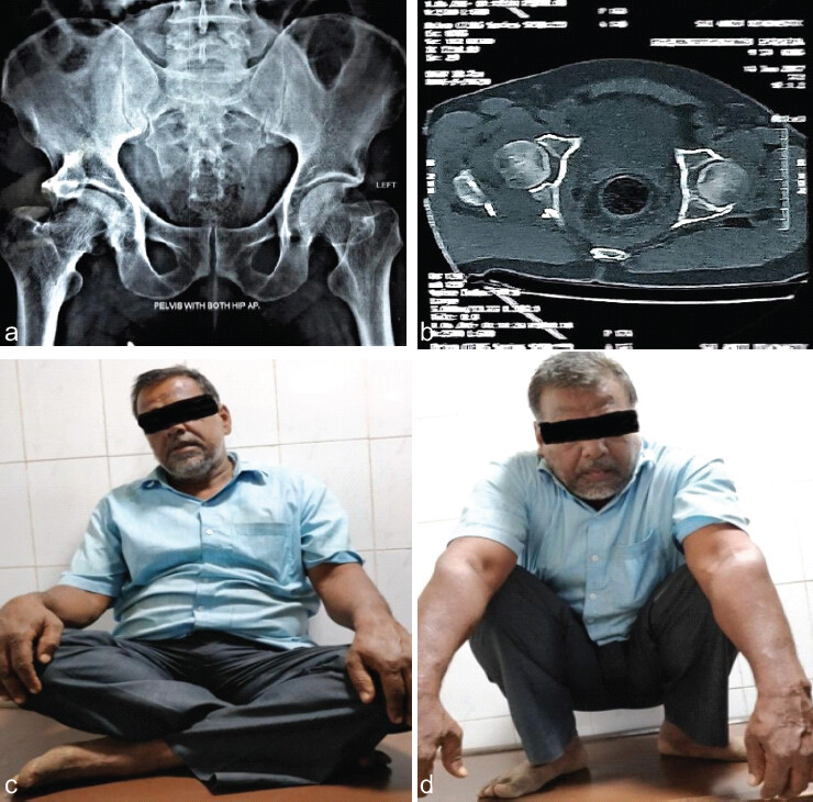
Among the associated fractures good clinical results were seen in four cases, fair in one case, and poor result in one case.
Rowe and Lowell [28] stated that “clinical and roentgenographic findings in the affected hips 1 year after injury were found to be a most reliable guide to the ultimate prognosis of the hip, since definite changes were noted at 1 year in those hips that deteriorated.”
The functional outcome in our series was comparable to those of various studies in the literature [2,10,29,35,38,39]. Excellent to good functional outcome were found in around 70–80% in these studies. The results in our study show that above the mean age of 40 years, the functional outcome deteriorates but this result was not statistically significant.
Posterior dislocations were seen in 11 cases. The dislocation per se does not have any influence on the outcome but the time of reduction of dislocation has a strong influence on the viability of femoral head and thereby the clinical outcome. Hip dislocation has detrimental effect on the eventual functional outcome; it not only compromises vascularity of the femoral head but also makes the surgical reconstruction of the acetabulum more challenging. Hip dislocation with acetabular fracture indicates a high velocity injury. Fracture comminution, articular impaction, and cartilage damage and associated soft-tissue disruptions also contribute to poor outcome in such scenario. Adequacy of surgical reduction determines the long-term outcome of surgically managed posterior hip dislocations associated with posterior wall [5,9].
Acetabular fracture [40] hence, one of the factors affecting clinical results of acetabular fractures was hip dislocation and time of reduction [36]. Although, literature says associated concurrent injuries have an adverse effect on the functional outcome [31,41], this correlation was not found to be significant in our series.
Two cases of sciatic nerve palsy were present in our series. The peroneal component was involved which recover by 6–8 months post-operative. Various studies report the rate of iatrogenic sciatic nerve palsy somewhere between 5% and 10% [31,36,42]. One case of post-traumatic AVN femoral head was found in our series. The rate of AVN of femoral head is roughly reported to be 3–6% [2]. No case of heterotopic ossification has been encountered in our study. Heterotopic ossification was reported to be as high as 37% in posterior approaches in a study by Guo et al. They also concluded that the use of the anterior approach may result in a higher risk for heterotopic ossification and the posterior approach may result in a higher risk for AVN. Based on various studies, we prescribed indomethacin to patients for 6 weeks as prophylaxis for heterotopic ossification in susceptible cases only. Although there has not been a single consensus on the use of indomethacin for prophylaxis and treatment of heterotopic ossification, early surgical resection combined with radiation and indomethacin for severe cases of heterotopic ossification after the ORIF of acetabular fractures has been an accepted widely. One case of superficial infection was observed in our series. Wound was debrided well and culture specific antibiotics were administered which resulted in healing. Literature reports the incidence of infection after operative management of acetabular fractures to be <5% in most studies [22,25,32,36,43].
The strengths of this study include its prospective design, standardized pre-operative radiological assessment, use of a uniform surgical approach and fixation technique, and assessment of both radiological reduction and functional outcome using established criteria. Patients were followed for up to 18 months, allowing assessment of early post-operative complications and functional recovery [4,25] (Fig. 2a,b,cd).
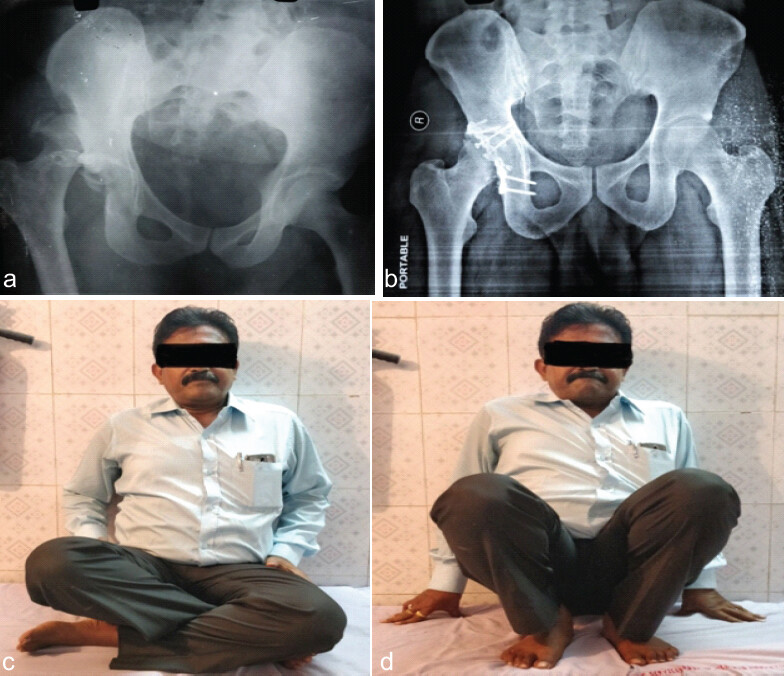
However, this study has several limitations. It was a single-center observational case series with a small sample size of 20 patients and no non-operative or comparative control group. Therefore, the findings cannot establish superiority over conservative management or determine causal relationships. The inclusion of different fracture patterns and associated injuries may also have introduced clinical heterogeneity. Furthermore, the follow-up period was insufficient to assess long-term complications such as post-traumatic osteoarthritis, AVN, and late functional deterioration. Larger multicenter comparative studies with longer follow-up are required to validate these findings [25,43].
CONCLUSION
Surgical management of posterior wall acetabular fractures with ORIF resulted in good-to-excellent functional outcomes in 70% of patients. Earlier surgical intervention and anatomical reduction were significantly associated with better modified HHSs. Associated posterior column fractures and posterior hip dislocation did not show a statistically significant association with the final functional outcome. These findings highlight the importance of timely surgery, accurate reduction, stable fixation, and structured rehabilitation in optimizing post-operative recovery. Larger multicenter studies with longer follow-up are required to confirm these findings and assess long-term complications.
Clinical Message
Management of posterior wall fractures involving the acetabulum requires systematic pre-operative assessment, prompt surgical intervention, and rigid internal fixation to achieve anatomical alignment. Fixation performed early by skilled surgical teams at tertiary trauma centers ensures improved functional results while mitigating the risk of late sequelae such as post-traumatic osteoarthritic changes and femoral head necrosis.
Conflict of Interest:
Nil
Source of Support:
Nil
Consent:
The authors confirm that informed consent was obtained from the patient for publication of this article
References
- Letournel E, Judet R Fractures of the Acetabulum New York Springer-Verlag 1993 2. [Google Scholar] | [PubMed]
- Matta JM. Fractures of the acetabulum: Accuracy of reduction and clinical results in patients managed operatively within three weeks after the injury J Bone Joint Surg Am 1996;78:1632-45. [Google Scholar] | [PubMed]
- Cutrera NJ, Pinkas D, Toro JB. Surgical approaches to the acetabulum and modifications in technique J Am Acad Orthop Surg 2015;23:592-603. [Google Scholar] | [PubMed]
- Albrektsson M, Möller M, Sundfeldt M, Wennergren D, Wolf O, Bergdahl C. Patient-reported outcome following an acetabular fracture: An observational study of 385 patients from the Swedish fracture register Acta Orthop 2024;95:695-700. [Google Scholar] | [PubMed]
- Li R, Zhao P, Guan J, Wang X, Liu L, Wu M. Combined pelvic and acetabular injuries: Clinical features and treatment strategies of a unique injury pattern J Orthop Surg Res 2023;18:415. [Google Scholar] | [PubMed]
- Judet R, Letournel E. Fractures of the acetabulum: Classification and surgical approaches for open reduction. Preliminary report J Bone Joint Surg Am 1964;46:1615-46. [Google Scholar] | [PubMed]
- Roberts CS, Pape HC, Jones AL, Malkani AL, Rodriguez JL, Giannoudis PV. Damage control orthopaedics: Evolving concepts in the treatment of patients who have sustained orthopaedic trauma Instr Course Lect 2005;54:447-62. [Google Scholar] | [PubMed]
- American College of Surgeons. Advanced Trauma Life Support for Doctors Student Course Manual 7th ed Chicago American College of Surgeons 2004 [Google Scholar] | [PubMed]
- Puchwein P, Sandersjöö G, Lindahl J, Eibinger N. Combined pelvic ring and acetabular fractures - strategies and sequence of surgery State of the art. Arch Orthop Trauma Surg 2024;144:4577-86. [Google Scholar] | [PubMed]
- Kim HT, Ahn JM, Hur JO, Lee JS, Cheon SJ. Reconstruction of acetabular posterior wall fractures Clin Orthop Surg 2011;3:114-20. [Google Scholar] | [PubMed]
- Hoge S, Chauvin BJ. Acetabular fractures In: StatPearls Treasure Island, FL StatPearls Publishing 2024 [Google Scholar] | [PubMed]
- Lin SY, Ho CJ, Liu WC, Chen JK, Tu HP, Lee TC, et al. Predicting the poor clinical and radiographic outcomes after the anatomical reduction and internal fixation of posterior wall acetabular fractures: A retrospective analysis J Clin Med 2022;11:3244. [Google Scholar] | [PubMed]
- Kavak M, Çeliksöz AH, Tokmak B, Inan U. Analysis of predictive factors for post-traumatic osteoarthritis and poor outcomes in acetabular fractures treated surgically Acta Orthop Traumatol Turc 2023;57:141-7. [Google Scholar] | [PubMed]
- Dissaneewate K, Khongkanin T, Jarayabhand R. Delayed fixation of acetabular fractures affects functional outcomes and health-related quality of life, not just quality of reduction Indian J Orthop 2024;58:722-31. [Google Scholar] | [PubMed]
- Zhong Z, Feng Z, Tong K, Lin S, Yang Y, Wang G, et al. Posterior wall fragments in acetabular both-column fractures: Morphology, type, and the significance of its projection Orthop Surg 2024;16:1955-62. [Google Scholar] | [PubMed]
- Sullivan MP, Telgheder ZL, Kleweno CP. Three-dimensional computed tomography posterior iliac oblique images enhance preoperative planning for acetabular fracture surgery J Surg Orthop Adv 2021;30:50-4. [Google Scholar] | [PubMed]
- Jindal K, Aggarwal S, Kumar P, Kumar V. Complications in patients of acetabular fractures and the factors affecting the quality of reduction in surgically treated cases J Clin Orthop Trauma 2019;10:884-9. [Google Scholar] | [PubMed]
- Hanschen M, Pesch S, Huber-Wagner S, Biberthaler P. Management of acetabular fractures in the geriatric patient SICOT J 2017;3:37. [Google Scholar] | [PubMed]
- Zhang H, Guo HP, Xu RD, Duan SY, Liang HR, Cai ZC. Surgical treatment outcomes of acetabular posterior wall and posterior column fractures using 3D printing technology and individualized custom-made metal plates: A retrospective study BMC Surg 2024;24:157. [Google Scholar] | [PubMed]
- Aprato A, Caruso R, Reboli M, Giachino M, Massè A. Surgical technique: Arthroscopic reduction and fixation of partial posterior wall acetabular fractures Arthrosc Tech 2024;13:102932. [Google Scholar] | [PubMed]
- Buddhist H, Sinha S, Maurya R, Ansari MW, Kumar K, Panda A, et al. Correlation between radiological and functional outcomes following operative and nonoperative management of acetabular fractures: A prospective observational study Cureus 2024;16:e58146. [Google Scholar] | [PubMed]
- Timmer RA, Mostert CQ, Krijnen P, Meylaerts SA, Schipper IB. The relation between surgical approaches for pelvic ring and acetabular fractures and postoperative complications: A systematic review Eur J Trauma Emerg Surg 2023;49:709-22. [Google Scholar] | [PubMed]
- De Mauro D, Rovere G, Are L, Smakaj A, Aprato A, Mezzadri U, et al. Spring plates as a valid additional fixation in comminuted posterior wall acetabular fractures: A retrospective multicenter study J Clin Med 2023;12:576. [Google Scholar] | [PubMed]
- umar D, Raghuwanshi P, Sriwastava A, Mohan R, Singh A, Kumar A. Outcomes of posterior wall acetabular fractures with marginal impaction using greater trochanter autografting: A prospective cohort study J Clin Orthop Trauma 2025;65:103029. [Google Scholar] | [PubMed]
- Albrektsson M, Möller M, Sundfeldt M, Wennergren D, Wolf O. Secondary surgery and mortality following primary treatment for acetabular fractures - an observational study from Swedish national quality registers J Orthop Surg Res 2025;20:465. [Google Scholar] | [PubMed]
- Moed BR, WillsonCarr SE, Watson JT. Results of operative treatment of fractures of the posterior wall of the acetabulum J Bone Joint Surg Am 2002;84:752-8. [Google Scholar] | [PubMed]
- Aho AJ, Isberg UK, Katevuo VK. Acetabular posterior wall fracture 38 Cases followed for 5 years Acta Orthop Scand 1986;57:101-5. [Google Scholar] | [PubMed]
- Rome CR, Lowell JD. Prognosis of fractures of the acetabulum J Bone Joint Surg 1961;43:30-92. [Google Scholar] | [PubMed]
- Ebraheim NA, Patil V, Liu J, Sanford CG Jr, Haman SP. Reconstruction of comminuted posterior wall fractures using the buttress technique: A review of 32 fractures Int Orthop 2007;31:671-5. [Google Scholar] | [PubMed]
- atta JM, Mehne DK, Roffi R. Fractures of the acetabulum. Early results of a prospective study Clin Orthop Relat Res 1986;205:241-50. [Google Scholar] | [PubMed]
- Li XG, Tang TS, Sun JY. Results after surgical treatment of transtectal transverse acetabular fractures Orthop Surg 2010;2:7-13. [Google Scholar] | [PubMed]
- Petsatodis G, Antonarakos P, Chalidis B, Papadopoulos P, Christoforidis J, Pournaras J. Surgically treated acetabular fractures via a single posterior approach with a follow-up of 2-10 years Injury 2007;38:334-43. [Google Scholar] | [PubMed]
- Letournel E, Judet R Fractures of the Acetabulum 1st ed Berlin Springer-Verlag 1981 [Google Scholar] | [PubMed]
- Karunakar MA, Sen A, Bosse MJ, Sims SH, Goulet JA, Kellam JF. Indometacin as prophylaxis for heterotopic ossification after the operative treatment of fractures of the acetabulum J Bone Joint Surg Br 2006;88:1613-7. [Google Scholar] | [PubMed]
- Giannoudis PV, Tzioupis C, Moed BR. Two-level reconstruction of comminuted posterior-wall fractures of the acetabulum J Bone Joint Surg Br 2007;89:503-9. [Google Scholar] | [PubMed]
- Zhi C, Li Z, Yang X, Fan S. Analysis of result and influence factors of operative treatment of acetabular fractures Zhongguo Xiu Fu Chong Jian Wai Ke Za Zhi 2011;25:21-5. [Google Scholar] | [PubMed]
- Brueton RN. A review of 40 acetabular fractures: The importance of early surgery Injury 1993;24:171-4. [Google Scholar] | [PubMed]
- Im GI, Chung WS. Fractures of the posterior wall of the acetabulum: Treatment using cannulated screws Injury 2004;35:782-6. [Google Scholar] | [PubMed]
- Gänsslen A, Steinke B, Krettek C. Internal fixation of acetabular posterior wall fractures Oper Orthop Traumatol 2009;21:283-95. [Google Scholar] | [PubMed]
- Mitsionis GI, Lykissas MG, Motsis E, Mitsiou D, Gkiatas I, Xenakis TA, et al. Surgical management of posterior hip dislocations associated with posterior wall acetabular fracture: A study with a minimum follow-up of 15 Years J Orthop Trauma 2012;26:460-5. [Google Scholar] | [PubMed]
- Magill P, McGarry J, Queally JM, Morris SF, McElwain JP. Minimum ten-year follow-up of acetabular fracture fixation from the Irish tertiary referral centre Injury 2012;43:500-4. [Google Scholar] | [PubMed]
- Giannoudis PV, Nikolaou VS. Surgical techniques-how do i do it? Open reduction and internal fixation of posterior wall fractures of the acetabulum Injury 2008;39:1113-8. [Google Scholar] | [PubMed]
- Shaker F, Esmaeili S, Nakhjiri MT, Azarboo A, Shafiei SH. The outcome of conversion total hip arthroplasty following acetabular fractures: A systematic review and meta-analysis of comparative studies J Orthop Surg Res 2024;19:83. [Google Scholar] | [PubMed]
© 2026 Journal of Orthopaedic Case Reports - Published by Indian Orthopaedic Research Group
About the Authors
-

Dr. Rajesh Senapati
-

Dr. Alok Sahu
-

Dr. Debashish Meher
-

Dr. Hemanth Raj
Related Articles in Journal of Orthopaedic Case Reports
 June 1, 2026 Functional Outcomes of Total Hip Arthroplasty using Modified Harris Hip Score and Oxford Hip Score: A Prospective Study
 October 1, 2026 A Study of Spinopelvic Relationship and its Impact on Functional Outcome of Total Hip Arthroplasty
 October 1, 2026 Association between Coronal Mechanical Axis Correction and Early Functional Outcomes after Primary Total Knee Arthroplasty: A Retrospective Observational Study
 October 1, 2026 Minimally Invasive J-Nail Fixation for Neer Type 2, 3, and 4 Proximal Humerus Fractures in Elderly Patients: A Prospective Clinical and Radiological Outcome Study