


Introduction
Spinal tuberculosis is the most common form of musculoskeletal tuberculosis and remains a major diagnostic and therapeutic challenge, particularly in developing countries. Musculoskeletal tuberculosis accounts for 10–15% of extrapulmonary tuberculosis cases, with spinal tuberculosis (Pott’s spine) constituting 50–60% of musculoskeletal involvement [1,2]. Although the thoracic and lumbar spine are most commonly affected, cervicodorsal junction involvement is rare [1,3].
The cervicodorsal junction (C7–T1) represents a biomechanically complex transition between the mobile cervical and rigid thoracic spine, making lesions at this level highly unstable and surgically challenging [2,4]. The narrow spinal canal and proximity to vital neurovascular and mediastinal structures increase the risk of severe neurological compromise. Tuberculosis at this level may result in vertebral destruction, epidural abscess formation, deformity, and spinal cord compression. Acute neurological deterioration without trauma is uncommon and suggests severe cord compression requiring urgent intervention [1,3].
Because early symptoms are often non-specific, diagnosis is frequently delayed, leading to progressive neurological deficits. Magnetic resonance imaging (MRI) is essential for early diagnosis, accurately demonstrating vertebral destruction, epidural abscesses, cord compression, and soft tissue extension [2,3]. Prompt antitubercular therapy and timely surgical decompression are critical to prevent irreversible neurological deficits and optimize functional recovery.
Management of cervicodorsal spinal tuberculosis remains challenging due to regional anatomical complexity and the lack of consensus regarding the optimal surgical approach. Surgery is indicated in patients with neurological deficits, spinal instability, significant deformity, large abscesses, or failure of conservative treatment. Various anterior, posterior, and combined approaches have been described, each with distinct technical challenges and potential complications.
Case Report
A 13-year-old male from Maharashtra presented to the emergency medical services with sudden-onset weakness of both lower limbs. He had a history of insidious-onset neck pain for 6 months which was gradually progressive and radiating to both upper limbs. There was no history of trauma, fall, fever, weight loss, or previous history of tuberculosis. Bowel and bladder functions were intact.
On examination, the patient was conscious, oriented, and hemodynamically stable. No spinal deformity or gibbus was noted. Neurological examination revealed normal motor power (MRC grade 5/5) in both upper limbs at the shoulder, elbow, and wrist joints, with bilateral weakness of finger grip. Lower-limb examination showed complete motor paralysis, with MRC grade 0/5 power at the hips, knees, ankles, and toes bilaterally. Muscle tone was flaccid in both lower limbs. Deep tendon reflexes and plantar responses were absent. Sensory examination was intact in all dermatomes. Bowel and bladder control was preserved, and clonus was absent.
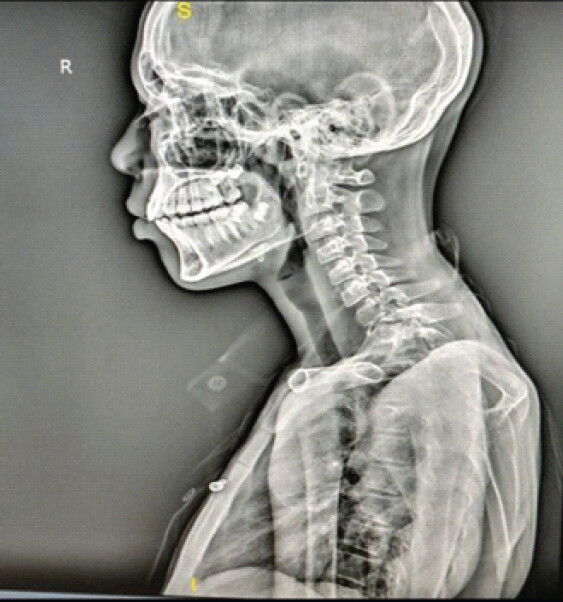
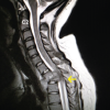
Plain radiographs of the cervical spine demonstrated destruction at the C7–T1 vertebral level with reduction of destruction of T1 and anterolisthesis and prevertebral soft tissue shadow (Fig. 1). Laboratory investigation revealed elevated erythrocyte sedimentation rate and C-reactive protein levels suggestive of active inflammatory pathology. MRI of the whole spine revealed near-total destruction of the vertebral body with a large pre- and paravertebral abscess extending from C6 to T3 and epidural extension causing severe spinal canal stenosis with spinal cord compression and cord signal changes suggestive of compressive myelopathy (Fig. 2a and b).
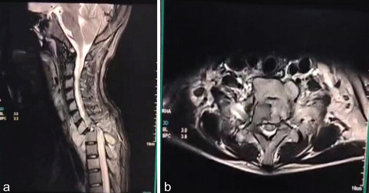
A diagnosis of cervicodorsal Pott’s spine involving the C7–T1 junction with acute-onset paraplegia was made. Based on radiological findings suggestive of spinal tuberculosis, the patient was started on standard first-line anti-tubercular therapy pre-operatively. In view of the acute neurological deterioration, spinal instability, and severe cord compression, urgent surgical intervention was planned under Antitubercular therapy coverage.
Multimodal management was chosen in the form of Middle Path Regimen preoperatively, followed by surgery by anterior approach [5].
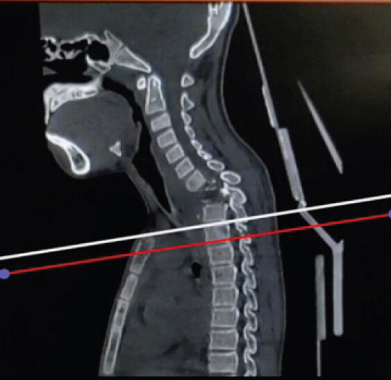
Using the normal anterior cervical approach, surgeons can usually reach up to the C6-C7 vertebral level, and sometimes, a posterior approach may be considered for lower levels. However, by applying the Kari-Kari method, the estimated reachable level in this patient was up to T2 (Fig. 3), so the anterior approach was chosen instead of the posterior approach [6,7].
Surgical management and operative details
Surgery was performed under general anesthesia with the patient in the supine position using a left-sided Smith–Robinson anterior cervical approach. Extensive inflammatory granulation tissue, caseous necrosis, and destruction of the cervicodorsal junction were encountered intraoperatively. As standard anterior exposure, we could reach up to lower T1 and upper T2; a partial manubriotomy was performed for reaching up to T3.
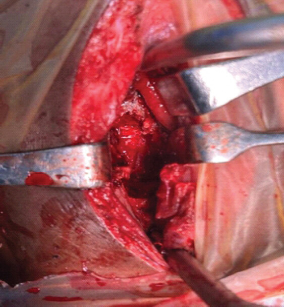
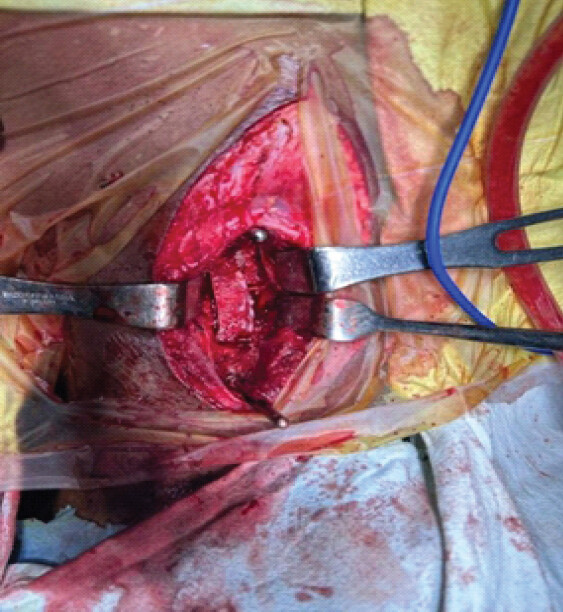
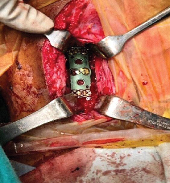
T1–T2 corpectomy with thorough debridement of necrotic vertebral tissue, infected disc material, and epidural granulation tissue was undertaken. A large epidural abscess causing severe spinal cord compression was evacuated, achieving direct anterior decompression (Fig. 4). The anterior column was reconstructed using a structural bone graft, followed by anterior plate fixation from C7 to T3 to restore stability across the cervicodorsal junction (Figs. 5 and 6).
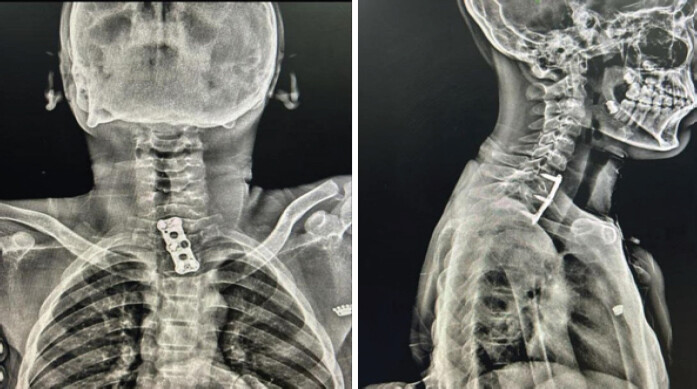
Granulation tissue, pus, and necrotic bone samples were sent for histopathological examination, Ziehl–Neelsen staining, Cartridge-based nucleic acid amplification test (CBNAAT), mycobacterial culture, and drug susceptibility testing. CBNAAT was positive, with no rifampicin resistance detected. Post-operative cervical spine X-ray done showing anterior cervical plate fixation (Figs. 7 and 8). Postoperatively, antitubercular therapy was continued, and the patient underwent supervised rehabilitation.
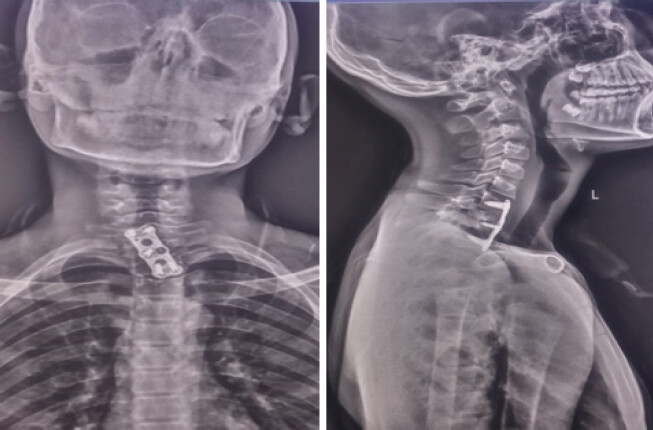
Serial radiographs obtained immediately postoperatively and at 6 weeks, 3 months, 6 months, and 1 year assessed graft incorporation, sagittal alignment, implant stability, and fusion. Maintained alignment, stable graft position, absence of implant-related complications, and bridging trabeculae across the graft–endplate interface indicated successful fusion. Computed tomography was used selectively for detailed evaluation of fusion and implant positioning.
The patient subsequently underwent neurorehabilitation with progressive mobilization and gait training. Serial clinical, neurological (Table 1), and radiological follow-up demonstrated gradual neurological recovery and disease resolution, while antitubercular therapy was continued until satisfactory clinical and radiological healing was achieved.
Serial neurological assessment (MRC power grades) from presentation to 1 year post-operative follow-up
| N/E | Presentation right | Presentation left | 1 Month right | 1 Month left | 3 Months right | 3 Months left | 12 Months right | 12 Months left |
|---|---|---|---|---|---|---|---|---|
| SAB | 5 | 5 | 5 | 5 | 5 | 5 | 5 | 5 |
| EF | 5 | 5 | 5 | 5 | 5 | 5 | 5 | 5 |
| EE | 5 | 5 | 5 | 5 | 5 | 5 | 5 | 5 |
| WF | 5 | 5 | 5 | 5 | 5 | 5 | 5 | 5 |
| WE | 5 | 5 | 5 | 5 | 5 | 5 | 5 | 5 |
| FG | Weak | Weak | Weak | Weak | 5 | 5 | 5 | 5 |
| HF | 0 | 0 | 2 | 2 | 5 | 5 | 5 | 5 |
| KF | 0 | 0 | 3 | 3 | 5 | 5 | 5 | 5 |
| KE | 0 | 0 | 3 | 3 | 5 | 5 | 5 | 5 |
| ADF | 0 | 0 | 3 | 3 | 3 | 5 | 4 | 5 |
| APF | 0 | 0 | 3 | 3 | 3 | 5 | 4 | 5 |
| EHL | 0 | 0 | 3 | 3 | 3 | 5 | 4 | 5 |
| Tone | Flaccid | Flaccid | Normal | Normal | Normal | Normal | Normal | Normal |
| DTR | Absent | Absent | Absent | Absent | ++ | ++ | ++ | ++ |
| Plantar | Absent | Absent | Absent | Absent | Flexor | Flexor | Flexor | Flexor |
| Sensory | Intact | Intact | Intact | Intact | Intact | Intact | Intact | Intact |
| B/BL | Intact | Intact | Intact | Intact | Intact | Intact | Intact | Intact |
| ASIA Grade | C | C | D | D |
N/E: Neurological examination, MRC: Medical research council power grading scale (0–5), SAB: Shoulder abduction, EF: Elbow flexion, EE: Elbow extension, WF: Wrist flexion, WE: Wrist extension, FG: Finger grip, HF: Hip flexion, KF: Knee flexion, KE: Knee extension, ADF: Ankle dorsiflexion, APF: Ankle plantar flexion, EHL: Extensor hallucis longus, DTR: Deep tendon reflexes, B/BL: Bowel/bladder function, ASIA: American spinal injury association
Discussion
The optimal surgical approach for cervicodorsal spinal tuberculosis remains controversial and should be individualized based on the location of pathology, degree of vertebral destruction, neurological status, and spinal stability. Various authors have reported favorable outcomes using anterior, posterior, and combined approaches depending on disease characteristics.
Tarantino et al. reported that anterior approaches combined with manubriotomy provide excellent exposure of the cervicothoracic junction with a low complication rate, allowing direct decompression, debridement, and reconstruction of anterior pathology [4]. They concluded that manubriotomy facilitates adequate visualization of the cervicothoracic junction while avoiding the morbidity associated with more extensive approaches. In our case, the presence of severe anterior vertebral destruction, a large epidural abscess, and significant ventral spinal cord compression made the transmanubrial anterior approach particularly suitable, enabling complete decompression and stable anterior reconstruction through a single-stage procedure.
Jain et al., in their series on cervicodorsal spinal tuberculosis, emphasized that the choice of surgical approach should be guided by the extent and location of disease [2]. They reported satisfactory outcomes with posterior stabilization in patients with multilevel involvement and deformity but advocated anterior or extended anterior approaches when significant anterior vertebral body destruction and ventral neural compression were present. Consistent with their recommendations, our patient had predominant anterior pathology at the C7–T1 junction, making direct anterior decompression and reconstruction a more appropriate strategy than a posterior-only procedure.
Rajasekaran highlighted the role of combined anterior and posterior procedures in patients with severe kyphotic deformity and extensive vertebral destruction requiring circumferential stabilization [8–11]. Although combined approaches provide robust biomechanical stability, they are associated with longer operative time, increased blood loss, and greater surgical morbidity. In contrast, our patient achieved satisfactory decompression, anterior column reconstruction, and stabilization through a single anterior transmanubrial approach, thereby avoiding the potential morbidity associated with circumferential surgery.
Furthermore, Jia et al. demonstrated that early surgical decompression combined with antitubercular chemotherapy results in favorable neurological recovery in patients with spinal tuberculosis presenting with neurological deficits [9]. Similar to their findings, urgent surgical intervention in our patient led to significant neurological improvement during follow-up rehabilitation.
The present case suggests that, in selected patients with predominant anterior cervicodorsal disease, severe ventral spinal cord compression, and limited posterior column involvement, an extended anterior transmanubrial approach may offer distinct advantages by enabling direct neural decompression, complete debridement of infective tissue, restoration of anterior column support, and stable fixation through a single-stage procedure while avoiding the additional morbidity associated with combined anterior-posterior surgeries.
Conclusion
Cervicodorsal Pott’s spine is a rare but potentially devastating form of spinal tuberculosis due to the complex anatomy and biomechanical instability of the cervicothoracic junction. Acute paraplegia may occur from vertebral destruction, epidural abscess formation, instability, and severe cord compression, even without trauma or constitutional symptoms. Early MRI diagnosis and prompt initiation of antitubercular therapy are essential to prevent irreversible neurological deficits. Surgical intervention is indicated in patients with acute neurological deterioration, significant cord compression, vertebral collapse, deformity, or instability.
When pathology is predominantly anterior, anterior decompression with corpectomy and stabilization is preferred, as it enables direct access to diseased vertebrae, effective abscess evacuation, adequate neural decompression, restoration of anterior column support, and immediate spinal stability. The transmanubrial extended anterior approach provides safe and sufficient exposure of the cervicodorsal junction, particularly at the T2–T3 level, while avoiding the morbidity of combined staged procedures. Early surgical decompression, combined with prolonged antitubercular chemotherapy and rehabilitation, can result in significant neurological recovery and favorable functional outcomes, especially in pediatric and adolescent patients.
Clinical Message
Acute paraplegia in cervicodorsal junction tuberculosis requires urgent MRI evaluation and early decompression to prevent irreversible spinal cord injury. The transmanubrial anterior approach provides adequate exposure for safe decompression and stable reconstruction in lesions extending to the upper thoracic spine.
Conflict of Interest:
Source of Support:
Nil
Consent:
The authors confirm that informed consent was obtained from the patient for publication of this article
References
- Tuli SM..Tuberculosis of the Skeletal System. 5th ed. New Delhi: Jaypee Brothers Medical Publishers; 2016. [Google Scholar] | [PubMed]
- Jain AK, Dhammi IK, Arora R, Gain A.Cervicodorsal spine tuberculosis -- surgical approach. J Clin Orthop Trauma 2024;52:102420. [Google Scholar] | [PubMed]
- Garg RK, Somvanshi DS.Spinal tuberculosis: A review. J Spinal Cord Med 2011;34:440-54. [Google Scholar] | [PubMed]
- Tarantino R, Donnarumma P, Marruzzo D, Landi A, De Giacomo T, Delfini R.Anterior surgical approaches to the cervicothoracic junction: when to use the manubriotomy?. Spine J 2013;13:1064-8. [Google Scholar] | [PubMed]
- Tuli SM.Results of treatment of spinal tuberculosis by “middle-path” regime. J Bone Joint Surg Br 1975;57:13-23. [Google Scholar] | [PubMed]
- Karikari IO, Powers CJ, Isaacs RE.Simple method for determining the need for sternotomy/manubriotomy with the anterior approach to the cervicothoracic junction. Neurosurgery 2009;656 SupplE165-6. [Google Scholar] | [PubMed]
- Darling GE, McBroom R, Perrin R.Modified anterior approach to the cervicothoracic junction. Spine (Phila Pa 1976) 1995;20:1519-21. [Google Scholar] | [PubMed]
- Okwudire EG, Ezenwugo UM, Ugwoegbu JU, Okoro CA, Isiozor I.Atypical presentation of cervical Pott's disease: A case report. J Tuberc Res 2021;9:211-8. [Google Scholar] | [PubMed]
- Jia CG, Gao JG, Liu FS, Li Z, Dong ZL, Yao LM.Efficacy, safety and prognosis of treating neurological deficits caused by spinal tuberculosis within 4 weeks' standard anti-tuberculosis treatment: A single medical center's experience. Exp Ther Med 2019;19:519-52. [Google Scholar] | [PubMed]
- Singh J, Gupta P, Singh A, Singh A.Atypical cervical pott's spine with multiple level epidural collection: A case series of three patients. J Orthop Case Rep 2023;13:115-20. [Google Scholar] | [PubMed]
- Rajasekaran S.The problem of deformity in spinal tuberculosis. Clin Orthop Relat Res 2002;398:85-92. [Google Scholar] | [PubMed]
© 2026 Journal of Orthopaedic Case Reports - Published by Indian Orthopaedic Research Group
About the Authors
-

Dr. Manish Kawade
-

Dr. Dilipkumar Rajeshkumar Singh
-

Dr. Akshay B Ingale
-

Dr. Nilesh S Sakhare
-

Dr. Tejas Vinayak Vatkar
Related Articles in Journal of Orthopaedic Case Reports
 May 1, 2026 Surgical Management of Pott’s Paraplegia in a 27-Week Pregnant Woman with Continuation of Pregnancy: A Case Report
 April 1, 2026 Multifocal Non-contiguous Spinal Tuberculosis: A Report of 3 Surgically-Treated Patients
 March 1, 2026 Silently Gradually Progressing Paraparesis Pott’s Spine Lumbar Vertebra with Bilateral Psoas Abscess
 December 1, 2024 Management of Thoracolumbar Spinal Tuberculosis by Decompression and Posterior Stabilization with Pedicle Screw Fixation