


Introduction
Torticollis is an abnormal head posture characterized by ipsilateral head tilt and contralateral chin rotation. Its causes include musculoskeletal, neurological, ophthalmic, auditory, and traumatic conditions [1]. Posterior sternoclavicular joint (SCJ) dislocation is rare and potentially life-threatening because of the close relationship of the medial clavicle to the trachea, esophagus, and major vessels [2]. Although SCJ dislocations may cause pain, shoulder dysfunction, dysphagia, respiratory symptoms, or neurovascular complications, torticollis has not been described as a presenting feature [3, 4]. We report a rare case of posterior SCJ dislocation in a young kabaddi player presenting primarily with acute torticollis.
Case Report
A 16-year-old teenager presented to the emergency department with complaints of neck tilt and pain in the left upper limb following a fall during a kabaddi match 3 days ago. The injury occurred after being tackled, with a brief episode of giddiness during a kabaddi session. The patient could not recall the precise mechanism of injury.
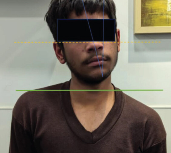
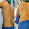
He reported right-sided head tilt and pain exacerbated by movement. He denied any breathing difficulty, dysphagia, or hoarseness. No visible external injuries or neurological deficits were noted. Examination revealed: Torticollis with head tilted to the right, left shoulder positioned lower than the right, and tenderness over the left SCJ (Fig. 1).
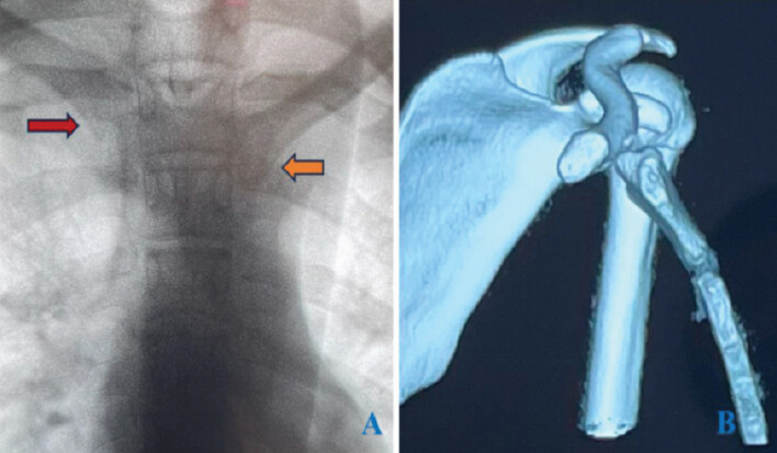
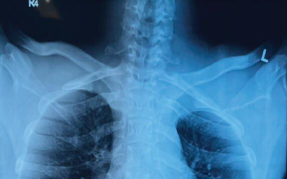
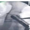
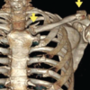
Initial X-ray and non-contrast computed tomography (CT) revealed a posterior dislocation of the left SC joint. Given the proximity of large vessels to the injury site, CT angiography was performed, which revealed large vessels abutting the medial end of the clavicle (Fig. 2). A multidisciplinary management plan was discussed among orthopedic, vascular surgery, and anesthetic teams. The agreed plan was to attempt manipulation under anesthesia as the first step. On the 4th day post-injury, the patient was taken to the operating room. The patient was positioned supine with a sandbag between the scapulae. Closed reduction was performed using the abduction-traction-extension technique under fluoroscopic guidance [5]. A sheet was used around the upper torso to give counter-traction by the assistant. The C-arm was positioned over the SCJ area with a 45° cephalad tilt to obtain serendipity views. Reduction was appreciated with the clunk. Post-reduction serendipity views confirmed symmetrical clavicular alignment over the SCJ (Fig. 3). Distal vascularity of the upper limb was assessed at wrist level for 30 min continuously and was intact. Intraoperative assessment of joint stability on shoulder range of motion was satisfactory. In the post-operative recovery room, torticollis deformity was seen fully reversed. Left upper limb was kept in the shoulder sling.
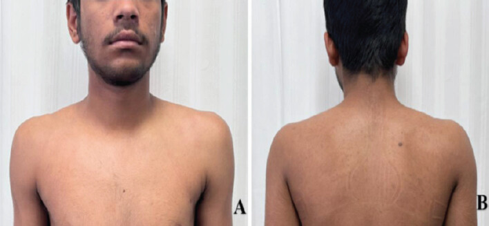
The patient was discharged the next day with advice on immobilization for 2 weeks and gradual mobilization. At 6-week follow-up, he regained full, pain-free range of motion without any instability. At 2-year follow-up, the patient was continuing with his sports activity with no residual pain or deformity (Fig. 4).
Discussion
Posterior SCJ dislocation is rare, accounting for approximately 3% of shoulder-girdle injuries and <1% of all joint dislocations [6]. Its infrequency makes diagnosis and management challenging, while posterior displacement carries additional risk because of the adjacent mediastinal structures [7]. Indirect force transmitted through the clavicle, particularly during contact sports, is a common mechanism [2].
Because of the risk of vascular, airway, or esophageal injury, prompt diagnosis and careful planning are essential. CT, preferably with angiography when vascular involvement is suspected, provides accurate assessment of the dislocation and adjacent structures [8]. In stable patients without vascular compromise, closed reduction is generally the initial treatment, whereas open reduction and stabilization are considered when closed reduction fails, instability persists, or mediastinal complications are present [2, 3]. In our patient, closed reduction remained successful despite presentation 3 days after injury.
Damschen et al. reported favorable return-to-sport outcomes after closed reduction of acute traumatic posterior SCJ dislocations, with 23 of 31 patients returning to pre-injury activity and a mean return-to-sport time of 3.1 months [9]. Our patient returned to sports at approximately 2.5 months without functional limitation. Relevant pediatric and adolescent cases are summarized in Table 1.
Reported pediatric and adolescent posterior sternoclavicular joint dislocations
| Study | Age/sex | Mechanism/sport | Presentation | Treatment | Outcome |
|---|---|---|---|---|---|
| Galanis et al., 2014 [10] | 12-year-old boy | Judo | SCJ pain; no torticollis reported | Closed reduction under GA | Full ROM at 2 years |
| Ishii et al., 2018 [11] | 14-year-old boy | Sports injury | Posterior SCJ injury; no torticollis reported | Temporary K-wire SCJ stabilization | Successful stabilization |
| El Khassoui et al., 2023 [12] | 12-year-old boy | Kickboxing | Shoulder/clavicular symptoms; no torticollis reported | Closed reduction | No complications at 24 months |
| Gurcinar et al., 2023 [13] | 13-year-old boy | Football | Pain, deformity, upper-limb numbness; no torticollis reported | Closed reduction under sedation | Full ROM and strength by 8 weeks |
| Kralj et al., 2023 [14] | 15-year-old boy | Trauma | Posterior SCJ dislocation; no mediastinal injury | Open reduction and cerclage | Asymptomatic follow-up |
| Ngatchou et al., 2024 [4] | Young male | Trauma | Brachiocephalic artery and esophageal compression; no torticollis reported | Open reduction and wire cerclage | Good post-operative outcome |
| Present case | 16-year-old boy | Kabaddi tackle | Acute torticollis with left SCJ tenderness | Closed reduction under fluoroscopy | Full ROM; returned to sports; 2-year symptom-free follow-up |
SCJ: Sternoclavicular joint, ROM: Range of motion, GA: General anesthesia
Torticollis appears to be an exceptionally uncommon presentation of SCJ dislocation. Galanis et al. [10] reported a 12-year-old judo athlete with posterior SCJ dislocation who had severe SCJ pain and other local symptoms but no torticollis. Other reported pediatric and adolescent cases similarly describe pain, deformity, or upper-limb symptoms rather than abnormal neck posture. Thus, the present case appears distinctive because torticollis was the dominant clinical manifestation.
The postural abnormality in our patient may be explained by altered sternocleidomastoid (SCM) mechanics. Torticollis is usually produced by asymmetric SCM activity, with contraction of one SCM producing ipsilateral head tilt and contralateral rotation. In this case, the head tilted away from the side of the posterior SCJ dislocation, suggesting relative overactivity of the contralateral SCM.
We hypothesize that posterior displacement of the medial clavicle shortened the ipsilateral SCM’s effective resting length by reducing the distance between its clavicular attachment and the mastoid process [15, 16]. This may have reduced ipsilateral SCM tension, allowing the relatively normal contralateral SCM to predominate and produce the observed contralateral head tilt. This mechanism remains hypothetical and requires biomechanical validation, but it provides a plausible explanation for the unusual postural presentation.
This case highlights the importance of including posterior SCJ dislocation in the differential diagnosis of acute post-traumatic torticollis, particularly in young athletes. CT angiography and multidisciplinary preparation are important because reduction may endanger adjacent vascular structures. In a stable patient without vascular compromise, a carefully planned attempt at closed reduction can be successful even several days after injury.
Conclusion
Posterior SCJ dislocation can rarely manifest as acute torticollis. Recognition of this atypical presentation should prompt appropriate cross-sectional imaging and assessment for mediastinal injury. In stable patients without vascular compromise, carefully planned closed reduction can provide excellent functional recovery, even after a short delay.
Clinical Message
Posterior SCJ dislocation should be considered in acute post-traumatic torticollis, particularly in young athletes. CT angiography and multidisciplinary planning facilitate safe diagnosis and reduction.
Conflict of Interest:
Source of Support:
Nil
Consent:
The authors confirm that informed consent was obtained from the patient for publication of this article
References
- Peyrou P, Moulies D.Le torticolis de l'enfant: Démarche diagnostique. Arch Pediatr 2007;14:1264-70. [Google Scholar] | [PubMed]
- Sernandez H, Riehl J.Sternoclavicular joint dislocation: A systematic review and meta-analysis. J Orthop Trauma 2019;33:e251-5. [Google Scholar] | [PubMed]
- Groh GI, Wirth MA, Rockwood CA Jr.Treatment of traumatic posterior sternoclavicular dislocations. J Shoulder Elbow Surg 2011;20:107-13. [Google Scholar] | [PubMed]
- Ngatchou W, Fosso MN, Djumegue VG, Surdeanu IR, Jissendi P, Towo PY.Posterior sternoclavicular joint dislocation in a young male: A case report. Pan Afr Med J 2024;47:138. [Google Scholar] | [PubMed]
- Garcia JA, Arguello AM, Momaya AM, Ponce BA.Sternoclavicular joint instability: Symptoms, diagnosis and management. Orthop Res Rev 2020;12:75-87. [Google Scholar] | [PubMed]
- Moyal AJ, Burkhart RJ, Adelstein JM, Voos JE, Apostolakos JM, Calcei JG.Acromioclavicular and sternoclavicular joint injuries in contact sports: A narrative review of conservative and surgical treatments. Ann Jt 2025;10:31. [Google Scholar] | [PubMed]
- Obremskey WT, Rodriguez-Baron EB, Tatman LM, Pesantez RF.Acute dislocations of the sternoclavicular joint: A review article. J Am Acad Orthop Surg 2022;30:148-54. [Google Scholar] | [PubMed]
- Ingoe HM, Mohammed K, Malone AA, Beadle G, Sharpe T, Cockfield A.Traumatic posterior sternoclavicular joint dislocation - current aspects of management. Injury 2023;54:110983. [Google Scholar] | [PubMed]
- Damschen J, Nowak M, Murphy A, Park S, Li X, Galvin J.Return to sports after closed reduction of acute traumatic posterior sternoclavicular joint dislocations: A systematic review. Am J Sports Med 2023;51:3076-83. [Google Scholar] | [PubMed]
- Galanis N, Anastasiadis P, Grigoropoulou F, Kirkos J, Kapetanos G.Judo-related traumatic posterior sternoclavicular joint dislocation in a child. Clin J Sport Med 2014;24:271-3. [Google Scholar] | [PubMed]
- Ishii N, Matsumura N, Iwamoto T, Sato K, Nakamura M, Matsumoto M.Temporary sternoclavicular stabilization using an innovative blocking technique with Kirschner wires for the treatment of posterior sternoclavicular joint injury in adolescent patients: A report of two cases. JBJS Case Connect 2018;8:e67. [Google Scholar] | [PubMed]
- El Khassoui A, Idrissi Bahre MN, Tahiri D, Salama T, Aghoutane EM, Fezzazi RE.Posterior sternoclavicular dislocation in children: A case report. J Orthop Case Rep 2023;13:98-102. [Google Scholar] | [PubMed]
- Gurcinar MG, Ozer M, Afacan MY, Ustundag S.Management of posterior sternoclavicular joint dislocation in a teenager after a direct elbow strike to his clavicle: A case report. Cureus 2023;15:e49916. [Google Scholar] | [PubMed]
- Kralj R, Bumči B, Pejić J, Bumči I, Višnjić S, Žic R.Posterior sternoclavicular dislocation in a 15-year old boy: Case report. Paediatr Croat 2023;67:31-4. [Google Scholar] | [PubMed]
- Saha A, Mandal S, Chakraborty S, Bandyopadhyay M.Morphological study of the attachment of sternocleidomastoid muscle. Singapore Med J 2014;55:45-7. [Google Scholar] | [PubMed]
- Byrd JJ, McCumber TL, Snow EL.Cadaveric case report and biomechanical analysis of an accessory clavicular head to the sternocleidomastoid. Transl Res Anat 2022;28:100215. [Google Scholar] | [PubMed]
© 2026 Journal of Orthopaedic Case Reports - Published by Indian Orthopaedic Research Group
About the Authors
-

Dr. Kushagr Katyal
-

Dr. Jeevan Kumar Sharma
-

Dr. Sunil Kumar Choudhary
Related Articles in Journal of Orthopaedic Case Reports
 October 1, 2026 Recurrent Ventral Scapular Osteochondroma Presenting with Pseudo-winging in an Adolescent: A Case Report and Follow-up of a Previously Published Case
 August 1, 2026 Beyond Pipkin IV: Management of Femoral Head-neck Fracture-dislocation with Posterior Acetabular Wall and Peritrochanteric Fracture in an Adolescent Using a Modified Kocher-Langenbeck Approach
 June 1, 2026 Bipolar Clavicle Fracture: A Rare Entity of the Commonest Fracture – A Case Report
 June 1, 2026 Adolescent Paraspinal Lipoblastoma: A Case Report