


Introduction
Melorheostosis, also known as Leri disease, is a rare, non-hereditary sclerosing bone dysplasia characterized by cortical hyperostosis with a characteristic flowing appearance along the bone surface [1]. The term “melorheostosis” is derived from the Greek words “melos” (limb), “rhein” (to flow), and “ostosis” (bone formation), reflecting the classical radiographic appearance often described as resembling “dripping candle wax” [2,3]. The disease typically affects long bones of the appendicular skeleton, most commonly the lower limbs, and may occur in monostotic, polyostotic, or rarely axial forms [3].
The etiology remains unclear. Recent studies have suggested somatic mosaic mutations involving the MAP2K1 gene [1, 3, 4]. Other proposed mechanisms include sclerotomal distribution abnormalities, vascular malformations, and mesenchymal dysregulation [5].
Clinical presentation is highly variable, ranging from asymptomatic radiological findings to progressive limb pain, deformity, joint contractures, stiffness, and functional impairment [6]. Because of overlapping radiographic features with other sclerosing bone dysplasias and neoplastic conditions, accurate diagnosis requires careful correlation of clinical, radiologic, and histopathologic findings [7].
We present a case of polyostotic melorheostosis involving the right tibia, femur, and foot bones in a young adult male presenting with progressive painful swelling, highlighting the diagnostic considerations and management challenges.
Case Report
A 28-year-old male manual laborer presented to the outpatient department with complaints of swelling and pain in the right lower limb for 2 months. The swelling extended from the knee to the foot and was insidious in progression. The pain was initially sudden in onset, gradually progressive in intensity, and persistent in nature. It interfered significantly with ambulation and occupational activities.
There were no identifiable aggravating or relieving factors. No history of trauma, fever, weight loss, constitutional symptoms, prior fractures, morning stiffness, or similar swellings elsewhere was reported. There was no history of chronic illness, metabolic bone disease, or malignancy. Family history was non-contributory.
Clinical examination
On local examination:
-
Diffuse swelling extending from the knee to the entire foot
-
Irregular surface
-
Hard consistency
-
Irregular margins
-
Fixed to underlying bone
-
Overlying skin freely mobile
-
No warmth or erythema
-
No visible deformity
-
No limb length discrepancy
-
Full range of motion at knee and ankle joints.
Neurovascular examination of the limb was normal. No lymphadenopathy was detected. Systemic examination was unremarkable.
Differential diagnosis
Based on clinical presentation, the following differentials were considered:
-
Osteoma
-
Myositis ossificans
-
Progressive diaphyseal dysplasia (Camurati–Engelmann disease)
-
Hereditary multiple diaphyseal sclerosis
-
Sclerosing variants of osteosarcoma
-
Chronic osteomyelitis.
Investigations
-
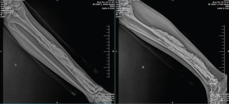
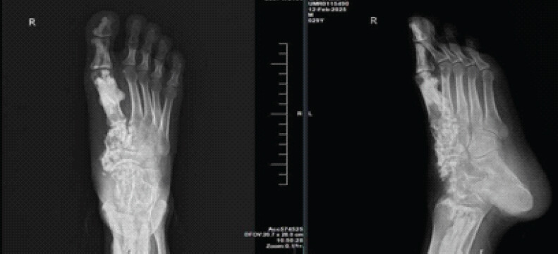
X-ray revealed irregular cortical thickening with dense hyperostosis along the medial cortex of the tibia, demonstrating the classical “dripping candle wax” appearance. There was involvement of the entire tibial shaft. Similar sclerotic changes were noted in the lower end of the femur and multiple tarsal bones, including the great toe.
-
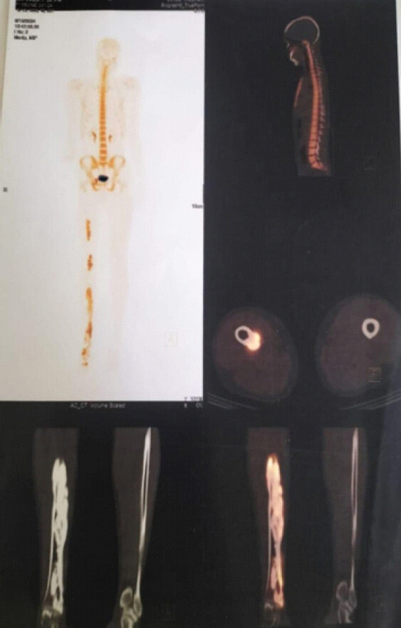
Bone scan (Fig. 3):
-
Demonstrated increased tracer uptake throughout the affected regions, indicating metabolically active sclerotic bone formation.
-
Magnetic resonance imaging (MRI):
-
MRI showed nodular, thick periosteal and endosteal cortical thickening and sclerosis along the entire length of the tibia. Mild similar changes were observed in the distal femur and tarsal bones. No soft-tissue mass or aggressive periosteal reaction was noted.
-
Histopathology:
-
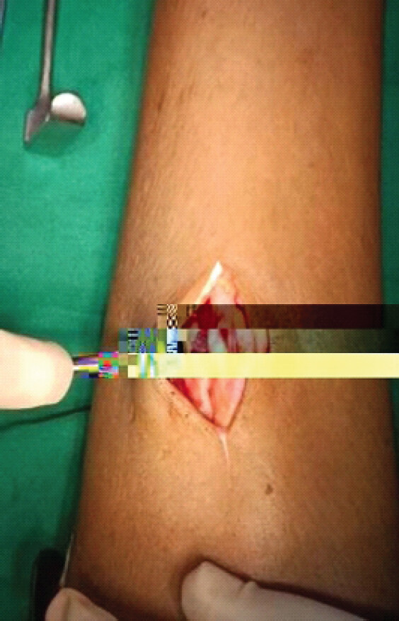
An intraoperative biopsy (Fig. 4) was taken and revealed scattered lamellar bone with osteoblastic rimming. Areas of dense sclerosis were present. Skeletal muscle tissue showed fibrocollagenous stroma with focal vascular proliferation and areas of hyalinization. No cellular atypia or malignant features were identified.
Final diagnosis
Based on characteristic radiographic findings, metabolic activity on bone scan, histopathological features, and clinical presentation, a diagnosis of polyostotic melorheostosis involving the right tibia, distal femur, and foot was established.
Management and follow-up
Management was primarily conservative and symptomatic. The patient was started on a multimodal analgesic regimen, including non-steroidal anti-inflammatory drugs and supportive measures.
Given the absence of deformity, joint restriction, limb discrepancy, or neurovascular compromise, surgical intervention was not indicated.
The patient was followed up over 4–5 outpatient visits. He remained hemodynamically stable with no progression of swelling or development of deformity. Pain scores reduced progressively from severe at presentation to mild discomfort on prolonged standing or walking by the final follow-up.
Discussion
Melorheostosis is a rare sclerosing bone dysplasia characterized by cortical hyperostosis that often follows a sclerotomal distribution [1, 8]. The disease predominantly affects long bones of the lower extremities and may extend into adjacent soft tissues.
Pathogenesis
The exact pathogenesis remains unclear. Current evidence supports somatic mosaic mutations affecting the MAP2K1 gene, leading to abnormal osteoblastic activity and localized cortical overgrowth [3, 4, 5, 9]. The mosaic nature explains the segmental distribution of lesions. Earlier theories proposed embryologic sclerotomal distribution abnormalities or vascular malformations as contributing factors [5].
Radiologic features
Radiographic patterns of melorheostosis include:
-
Classic “dripping candle wax” cortical hyperostosis
-
Osteoma-like pattern
-
Myositis ossificans-like pattern
-
Osteopathia striata-like pattern.
The classical flowing hyperostosis along one side of the bone is considered pathognomonic [10]. MRI helps assess soft-tissue involvement, whereas bone scintigraphy demonstrates increased metabolic activity [4, 6, 7].
Differential diagnosis
Important differentials include:
-
Camurati–Engelmann disease
-
Hereditary multiple diaphyseal sclerosis
-
Erdheim–Chester disease, chronic osteomyelitis
-
Parosteal osteosarcoma.
Unlike hereditary sclerosing dysplasias, melorheostosis is usually sporadic and asymmetrical [7]. Absence of systemic manifestations and characteristic imaging features aid differentiation.
Clinical course
Clinical manifestations vary from asymptomatic to severe pain, deformity, contractures, limb shortening, and joint stiffness [1, 6]. Soft-tissue fibrosis may lead to restricted joint mobility [2]. Polyostotic involvement, as seen in this case, is less common than monostotic disease [10].
Management
There is no definitive cure. Management is symptomatic and individualized:
-
Non-steroidal anti-inflammatory drug for pain
-
Bisphosphonates in selected cases
-
Physiotherapy to prevent contractures
-
Surgical intervention for deformity correction, limb length discrepancy, or neurovascular compression [5].
Our patient demonstrated symptomatic improvement with conservative management, and no progression requiring surgical intervention during follow-up.
Conclusion
Polyostotic melorheostosis is a rare sclerosing bone dysplasia that should be considered in young adults presenting with unilateral limb pain and cortical thickening on imaging. The classical “dripping candle wax” radiographic appearance is highly suggestive and may obviate the need for extensive invasive investigations.
Accurate diagnosis is essential to avoid unnecessary aggressive interventions, as management is primarily supportive unless functional impairment or deformity develops. Early recognition and periodic follow-up are important to monitor progression and preserve limb function.
Clinical Message
Recognition of the characteristic “dripping candle wax” cortical hyperostosis on imaging is key to diagnosing melorheostosis. Early identification prevents misdiagnosis as malignancy or infection and avoids unnecessary aggressive interventions.
Conflict of Interest:
Nil
Source of Support:
Nil
Consent:
The authors confirm that informed consent was obtained from the patient for publication of this article
References
- Deshmukh NS, Phansopkar P. Melorheostosis: A systematic review of clinical manifestations, diagnostic challenges, therapeutic strategies, and physiotherapeutic interventions. Cureus 2025;17:e80407. [Google Scholar] | [PubMed]
- Dhillon MS, Saibaba B. Melorheostosis-foot and ankle perspective. Foot (Edinb) 2017;31:44-8. [Google Scholar] | [PubMed]
- Iordache S, Cursaru A, Serban B, Costache M, Spiridonica R, Cretu B. Melorheostosis: A review of the literature and a case report. Medicina (Kaunas) 2023;59:869. [Google Scholar] | [PubMed]
- Bhattacharyya T. Unraveling melorheostosis: Insights into clinical features, diagnosis, and treatment. JBMR Plus 2024;9:zia.e163. [Google Scholar] | [PubMed]
- Tladi M. Clinics in surgery melorheostosis: A review article open access. Clinics 2020;2020:2926. [Google Scholar] | [PubMed]
- Teoh CC, Teoh SR, Chieng DC, Ang XY, Hassim MH. Polyostotic melorheostosis: Approach to clinical evaluation and management. Open J Orthop 2019;9:137-44. [Google Scholar] | [PubMed]
- Bahk WJ. Melorheostosis. In: Diagnosis and Management of Primary Bone Tumors. Springer Nature Singapore Private Ltd.; 2025. 65-71. [Google Scholar] | [PubMed]
- Ruggiero M, Conforti A, Culcasi A, Mazzanti C, Sibahi G, Rani N. A focus on melorheostosis disease: A literature review and case report of femoral-acetabular impingement due to melorheostosis treated with surgical hip osteoplasty. Reumatismo 2024;76:(1)doi: 10.4081/reumatismo.2024.1621. [Google Scholar] | [PubMed] | [CrossRef]
- Koca E, Kavak Ş, Demir B, Comunoglu N, Kantarci F, Koca O. Melorheostosis: Clinical, radiological, and histopathological features with a literature review. Physiol Rep 2025;13:e70652. [Google Scholar] | [PubMed]
- Pandey DN, Singh HP, Singh C, Kumar S, Prakash A. Melorheostosis: A rare case report with pelvic and lumbar involvement. J Orthop Case Rep 2025;15:213-6. [Google Scholar] | [PubMed]
© 2026 Journal of Orthopaedic Case Reports - Published by Indian Orthopaedic Research Group
About the Authors
-

Dr. Aditi Shukla
-

Dr. Deepthi Marri
-

Dr. Palagiri Daksha Sanjana
-

Dr. Putha Harshitha
-

Dr. Yallanti Pavithra
-

Dr. Nallam Venkata Sai Deekshitha
Related Articles in Journal of Orthopaedic Case Reports
 October 1, 2026 Development of a Non-ossifying Fibroma in the Same Femur 6 Years after Limb-Salvage Surgery for Osteosarcoma: A Case Report
 October 1, 2026 Recurrent Ventral Scapular Osteochondroma Presenting with Pseudo-winging in an Adolescent: A Case Report and Follow-up of a Previously Published Case
 October 1, 2026 Staged Bilateral Total Elbow Arthroplasty in Advanced Rheumatoid Elbow Disease: A Case Report
 October 1, 2026 Posterior Fibular Entrapment in a Bosworth Fracture-Dislocation: A Case Report and Review of Diagnostic and Operative Considerations