


Introduction
Osteoblastoma is a rare benign bone-forming neoplasm accounting for approximately 1% of all primary bone tumors [1,2]. It predominantly affects adolescents and young adults and most commonly involves the vertebral column and long bones [2,3]. Involvement of the radius is uncommon, with only a limited number of cases reported in the literature [4]. The diagnosis of osteoblastoma may be challenging because its clinical, radiological, and histopathological features can overlap with those of low-grade osteogenic malignancies, particularly parosteal osteosarcoma [2,5,6]. Accurate diagnosis therefore requires careful clinicoradiological correlation and histopathological evaluation [5, 7].
We report a rare case of osteoblastoma involving the radial shaft that was initially suspected to be parosteal osteosarcoma based on imaging and biopsy findings. The lesion was managed with wide excision and biological reconstruction using an ipsilateral fibular autograft.
Case Report
A 17-year-old male presented with progressive pain and swelling over the middle third of the right forearm for the preceding 6 months. Clinical examination revealed a firm swelling over the radial aspect of the forearm without distal neurovascular deficit.
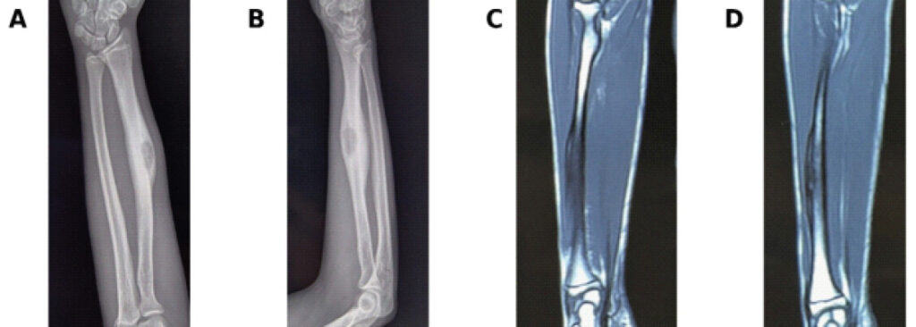
Plain radiographs demonstrated a surface-based lesion involving the radial shaft associated with cortical thickening and periosteal reaction. Magnetic resonance imaging revealed cortical involvement with surrounding soft-tissue extension, raising suspicion for a surface osteogenic malignancy (Fig. 1).
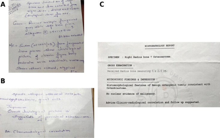
A core needle biopsy was performed and was suggestive of parosteal osteosarcoma (Fig. 2). Considering the clinicoradiological findings and biopsy report, wide excision was planned.
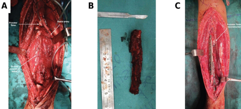
Through a Henry approach, the involved segment of the radius was excised en bloc. The pronator quadratus was excised along with the lesion to obtain adequate margins. Reconstruction of the resulting bony defect was performed using an ipsilateral non-vascularized fibular autograft stabilized with locking plate fixation. To restore forearm function, the pronator teres tendon was reattached to the fibular graft using a 2.8-mm suture anchor (Fig. 3).
Histopathological examination of the resected specimen demonstrated histomorphological features of a benign osteogenic tumor consistent with osteoblastoma. No nuclear evidence of malignancy was identified. The pathologist advised clinicoradiological correlation and follow-up.
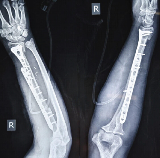
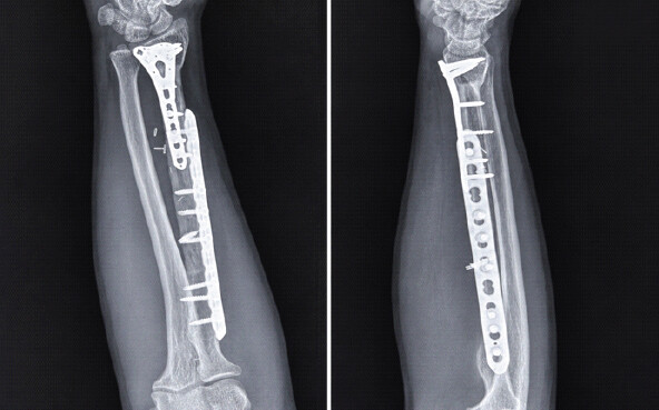
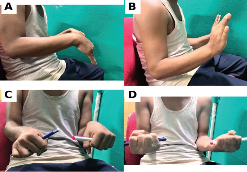
The post-operative period was uneventful(Fig. 4). At 12-month follow-up, radiographs demonstrated maintained alignment and progressive graft incorporation without evidence of local recurrence (Fig. 5) . Clinically, the patient achieved wrist flexion of 75°, wrist extension of 70°, forearm pronation of 80°, and forearm supination of 85°. The patient had satisfactory functional recovery and returned to activities of daily living without significant limitation(Fig. 6) .
Discussion
Osteoblastoma is a rare benign osteogenic tumor that may occasionally mimic malignant bone lesions both radiologically and histologically [2,5,6]. Distinguishing osteoblastoma from parosteal osteosarcoma can be particularly challenging because both lesions may demonstrate osteoid production, cortical involvement, and varying degrees of mineralization [5, 6, 7, 8].
In the present case, imaging findings and core needle biopsy favored a diagnosis of parosteal osteosarcoma. However, final histopathological examination of the excised specimen demonstrated histomorphological features of a benign osteogenic tumor consistent with osteoblastoma, with no nuclear evidence of malignancy. This discrepancy highlights the limitations of small biopsy samples and emphasizes the importance of correlating clinical, radiological, and pathological findings before establishing a definitive diagnosis [2, 5]. Sampling error remains a recognized limitation of core needle biopsy in surface osteogenic lesions, particularly when benign and low-grade malignant entities share overlapping histological features.
The rarity of osteoblastoma involving the radius further contributed to the diagnostic challenge. Bertoni et al. reported that involvement of the radius and ulna is uncommon compared with the more frequently affected vertebral column and long bones [4]. Because of the suspicious clinicoradiological features and biopsy findings in our patient, an oncological approach with wide excision was considered appropriate to avoid undertreatment of a potentially malignant lesion. The differential diagnosis of osteoblastoma includes osteoid osteoma, aggressive osteoblastoma, and low-grade osteosarcoma, as described in standard orthopaedic oncology literature [9,10].
Management of osteoblastoma ranges from intralesional curettage to en bloc excision depending on tumor location, biological behavior, and suspicion of malignancy [2,11]. In lesions where malignancy cannot be confidently excluded, wide excision remains a reasonable treatment option. In the present case, complete excision allowed definitive diagnosis and reduced the risk of local recurrence.
Reconstruction of segmental radial defects following tumor excision remains technically demanding because restoration of forearm stability and rotation is essential for optimal upper-limb function [12,13]. Autologous fibular graft reconstruction has been widely used following tumor resection because it provides biological incorporation, long-term structural support, and satisfactory functional outcomes [14,15]. In our patient, an ipsilateral non-vascularized fibular autograft successfully restored skeletal continuity and demonstrated progressive incorporation during follow-up.
A unique aspect of the present case was reattachment of the pronator teres to the fibular graft using a 2.8-mm suture anchor following excision of the pronator quadratus with the tumor. This technique was intended to preserve dynamic forearm pronation and optimize functional recovery. At 12-month follow-up, the patient achieved wrist flexion of 75°, wrist extension of 70°, forearm pronation of 80°, and forearm supination of 85°, with no evidence of local recurrence. These findings suggest that biological reconstruction using a non-vascularized fibular autograft, combined with tendon reattachment, can provide satisfactory functional and oncological outcomes in selected patients with radial shaft tumors.
This case highlights the importance of maintaining a broad differential diagnosis when evaluating surface lesions of the radius. It also demonstrates that biological reconstruction with an ipsilateral fibular autograft, combined with tendon reattachment techniques, can provide satisfactory oncological, radiological, and functional outcomes following excision of complex radial lesions.
Conclusion
Osteoblastoma of the radial shaft is a rare entity and may closely mimic parosteal osteosarcoma on imaging and biopsy. Definitive diagnosis may only become evident following examination of the excised specimen. Wide excision followed by ipsilateral fibular autograft reconstruction and pronator teres reattachment can provide satisfactory functional and radiological outcomes.
Clinical Message
Radial shaft osteoblastoma may present with clinicoradiological features suggestive of parosteal osteosarcoma. Accurate diagnosis requires careful clinicopathological correlation, and biological reconstruction using an ipsilateral fibular autograft can provide excellent limb preservation and functional recovery.
Conflict of Interest:
Nil
Source of Support:
Nil
Consent:
The authors confirm that informed consent was obtained from the patient for publication of this article
How to Cite this Article
Dhabal H, Barman S, Mallick SK, Kumar S. Osteoblastoma of the Radial Shaft Mimicking Parosteal Osteosarcoma: A Case Report. Journal of Orthopaedic Case Reports 2026 October;16(10): 193-197.
References
- Jaffe HL, Mayer L. An osteoblastic osteoid tissue-forming tumor of a metacarpal bone. Arch Surg 1932;24:550-64. [Google Scholar] | [PubMed]
- Lucas DR, Unni KK, McLeod RA, O'Connor MI, Sim FH. Osteoblastoma: Clinicopathologic study of 306 cases. Hum Pathol 1994;25:117-34. [Google Scholar] | [PubMed]
- Berry M, Mankin H, Gebhardt M, Rosenberg A, Hornicek F. Osteoblastoma: A 30-year study of 99 cases. J Surg Oncol 2008;98:179-83. [Google Scholar] | [PubMed]
- Bertoni F, Unni KK, McLeod RA, Dahlin DC. Osteoblastoma of the radius and ulna: Clinicopathologic study of uncommon locations. Skeletal Radiol 1985;14:123-9. [Google Scholar] | [PubMed]
- Lucas DR. Osteoblastoma. Arch Pathol Lab Med 2010;134:1460-6. [Google Scholar] | [PubMed]
- Kransdorf MJ, Stull MA, Gilkey FW, Moser RP Jr. Osteoblastoma. Radiographics 1991;11:671-81. [Google Scholar] | [PubMed]
- Greenspan A. Benign bone-forming lesions: Osteoma, osteoid osteoma, and osteoblastoma. Radiol Clin North Am 1993;31:237-60. [Google Scholar] | [PubMed]
- Okada K, Frassica FJ, Sim FH, Beabout JW, Bond JR, Unni KK. Parosteal osteosarcoma. A clinicopathological study. J Bone Joint Surg Am 1994;76:366-78. [Google Scholar] | [PubMed]
- Dorfman HD, Czerniak B. Bone Tumors. St Louis: Mosby; 1998. [Google Scholar] | [PubMed]
- Campanacci M. Bone and Soft Tissue Tumors. 2nd ed. Vienna: Springer-Verlag; 1999. [Google Scholar] | [PubMed]
- Unni KK. Dahlin's Bone Tumors: General Aspects And Data On 11,087 Cases. 5th ed. Philadelphia, PA: Lippincott-Raven; 1996. [Google Scholar] | [PubMed]
- Enneking WF. Musculoskeletal Tumor Surgery. New York: Churchill Livingstone; 1983. [Google Scholar] | [PubMed]
- Wuisman PI, Enneking WF. Reconstruction after resection of tumors of the forearm. Clin Orthop Relat Res 1992;275:203-11. [Google Scholar] | [PubMed]
- Ozaki T, Hillmann A, Bettin D, Wuisman P, Winkelmann W. Reconstruction of skeletal defects after tumor resection with autogenous fibula grafts. Clin Orthop Relat Res 1997;343:156-66. [Google Scholar] | [PubMed]
- Minami A, Kato H, Iwasaki N. Vascularized fibular grafts in reconstruction of skeletal defects after tumor resection. J Reconstr Microsurg 2000;16:197-203. [Google Scholar] | [PubMed]
© 2026 Journal of Orthopaedic Case Reports - Published by Indian Orthopaedic Research Group
About the Authors
-

Himadri Dhabal
-

Saptarshi Barman
-

Sunny Kumar Mallick
-

Sanjay Kumar
Related Articles in Journal of Orthopaedic Case Reports
 January 10, 2024 A Foot in the Right Direction: Metatarsal Osteoblastoma-A Rare Case and its Management
 October 1, 2026 A Solitary Large Calcaneal Osteochondroma in a Skeletally Mature Patient: Case Report
 September 1, 2026 Unnoticed Giant Cell Tumor of the Left Distal Tibia in a 52-Year-Old Male: A Rare Case Report
 September 1, 2026 Proximal Humerus Ewing Sarcoma: Technical Challenges and Strategies for Bone Defect Reconstruction