


Introduction
The infrapatellar fat pad, also known as Hoffa’s fat pad (HFP), first described by Hoffa [1], is an intracapsular but extra-synovial structure in the knee that plays several roles in knee joint function and pathology. It is a richly innervated structure receiving branches of the femoral, common peroneal, and saphenous nerves [2]. It is commonly injured, and the pathology of the fat pad is a common cause of anterior knee pain [3].
Apart from trauma and inflammation of the HFP, various intrinsic and extrinsic tumors or tumor-like lesions affecting the HFP have also been reported in the literature [4,5]. Tumors of the HFP can be classified as diffuse or solitary. In addition to the more common lesions of the HFP, such as pigmented villonodular synovitis, hemangioma, and ganglion cysts [5,6], rare lesions, like schwannomas, have also been reported in the literature [7].
A schwannoma, also known as a neurilemmoma or neurinoma, is a rare, usually encapsulated tumor of the peripheral nerve sheaths, composed of well-differentiated Schwann cells [8]. These tumors are typically benign; however, malignant transformation has been reported in the literature, particularly following radiation therapy [9]. Schwannomas of the peripheral nerve can be asymptomatic or usually present with a painful swelling associated with hypersensitivity or paresthesia in the affected area. Magnetic resonance imaging (MRI) helps distinguish a mass arising from a peripheral nerve or soft tissue. Histological examination reveals Antoni regions A and B, which are two distinct areas characterized by high and low cellularity, respectively. Immunohistochemical (IHC) studies demonstrate strong reactivity of schwannomas for S-100 protein [10].
Here, we present the case of a 42-year-old female who underwent surgical excision for recurrence of an HFP schwannoma, 20 years after the initial excision. Although cases of schwannoma of the HFP have been reported in the literature [8, 7, 11], this is the first reported case of local recurrence at this site, to the best of our knowledge.
Case Report
A 42-year-old female from South India, with no known comorbidities, presented to our outpatient department 3 years ago with complaints of a swelling over the anterior aspect of the left knee for the past 10 years, associated with pain in the same region for the past 6 months. The size of the swelling had gradually increased over a period of 10 years to attain its current size. There was no difficulty in performing day-to-day activities until the last 6 months, when she started experiencing anterior knee pain, which was insidious in onset and progressive in nature, along with a localized tingling sensation. Her symptoms aggravated with activity and were relieved with rest. She was mobilizing independently. There was no history of prior trauma.
On further evaluation of her history, it was noted that she had experienced similar complaints of swelling and pain over the left knee 20 years earlier, for which she had undergone surgical excision of the swelling at that time. Following the excision, there was complete resolution of symptoms for 10 years. Upon reviewing her previous histopathological reports, the primary lesion was identified as a schwannoma.
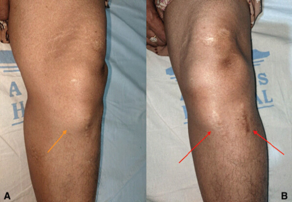
On clinical examination, there was a swelling over the anteromedial aspect of the left proximal tibia measuring 4 × 3 × 2 cm in size, with the proximal end of the swelling overlying the medial joint line. The swelling was round to oval in shape (Fig. 1a). There was no local warmth. The swelling was diffusely tender, soft in consistency, non-fluctuant, with a smooth surface, and normal overlying skin. There were two healed vertical scar marks present over the proximal tibia, each measuring approximately 4 cm, one over the swelling and the other about 3 cm lateral to it (Fig. 1b). The patient also had a limited extension of the knee joint. The Tinel’s sign was negative, and the remaining neurovascular examination was normal.
Plain radiographs of both knees showed the soft tissue shadow in the region of the tibial tuberosity on the left side (lateral view) with an otherwise normal knee joint (Fig. 2). An MRI of the left knee was performed, which demonstrated a well-circumscribed, juxta-articular multilobulated lesion in the infrapatellar fat pad measuring about 6.7 × 5.5 × 2.5 cm, extending inferiorly into the pretibial fat (Fig. 3a and b).
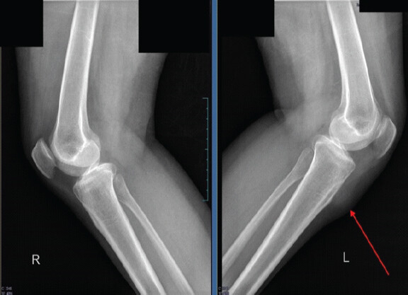
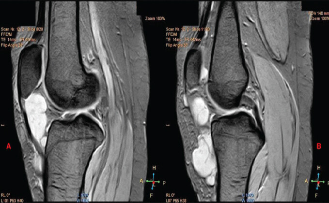
Fine-needle aspiration cytology of the lump revealed spindle cells embedded in the mesenchymal matrix with elongated hyperchromatic nuclei, suggesting a spindle cell lesion.
The patient was planned for surgical excision. Under spinal anesthesia, a midline incision of approximately 10 cm was made, and a medial parapatellar arthrotomy was performed. A multiloculated soft tissue lesion measuring 6 × 4 × 1.5 cm was found arising from the HFP beneath the patellar tendon, which was excised completely. The swelling was solitary, well-defined, and encapsulated, and grayish-yellow in color (Fig. 4). Minor wear of the medial femoral articular cartilage was noted. The anterior horns of the medial and lateral menisci were found to be normal and stable. The specimen was sent for histopathological examination and IHC analysis.
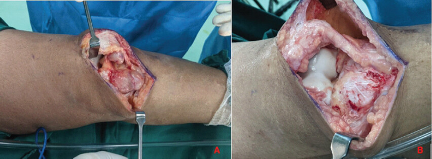
Standard post-operative protocol for analgesics and antibiotics was followed. The patient was mobilized on post-operative day 1 with full weight bearing, and physiotherapy was initiated. For osteoarthritis, she was started on non-operative management with lifestyle modification and quadriceps strengthening exercises. She was discharged on post-operative day 3.
On the cut surface of the specimen, the lesion was gray-white, trabeculated, and contained myxoid areas with intervening hemorrhagic space, giving it a firm to fibrous appearance. Histological examination revealed a cellular spindle cell lesion with hyper- and hypocellular areas. The spindle cells had eosinophilic fibrillary cytoplasm and elongate ovoid nuclei. These cells were arranged in intersecting fascicles and whorls. In the hypocellular areas, the cells were loosely arranged within an edematous stroma. Marked nuclear atypia was noted without evidence of mitoses or necrosis (Fig. 5a).
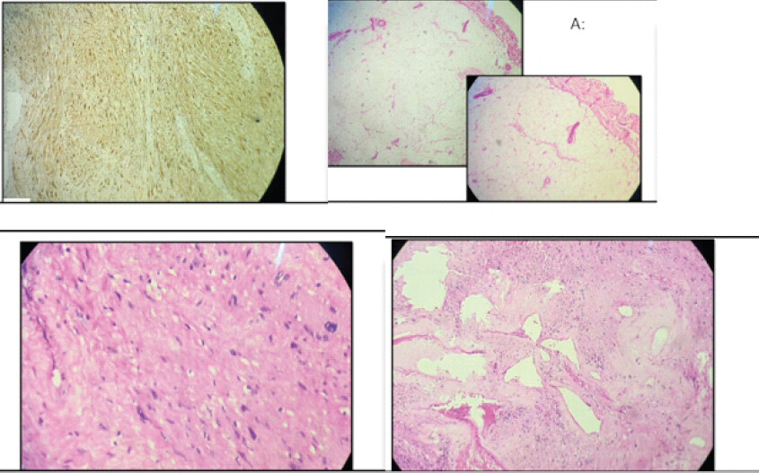
These findings suggested a spindle cell neoplasm, and IHC was performed. IHC showed strong positivity for S-100 protein, while epithelial membrane antigen was focally and weakly positive, and the Ki-67 proliferation index was 2-3%. The final diagnosis was spindle cell neoplasm consistent with schwannoma with degenerative changes (Fig. 5b).
At the 3-year follow-up, the patient reports symptoms only after prolonged standing or excessive walking. On clinical examination, there is no evidence of recurrence.
Discussion
Schwannoma, also known as neurilemmoma, is a benign encapsulated soft tissue tumor classified under the group of peripheral nerve sheath tumors [12]. It usually occurs in the fourth and fifth decades of life and affects men and women equally [13].
These tumors are often asymptomatic and are usually discovered incidentally owing to their slow growth. Apart from the long-standing swelling, patients may present with pain, paresthesia, and a tingling sensation, which may suggest an unfavorable postoperative outcome [10]. A Tinel’s sign along the course of the nerve is present in 4–76% of patients [10]. These symptoms develop when eccentrically placed lesions become large enough to compress the nerve.
Schwannomas can occur anywhere in the body and represent about 5% of all benign soft-tissue neoplasms [13]. To the best of the author’s knowledge, only two cases of intra-articular knee schwannomas have been reported in the literature, of which one was reported to originate from HFP. Recurrence of the Schwannoma itself is rare (<1%) [13]. To the best of the author’s knowledge, this is the first reported case of intra-articular recurrence of a schwannoma arising from the HFP.
The HFP is a richly innervated structure, receiving branches of the femoral, common peroneal, and saphenous nerves [2]. Since the tumor was located within the HFP, it is possible that it originated from one of these nerves. However, the exact nerve of origin or the specific branch could not be identified.
The principle of treatment for schwannomas involves complete surgical excision of the tumor through an open approach. Due to the encapsulating nature of schwannomas, en bloc excision is usually achievable. Careful dissection is essential to preserve the function of the involved nerve [14]. Because of the extra-synovial nature of HFP, open arthrotomy with complete excision is the recommended treatment for both cystic and solid tumors involving the HFP, as arthroscopic resection may not allow complete removal of the lesion [3]. In the present case, the patient underwent open arthrotomy and excision to achieve complete resection.
Conclusion
In summary, this case is a significant contribution to the literature as it is the first reported case of a recurrent intra-articular schwannoma involving the HFP. Given the scarcity of documented cases at this site, our findings emphasize the importance of considering schwannoma as a potential differential diagnosis for tumors originating in the HFP. Our contribution will facilitate early recognition, accurate diagnosis, and effective management of schwannomas in this anatomical location, ultimately improving patient outcomes and guiding future research endeavors in this area.
Clinical Message
Schwannoma, although rare, should be considered as a differential diagnosis for lesions arising from HFP, especially in patients presenting with long-standing swelling with recent onset of pain or neurological symptoms. Despite its benign nature, schwannoma can rarely recur even after complete excision, sometimes after a prolonged interval. A thorough clinical evaluation, supported by MRI and histopathological confirmation, is essential for accurate diagnosis. Complete surgical excision with meticulous dissection remains the treatment of choice to minimize recurrence and preserve function.
Conflict of Interest:
Nil
Source of Support:
Nil
Consent:
The authors confirm that informed consent was obtained from the patient for publication of this article
References
- Hoffa A. The influence of the adipose tissue with regard to the pathology of the knee joint. JAMA 1904;795:796. [Google Scholar] | [PubMed]
- Biedert RM, Sanchis-Alfonso V. Sources of anterior knee pain. Clin Sports Med 2002;21:335-47, vii. [Google Scholar] | [PubMed]
- Gallagher J, Tierney P, Murray P, O'Brien M. The infrapatellar fat pad: Anatomy and clinical correlations. Knee Surg Sports Traumatol Arthrosc 2005;13:268-72. [Google Scholar] | [PubMed]
- Helpert C, Davies AM, Evans N, Grimer RJ. Differential diagnosis of tumours and tumour-like lesions of the infrapatellar (Hoffa's) fat pad: Pictorial review with an emphasis on MR imaging. Eur Radiol 2004;14:2337-46. [Google Scholar] | [PubMed]
- Dean BJ, Reed DW, Matthews JJ, Pandit H, McNally E, Athanasou NA. The management of solitary tumours of Hoffa's fat pad. Knee 2011;18:67-70. [Google Scholar] | [PubMed]
- Albergo JI, Gaston CL, Davies M, Abudu AT, Carter SR, Jeys LM. Hoffa's fat pad tumours: What do we know about them? Int Orthop 2013;37:2225-9. [Google Scholar] | [PubMed]
- Ivanov S, Trizlov D. Painful anterior knee schwannoma: A case report. J IMAB 2023;29:4990-3. [Google Scholar] | [PubMed]
- Caballero Martel JR, Estévez Sarmiento S. Schwannoma: A rare Hoffa's fat pad tumor. Surg J (NY) 2019;5:e62-4. [Google Scholar] | [PubMed]
- Hosmann A, Kamdar V, Misra BK. Malignant transformation of vestibular schwannoma following radiosurgery-a case report and review of the literature. Acta Neurochir (Wien) 2024;166:52. [Google Scholar] | [PubMed]
- Karna MB, Kinanta PB, Aprilya D. Recurrent schwannoma of digital nerve on both hands: A very rare case report. Int J Surg Case Rep 2023;103:107915. [Google Scholar] | [PubMed]
- Fischer B, Munaretto F, Fritschy D, Peter RE. An unusual arthroscopic discovery: An intraarticular Schwannoma of the knee. Arthroscopy 1994;10:113-7. [Google Scholar] | [PubMed]
- Ferner RE, O'Doherty MJ. Neurofibroma and schwannoma. Curr Opin Neurol 2002;15:679-84. [Google Scholar] | [PubMed]
- Andrea A, Bevoni R, Biz C, Cerchiaro MC, Girolami M, Ruggieri P. Schwannoma of the foot: Report of four cases and literature review. Acta Biomed 2019;90:214-20. [Google Scholar] | [PubMed]
- Kang HJ, Shin SJ, Kang ES. Schwannomas of the upper extremity. J Hand Surg Br 2000;25:604-7. [Google Scholar] | [PubMed]
© 2026 Journal of Orthopaedic Case Reports - Published by Indian Orthopaedic Research Group
About the Authors
-

Dr. Vipin Mohan
-

Dr. Prince Agrawal
-

Mr. Evin Siby
-

Dr. Balu C Babu
-

Mr. Ikshu Jain
-

Dr. K K Chandrababu
Related Articles in Journal of Orthopaedic Case Reports
 July 1, 2026 Schwannoma Arising from the Deep Peroneal Nerve: A Case Report
 January 1, 2026 Anteromedial Knee Pain Unmasked: Schwannoma of the Infrapatellar Branch of the Saphenous Nerve – A Rare Case Report
 January 1, 2026 Recurrent Symptoms in Lumbar Disc Herniation with Concomitant Schwannoma: The Role of Tumor Microenvironment and Anti-Inflammatory Therapy
 August 1, 2025 Hidden in Plain Palm: A Rare Case of Incidental Thenar Schwannoma