


Introduction
Posterior shoulder and periscapular pain in young athletes are commonly approached as shoulder or soft-tissue problems. In softball and baseball players, the initial differential includes rotator cuff irritation, scapular dyskinesis, rhomboid strain, levator scapulae strain, labral pathology, and other overuse injuries. First rib stress fracture is much less common, but it should be considered because pain may localize to the scapular region rather than the chest wall.
Most published first rib stress fractures involve throwing athletes. Funakoshi et al. reported 24 first rib stress fractures in 23 overhead throwing athletes and identified acute posterior shoulder pain during throwing or batting as an important clinical clue [1]. The proposed mechanism involves repetitive muscular traction across the first rib, particularly from the scalene, serratus anterior, and intercostal muscles, with stress concentrating at vulnerable portions of the rib [1,2,3,4]. Batting-related cases have been reported, with repetitive traction and shear forces during the swing as the likely contributing mechanism [3].
The injury may be missed even when the athlete is evaluated and radiographs are obtained. Sheng et al. described three National Collegiate Athletic Association Division I athletes whose first rib stress fractures presented as vague rhomboid or interscapular pain [5]. Funakoshi et al. found that shoulder radiographs visualized only 46% of the first rib on average, compared with 97% on anteroposterior cervical spine radiographs [1]. For that reason, a first rib fracture may be present on initial imaging but overlooked when attention remains focused on the glenohumeral joint or periscapular soft tissue.
This diagnostic difficulty is not limited to youth athletes or settings with limited medical resources. New York Yankees outfielder Aaron Judge was diagnosed in March 2020 with a first rib stress fracture after several months of shoulder and pectoral symptoms. The injury was believed by the Yankees organization to have occurred during an attempted diving catch the previous September [6,7]. Although this publicly reported case is not medical evidence, it shows that a first rib stress fracture can present as shoulder-region pain and remain unrecognized even in an elite athlete with access to high-level sports medicine care.
We report a 10-year-old softball player whose first rib stress fracture was initially diagnosed as a periscapular muscle strain. The fracture was visible in retrospect on the original shoulder radiographs but was not identified until repeat imaging was obtained 4 months later. This case shows how familiar clinical presentation and limited visualization of the first rib can delay diagnosis.
Case Report
A 10-year-old female athlete who played competitive softball and basketball presented to a sports medicine clinic with right posterior shoulder pain. She was accompanied by her father, who reported that the pain began approximately 2 weeks earlier during a batting lesson. She had recently started light workouts twice weekly focused on agility and lower body strength. Two days before her symptoms started, she added upper body conditioning, including ring pull-ups and push-ups.
During the batting lesson, she felt a “pop” in her right shoulder on her third swing, but she completed two additional swings. The pain worsened after the lesson, and she stopped all activity for 1 week. Her symptoms improved with rest. When she resumed throwing 1 week later, she reported mild pain rated 2–3 out of 10. She participated in a basketball scrimmage without discomfort. During a subsequent batting lesson, the pain returned within 15 min, and she had difficulty getting out of bed the following morning.
The pain was localized to the posterior right shoulder and the medial scapular region. She had been using a tennis ball against the wall for self-massage, which she described as painful but temporarily helpful. Her father was concerned about a possible rotator cuff or ligament injury.
On examination, the shoulder appeared normal without swelling, ecchymosis, erythema, or deformity. Palpation demonstrated tenderness along the medial border of the scapula without glenohumeral joint tenderness. Forward flexion and abduction were both 170° and reproduced posterior pain. Internal rotation reached T10. Strength was 5 out of 5 with forward flexion, abduction, external rotation, and internal rotation, although resisted testing reproduced the pain. Neer, Hawkins, cross-body adduction, O’Brien, and empty can tests were negative. There was no scapular winging. Distal sensation and pulses were normal.
Right shoulder radiographs, anteroposterior, Grashey, and axillary views, were obtained and interpreted as normal. The initial diagnosis was a periscapular muscle strain, most likely involving the rhomboid musculature. She was treated with a 5-day course of oral prednisone and provided home physical therapy exercises. She was advised to avoid upper body, back, and chest exercises but was permitted to continue basketball and lower body or core exercises if these activities did not reproduce her pain.
Her symptoms improved with rest and activity modification. She participated in basketball throughout the winter without discomfort. Approximately 2 months later, she returned to softball and initially tolerated participation. As her batting volume increased, she developed recurrent pain while swinging. The pain returned to the superior and medial scapular region. Rest, ice, and anti-inflammatory medications provided partial relief, but symptoms persisted. She also developed pain with deep inspiration.
Four months after the initial presentation, she was reevaluated in a different clinic. There was no visible deformity. She had focal tenderness over the superior and medial scapula along the levator scapulae. Shoulder range of motion was full, and strength testing did not demonstrate weakness. Spurling test was negative. Provocative testing for rotator cuff, biceps, labral, and instability was negative. Sensation was grossly intact, and vascular examination was normal.
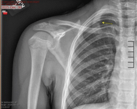
Repeat right shoulder radiographs were obtained with specific attention to the first rib and demonstrated a healing non-displaced right first rib fracture, which the radiology report stated, “likely explains the patient’s pain.”
Retrospective review of the initial radiographs confirmed that the non-displaced first rib fracture had been present but was not identified (Fig. 1). She was diagnosed with a first rib stress fracture and recurrent symptoms after returning to batting before the fracture had healed.
She was restricted from throwing, batting, and overhead activity for 6 weeks. Her Vitamin D was 32 ng/mL, and her serum calcium was 9.4 mg/dL. She was counseled about adequate dietary intake and was started on calcium and Vitamin D supplementation.
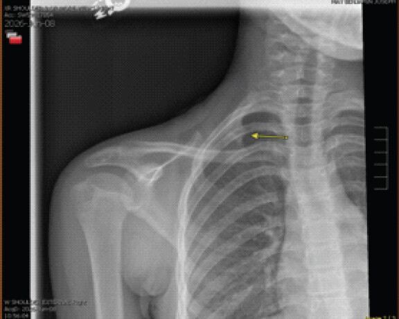
At a follow-up on June 8, 2026, she reported no pain during the previous 3–4 weeks. She had avoided throwing and batting and reported no new injuries. Examination showed no tenderness or deformity, full shoulder range of motion, normal strength, negative provocative shoulder testing, and intact neurovascular function.
Three-view right shoulder radiographs were compared with the April 2026 images and showed a healed right first rib fracture (Fig. 2). The fracture lucency was no longer visible, and no new fracture, dislocation, or other acute shoulder abnormality was identified.
Because she was asymptomatic and imaging confirmed complete healing, she was allowed to begin a gradual return to softball, including throwing and batting. She was advised to have 3–4 rest days/week during the 1st 3 weeks after resuming activity and to stop if pain or loss of motion developed. If she remained asymptomatic, she could then return to full participation as tolerated while continuing to incorporate at least 1 or 2 rest days each week.
The family was provided with a staged throwing program. They were advised that the throwing distances and number of repetitions had been designed for older baseball players and would need to be adjusted for her age and level of play. At her most recent primary care follow-up, she had returned to full softball participation and remained pain-free without recurrent symptoms.
Discussion
The initial presentation was consistent with a periscapular muscle injury. The patient had focal medial scapular tenderness, preserved shoulder strength, nearly full range of motion, no scapular winging, and negative provocative testing. The initial diagnosis of periscapular muscle strain was therefore reasonable. However, the mechanism was less typical for a routine muscle strain. Her pain began suddenly with a “pop” during batting and later returned when she resumed softball.
Pain with deep inspiration developed after the recurrence of symptoms. Although this finding is not specific, it suggested possible rib or chest wall involvement, particularly because the repeat shoulder examination remained reassuring. A retrospective review later showed that the fracture was visible on the original radiographs but was not recognized because the evaluation focused on the shoulder and periscapular soft tissue.
First rib stress fractures are rare and may be under-recognized. Funakoshi et al. reported 24 fractures in 23 overhead throwing athletes, many of whom presented with posterior shoulder or upper thoracic pain [1]. Nchinda et al. described pediatric first rib fractures occurring without major trauma and found that exercise was the most common cause, ipsilateral shoulder pain was the most common presenting symptom, most fractures were non-displaced, and all patients were treated without surgery [8]. These findings are similar to the presentation in this case.
Marcussen et al. described 14 isolated traumatic first rib fractures sustained during sports and concluded that these injuries may be more common than sports medicine clinicians realize [9]. Their series involved acute traumatic fractures rather than stress fractures, but it supports consideration of first rib injury in athletes with shoulder pain. The authors also found that return to sport after rehabilitation and resolution of symptoms appeared reasonable [9].
The first rib is exposed to opposing muscular forces. The scalene muscles act on the superior aspect of the rib, while the serratus anterior and the intercostal muscles apply force inferiorly. Repetitive contraction can create traction and bending stress, particularly near the subclavian groove [1,2]. Bat swinging may produce additional rotational and shear forces across the rib [3]. The recent addition of ring pull-ups and push-ups may have increased her training demands before symptoms began, although a direct causal relationship cannot be established.
Routine shoulder radiographs may not fully visualize the first rib, which can contribute to missed diagnoses. Funakoshi et al. reported that shoulder radiographs visualized 46% of the first rib on average, compared with 97% on anteroposterior cervical spine radiographs [1]. In this case, however, the fracture was visible on the original shoulder images but was not recognized because attention remained focused on the glenohumeral joint and periscapular soft tissue. When the history raises concern for a first rib injury, clinicians should specifically inspect the rib and consider additional radiographic views, an anteroposterior cervical spine radiograph, computed tomography, magnetic resonance imaging, or musculoskeletal ultrasound if the diagnosis remains uncertain.
Sheng et al. reported three Division I athletes with first rib stress fractures that presented as persistent rhomboid or interscapular pain. Ultrasound demonstrated cortical irregularity at the fracture site in each case [5]. Point-of-care ultrasound may be useful when the clinical suspicion remains high, although available evidence is limited, and the examination depends on operator experience.
Treatment is usually non-surgical and includes restrictions from painful activity, correction of training load, attention to nutrition, and gradual return to sport [1, 3, 5, 6, 7]. Funakoshi et al. reported healing in 71% of fractures at a mean follow-up of 7.5 months, whereas 29% developed non-union [1]. Persistent pain or neurovascular symptoms should prompt further evaluation because first rib non-union or associated anatomic changes may contribute to thoracic outlet compression [1, 10, 11].
Bone health and training factors should be considered in young athletes with stress fractures, including dietary intake, recent changes in training volume, prior stress injuries, and other risk factors for low energy availability [12,13,14]. She had not reached menarche, which may be developmentally appropriate at age 10 and should not itself be interpreted as menstrual dysfunction. In this patient, Vitamin D and calcium levels were 32 ng/mL and 9.4 mg/dL, respectively. She received nutrition counseling and was started on calcium and Vitamin D supplementation while the fracture healed.
This case is notable because the injury was mistaken for a periscapular muscle strain and was visible but overlooked on the initial radiographs. The delayed diagnosis reported in the case of Aaron Judge provides a recognizable example of the same clinical problem. It does not provide evidence regarding causality or recurrence, but it shows that the first rib stress fracture may present as shoulder or pectoral discomfort and remain unrecognized even in athletes receiving high-level medical care [6, 7].
Conclusion
First rib stress fracture should be considered in batting and overhead athletes who present with sudden posterior shoulder or periscapular pain, especially when symptoms recur with return to sport or are associated with pain during deep inspiration. Routine shoulder radiographs may incompletely visualize the first rib, and the rib should be specifically reviewed when the history remains concerning. In this patient, activity restriction led to resolution of symptoms and complete radiographic healing, followed by successful return to softball without recurrent pain.
Clinical Message
First rib stress fractures are an uncommon but important cause of posterior shoulder and periscapular pain in adolescent athletes and should be considered when symptoms persist or recur despite appropriate treatment for a presumed muscular injury.
Conflict of Interest:
Nil
Source of Support:
Nil
Consent:
The authors confirm that informed consent was obtained from the patient for publication of this article
References
- Funakoshi T, Furushima K, Kusano H, Itoh Y, Miyamoto A, Horiuchi Y. First-rib stress fracture in overhead throwing athletes. J Bone Joint Surg Am 2019;101:896-903. [Google Scholar] | [PubMed]
- Miller TL, Harris JD, Kaeding CC. Stress fractures of the ribs and upper extremities: Causation, evaluation, and management. Sports Med 2013;43:665-74. [Google Scholar] | [PubMed]
- Kawashima K, Terabayashi N, Miyagawa T, Tanaka R, Ogawa H, Takigami I. Stress fractures of the first rib related to swinging of a baseball bat: Two case reports. Clin J Sport Med 2016;26:e108-10. [Google Scholar] | [PubMed]
- Sakata T, Kimura Y, Hida T. First rib stress fracture in a sidearm baseball pitcher: A case report. J Sports Sci Med 2005;4:201-7. [Google Scholar] | [PubMed]
- Sheng DL, Burnham K, Boutin RD, Ray JW. Ultrasound identifies first rib stress fractures: A case series in national collegiate athletic association division I athletes. J Athl Train 2023;58:664-8. [Google Scholar] | [PubMed]
- Hoch B. Judge has Fractured Rib; Shut Down for 2 Weeks; 2020. Available from: https://www.com/mlb.com. [Google Scholar] | [PubMed]
- ESPN News Services. Yankees' Aaron Judge Says Rib Improving, Also Had Collapsed Lung India: ESPN; 2020. [Google Scholar] | [PubMed]
- Nchinda NN, Kamineni DP, Otjen JP, Rothstein DH, Javid PJ. Refining management of pediatric first rib fractures without major trauma. J Pediatr Surg 2025;60:162652. [Google Scholar] | [PubMed]
- Marcussen B, Negaard M, Hosey RG, Smoot MK. A case series and literature review: Isolated traumatic first rib fracture in athletes. Clin J Sport Med 2020;30:257-66. [Google Scholar] | [PubMed]
- Proffer DS, Patton JJ, Jackson DW. Nonunion of a first rib fracture in a gymnast. Am J Sports Med 1991;19:198-201. [Google Scholar] | [PubMed]
- Sheng GG, Duwayri YM, Emery VB, Wittenberg AM, Moriarty CT, Thompson RW. Costochondral calcification, osteophytic degeneration, and occult first rib fractures in patients with venous thoracic outlet syndrome. J Vasc Surg 2012;55:1363-9. [Google Scholar] | [PubMed]
- Gallant TL, Ong LF, Wong L, Sparks M, Wilson E, Puglisi JL. Low energy availability and relative energy deficiency in sport: A systematic review and meta-analysis. Sports Med 2025;55:325-39. [Google Scholar] | [PubMed]
- Angelidi AM, Stefanakis K, Chou SH, Valenzuela-Vallejo L, Dipla K, Boutari C. Relative energy deficiency in sport (REDs): Endocrine manifestations, pathophysiology and treatments. Endocr Rev 2024;45:676-708. [Google Scholar] | [PubMed]
- Weiss Kelly AK, Hecht S. Council on Sports Medicine and Fitness. The female athlete triad. Pediatrics 2016;138:e20160922. [Google Scholar] | [PubMed]
© 2026 Journal of Orthopaedic Case Reports - Published by Indian Orthopaedic Research Group
About the Authors
-

Dr. Burke McCarty
-

Dr. Benjamin May
Related Articles in Journal of Orthopaedic Case Reports
 June 1, 2026 T1Ρ and T2 Mapping Magnetic Resonance Imaging as Predictors of Cartilage Loss in Early Anterior Cruciate Ligament Injury
 October 10, 2016 Pneumothorax After Shoulder Arthroscopy: A Rare but Life-threatening Complication
 August 10, 2021 A Case Report of Bilateral Asymmetrical Traumatic Hip Dislocation – A Rare Presentation
 April 10, 2021 A Case Report of Proximal Migration of Fibular Head with Intact Peroneal Nerve in a Post-Operative Infected Tibia Gap Non-union Managed by Ilizarov Technique