


Introduction
Proximal femoral nailing (PFN) is widely used for the surgical management of intertrochanteric femur fractures. Nowadays, the commonly used implants in various hospital setups are PFN (conventional PFN), PFN antirotation, and InterTan, which are minimally invasive [1]. These nails have a greater trochanteric tip entry point because of their lateral bend in the coronal plane [2]. Thus, the surface landmark location of the greater trochanter (GT) tip is the most vital step at the beginning of the surgery using these implants, which helps in applying the appropriate skin incision and entry points. The localisation of the GT tip becomes difficult in edematous and obese patients, potentially resulting in a malpositioned entry point or an unnecessarily extended skin incision, which should ideally be placed 3–5 cm from the GT tip [3] and may be associated with other complications [2]. In this report, we describe a simple, reliable, reproducible surface marking, the “hand-palm technique,” to locate the tip of the GT, which is confirmed by C-arm images. A pilot observational study was additionally conducted to evaluate the clinical applicability, localization accuracy, reproducibility, and operative parameters associated with the technique.
Materials and Methods
Written informed consent (consent to participate and consent to publish) was obtained from all participants, and IEC approval was sought (AIIMS/IEC/M12/F379/2026). A prospective pilot observational study was performed in 41 patients undergoing PFN for intertrochanteric femur fractures between February and May 2026. Patient demographics, body mass index (BMI) distribution, localization accuracy, fluoroscopy usage, operative parameters, and reproducibility of the technique were evaluated. The marked point identified using the hand-palm technique was correlated with fluoroscopic confirmation of the GT tip in all patients.
Statistical analysis
Descriptive statistical analysis was performed using mean ± standard deviation, median, range, and frequency distribution. Subgroup analysis between obese and non-obese patients was performed using the Mann-Whitney U test. Additional BMI subgroup analysis was performed among underweight, normal BMI, overweight, and obese patients to evaluate validation parameters, reproducibility, and operative outcomes. A P < 0.05 was considered statistically significant.
Hand palm technique
The technique is outlined in two scenarios to ensure reproducible outcomes:
-
For surgeons using hand gloves size 7 or larger
-
For surgeons using gloves size 6.5 or smaller.
For a surgeon wearing gloves sized 7 and above
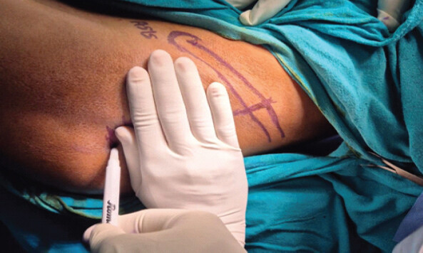
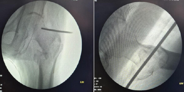
In this case, the patient was laid supine on the fracture table with traction applied. Following the closed reduction of the fracture, painting and draping procedures were performed. The anterior superior iliac spine (ASIS) was indicated with a skin marker. Subsequently, using the width of the surgeon’s four medial fingers (from index to little), a point was marked on the iliac crest (Fig. 1). From this marked point, the four fingers were aligned parallel to the iliac crest (Fig. 2). A point was then marked that indicates the precise location of the tip of the GT. This location was verified through direct palpation of the GT tip in the designated area and through fluoroscopic imaging (both anteroposterior and lateral views) utilizing a metal marker to confirm the technique (Fig. 3).
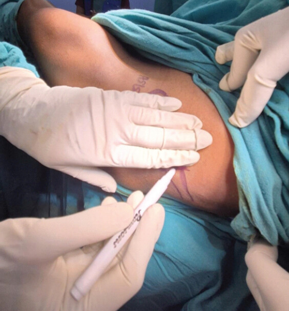
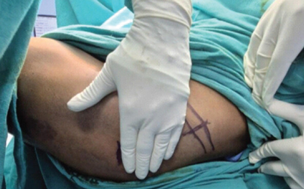
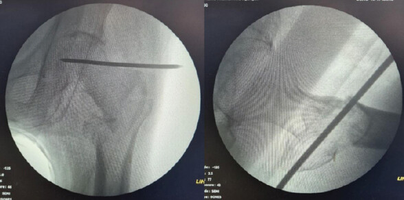
For a surgeon with glove size 6.5 and below
In a similar manner to the previous description, after the closed reduction of the fracture, painting and draping were conducted. The ASIS was marked. In this instance, the surgeon used the width of all fingers, including the thumb, thereby employing the entire palm. From the ASIS, a point was marked on the iliac crest, and then the palm was positioned parallel to the iliac crest. The marked point indicated the location of the GT tip, which was confirmed using a metal marker and C-arm imaging (Figs. 4, 5, 6).
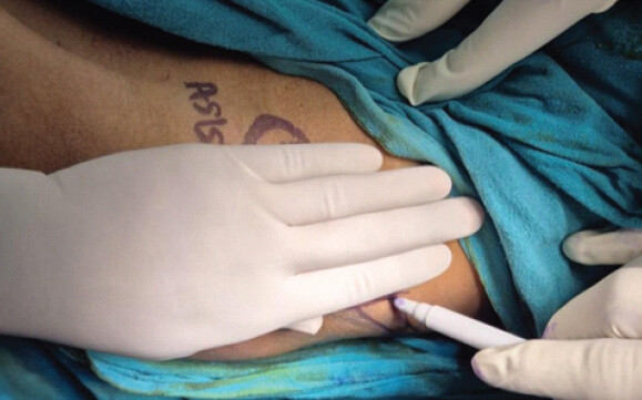
Results
A total of 41 patients were included in the pilot study. The mean age was 61.85 ± 9.70 years, and the mean BMI was 22.79 ± 3.01 kg/m2. Patient demographic characteristics and baseline parameters are summarized in Table 1. Fracture type distribution among the study population is shown in Table 1. Type A1 fractures were the most common pattern (60.9%), followed by type A2 (29.3%) and type A3 fractures (9.8%).
Demographic and baseline characteristics of patients
| Total (n) | Mean age (years)/range | Mean BMI (kg/m2)/Median/range | Male patients, n (%) | Female patients, n (%) | Obese patients (BMI ≥30), n (%) | Overweight (25–29.9), n (%) | Normal BMI (18.5–24.9), n (%) | Underweight (BMI <18.5), n (%) |
|---|---|---|---|---|---|---|---|---|
| 41 | 61.85±9.70 (42-80) | 22.79±3.01/22.3 5/(18.2–31.8) | 20 (48.8) | 21 (51.2) | 2 (4.9) | 6 (14.6) | 32 (78.0) | 1 (2.4) |
| Fracture type (AO type 31A) | Number of patients (n) | Percentage | ||||||
| Type A1 | 25 | 60.9 | ||||||
| Type A2 | 12 | 29.3 | ||||||
| Type A3 | 4 | 9.8 | ||||||
| Total | 41 | 100 | ||||||
Values are expressed as frequency and percentage of the total study population. Values are expressed as mean±standard deviation unless otherwise specified. BMI: Body Mass Index.
The hand-palm technique demonstrated satisfactory localization accuracy with a mean localization error of 2.19 ± 1.74 mm from the fluoroscopically confirmed GT tip. The mean number of fluoroscopic images required was 1.66 ± 0.62. Technique validation parameters are summarized in Table 2.
Technique validation parameters
| Parameter | Value |
|---|---|
| Mean localization error from fluoroscopic GT tip (mm)/range | 2.19±1.74 (0–5.4) |
| Median localization error (mm) | 2.1 |
| Mean fluoroscopic images required/range | 1.66±0.62 (1–3) |
| Median fluoroscopic images required | 2 |
| Cases requiring incision extension, n (%) | 3 (7.3) |
| Cases without incision extension, n (%) | 38 (92.7) |
Values are expressed as mean±standard deviation unless otherwise specified. GT: Greater trochanter
Operative parameters including operative time, blood loss, fluoroscopy time, and incision length were also analyzed and are summarized in Table 3.
Operative parameters
| Parameter | Value |
|---|---|
| Mean operative time (minutes)/range | 39.90±7.01 (25–54) |
| Median operative time (minutes) | 40 |
| Mean blood loss (mL)/range | 119.51±49.85 (50–200) |
| Median blood loss (mL) | 100 |
| Mean fluoroscopy time/range | 3.98±0.65 (3–5) |
| Median fluoroscopy time | 4 |
| Mean incision length (cm)/range | 5.61±0.40 (5–6.4) |
| Median incision length (cm) | 5.6 |
Values are expressed as mean±standard deviation unless otherwise specified
Subgroup analysis demonstrated a trend toward greater localization error and increased fluoroscopic image requirement in obese patients compared with non-obese patients, although the difference was not statistically significant (Table 4).
Subgroup analysis between obese and non-obese patients
| Variable | Obese patients (n=2) | Non-obese patients (n=39) | P-value* |
|---|---|---|---|
| Localization error (mm) | 3.75±2.33 | 2.11±1.71 | 0.258 |
| Fluoroscopic images required | 2.00±0.00 | 1.64±0.63 | 0.359 |
Values are expressed as mean±standard deviation.
*
Subgroup comparison performed using Mann-Whitney U test. A P<0.05 was considered statistically significant
BMI subgroup analysis demonstrated variations in localization accuracy, fluoroscopy requirement, reproducibility, and operative parameters among underweight, normal BMI, overweight, and obese patients. Obese patients demonstrated comparatively higher localization error and fluoroscopic image requirement, whereas overweight patients showed relatively higher operative time and blood loss. BMI-wise validation and operative parameters are summarized in Table 5. Incision extension was required in 3 patients (7.3%), including one patient each from the normal BMI, overweight, and obese categories.
BMI-wise validation, reproducibility, operative parameters
| BMI category | n | Localization error (mm) mean±SD | Fluoroscopic images mean±SD | Reproducibility Mean±SD | Operative time (min) mean±SD | Blood loss (mL) mean±SD | Fluoroscopy time Mean±SD | Incision length (cm) mean±SD |
|---|---|---|---|---|---|---|---|---|
| Underweight (<18.5) | 2 | 1.20±1.70 | 1.00±0.00 | 0.50±0.71 | 44.50±0.71 | 75.00±35.36 | 4.00±0.00 | 5.65±0.21 |
| Normal BMI (18.5–24.9) | 32 | 2.35±1.68 | 1.72±0.63 | 0.53±1.11 | 39.50±6.90 | 120.31±50.58 | 3.97±0.69 | 5.67±0.41 |
| Overweight (25–29.9) | 5 | 0.96±1.60 | 1.40±0.55 | 1.60±1.14 | 44.60±5.77 | 130.00±57.01 | 4.00±0.71 | 5.28±0.22 |
| Obese (>30) | 2 | 3.75±2.33 | 2.00±0.00 | 0.00±0.00 | 30.00±0.00 | 125.00±35.36 | 4.00±0.00 | 5.50±0.28 |
Values are expressed as mean±standard deviation. Localization error was calculated as the distance between the marked point and the fluoroscopically confirmed greater trochanter tip. Reproducibility was assessed using interobserver variation. Operative parameters were analyzed according to BMI subgroup distribution. BMI: Body mass index, SD: Standard deviation
Discussion
Intertrochanteric femur fractures are widely treated with PFNs with newer designs nowadays. The studies related to the surgical outcomes of these implants have documented less blood loss during surgery because of smaller surgical incisions and less operating time [1,4,5,6]. In a scenario of obese patients, an extended incision because of improper localization of the GT tip may lead to excessive blood loss; hence, proper identification of the tip of the GT is necessary. Furthermore, studies in the literature [4,5,7] have reported less use of fluoroscopy images while using newer designs of PFN. In the present pilot study, the mean number of fluoroscopic images required was low (1.66 ± 0.62), suggesting that the technique may help educe repeated C-arm localization attempts and associated radiation exposure. Although direct palpation techniques for identifying the GT tip have been described [3,7], palpation may be difficult in patients with increased soft-tissue thickness or obesity. Anatomical variability among patients, including differences in pelvic width, body habitus, obesity, soft tissue thickness, and skeletal proportions, may influence the exact surface relationship between the ASIS and the GT tip [2]. Potential inaccuracies may therefore occur in markedly short, tall, obese, or anatomically variant individuals, as obesity and altered body habitus may increase the difficulty of surface landmark identification and fluoroscopic localization during femoral nailing procedures [8]. BMI-wise subgroup analysis in the present pilot study demonstrated comparatively higher localization error and fluoroscopic image requirement among obese patients. However, operative parameters remained within acceptable limits across all BMI categories, suggesting that the technique may still be clinically applicable in patients with varying body habitus. Hence, the hand-palm technique may be considered an adjunctive surface localization aid, and fluoroscopic confirmation remains essential before definitive entry point creation. The technique may also help in positioning the correct entry point without damaging nearby structures such as abductors and tendons, the capsule of the hip joint, and the division of the medial circumflex femoral artery [2, 9]. Hence, the hand-palm technique appears to be a practical and reproducible surface landmarking aid for localization of the GT tip. The technique may also have potential utility in hip arthroplasty performed through a posterolateral approach, where the GT tip is used as a surface landmark for skin-incision placement [3], unless there is no proximal migration of the GT. Furthermore, the technique is useful for intra-articular hip injections in the outpatient clinic, where palpating the GT tip is a vital step for locating the landmark of the injection site [10,11,12]. It is easy to learn and apply, making it beneficial for trainees and in less-resourced settings..
Limitations
The present study has certain limitations. This was a pilot observational study with a relatively small sample size and without a comparative control group. Although operative parameters and localization accuracy were assessed, direct comparison with conventional palpation techniques was not performed. Larger prospective comparative studies are required to further validate reproducibility and clinical utility.
Conclusion
The hand-palm technique is a simple and practical surface landmarking method for approximate localization of the GT tip during PFN. The technique demonstrated satisfactory localization accuracy in this pilot study and may be particularly useful in obese or edematous patients, in whom direct palpation is difficult. However, fluoroscopic confirmation remains essential before definitive skin incision and entry point creation. Further prospective comparative studies are required for validation.
Clinical Message
The “hand-palm technique” is a practical surface landmarking aid for localization of the greater trochanter tip and demonstrates satisfactory localization accuracy in a pilot validation study.
Conflict of Interest:
Nil
Source of Support:
Nil
Consent:
The authors confirm that informed consent was obtained from the patient for publication of this article
How to Cite this Article
Mallya S, Puranik A. A Hand-Palm Technique for Localization of the Greater Trochanter Tip During Proximal Femoral Nailing: A Prospective Pilot Validation Study. Journal of Orthopaedic Case Reports 2026 October;16(10): 619-624.
References
- Shah MR, Shah MM, Shah IM, Shah KR. Surgical and functional outcomes of the results of conventional two-screw proximal femoral nail (PFN) versus helical-blade anti-rotation proximal femoral nail (PFNA2). Cureus 2023;15:e43698. [Google Scholar] | [PubMed]
- Farhang K, Desai R, Wilber JH, Cooperman DR, Liu RW. An anatomical study of the entry point in the greater trochanter for intramedullary nailing. Bone Joint J 2014;96-B:1274-81. [Google Scholar] | [PubMed]
- AO Foundation Surgery Reference. Nailing (PFN) for Intertrochanteric Fractures. AO Surgery Reference.. Available from: https://surgeryreference.aofoundation.org. [Last accessed on 15 Dec 2025]. [Google Scholar] | [PubMed]
- Dahuja A, Khatri K, Kaur R, Bansal K, Singh S. Comparative outcomes of PFN vs PFNA2 nailing for osteoporotic unstable intertrochanteric fractures in the elderly. Resultados comparativos del empleo de clavos PFN frente a PFNA2 para fracturas osteoporóticas inestables intertrocantéricas en ancianos. Acta Ortop Mex 2024;38:298-306. [Google Scholar] | [PubMed]
- Thusoo V, Nagpal BS, Kudyar S, Chakrapani AS, Saini ES, Alok KV. Comparison of twin screw derotation type versus single helical blade type cephalomedullary nail in the management of unstable intertrochanteric fractures. Cureus 2024;16:e61638. [Google Scholar] | [PubMed]
- Mallya S, Kamath SU, Madegowda A, Krishnamurthy SL, Jain MK, Holla R. Comparison of radiological and functional outcome of unstable intertrochanteric femur fractures treated using PFN and PFNA-2 in patients with osteoporosis. Eur J Orthop Surg Traumatol 2019;29:1035-42. [Google Scholar] | [PubMed]
- Mallya S, Kamath SU, Annappa R, Nazareth NE, Kamath K, Tyagi P. The results of unstable intertrochanteric femur fracture treated with proximal femoral nail antirotation-2 with respect to different greater trochanteric entry points. Adv Orthop 2020;2020:2834816. [Google Scholar] | [PubMed]
- Tucker MC, Schwappach JR, Leighton RK, Coupe K, Ricci WM. Results of femoral intramedullary nailing in patients who are obese versus those who are not obese: A prospective multicenter comparison study. J Orthop Trauma 2007;21:523-9. [Google Scholar] | [PubMed]
- Ricci WM, Gallagher B, Haidukewych GJ. Intramedullary nailing of femoral shaft fractures: Current concepts. J Am Acad Orthop Surg 2009;17:296-305. [Google Scholar] | [PubMed]
- Mu A, Peng P, Agur A. Landmark-guided and ultrasound-guided approaches for trochanteric bursa injection: A cadaveric study. Anesth Analg 2017;124:966-71. [Google Scholar] | [PubMed]
- Masoud MA, Said HG. Intra-articular hip injection using anatomic surface landmarks. Arthrosc Tech 2013;2:e147-9. [Google Scholar] | [PubMed]
- Tse S, Chut AK, Hutt J. Air arthrography: A safe technique for intra-articular hip injections. Hip Int 2025;35:4-8. [Google Scholar] | [PubMed]
© 2026 Journal of Orthopaedic Case Reports - Published by Indian Orthopaedic Research Group
About the Authors
-

Sharan Mallya
-

Ashok Puranik
Related Articles in Journal of Orthopaedic Case Reports
 October 1, 2026 Real-Time Coagulation Monitoring via Thromboelastography and Platelet Mapping in Total Hip Arthroplasty for a Patient with Factor XI Deficiency: A Case Report
 September 1, 2026 Iliopsoas Abscess as the Initial Manifestation of Periprosthetic Hip Joint Infection after Total Hip Arthroplasty: A Case Report and Literature Review
 June 1, 2026 The Evolving Role of Artificial Intelligence in Hip and Knee Arthroplasty: Indian Perspective and Indigenous Innovation
 February 1, 2026 Conventional Total Knee Arthroplasty in Severe Anterolateral Femoral Bowing: Lateralized Femoral Entry Point to Approach Navigation Level Alignment – A Case Report