


Introduction
The coccyx is located at the end of the vertebral column, also known as the tailbone. It is considered a vestigial part of the human body [1]. It is also involved in weight-bearing and is a dynamic structure that alters its position during sitting and standing. It also serves as a site for the attachment of various ligaments and structures in the pelvis. The study of the coccyx can be executed by studying cadavers, radiographs, computed tomography (CT), and magnetic resonance imaging (MRI) [2,3]. Coccydynia was initially described by Simpson in 1859 as severe pain in the coccyx region provoked by sitting or changing the position from sitting to standing [4]. Coccydynia can be due to various causes, and the majority (1/3rd of them) is idiopathic; other known causes include trauma after childbirth, long hours of sitting on hard surfaces, disc changes, inflammation, and even obesity and female gender [5,6].
The morphometry of the coccyx enhances the existing literature and aids in comprehending idiopathic coccydynia. There is a scarcity of data regarding the pattern and morphology in the Indian population. Consequently, this study conducted a morphological assessment of the coccyx in the eastern Indian population at a tertiary care center.
Materials and Methods
A cross-sectional study of coccyx morphometry was conducted on patients visiting the radiology department of a tertiary care hospital in eastern India. The study was started after approval by the Institutional Ethics Committee of AIIMS Bhubaneswar (T/IM-NF/Radiod/24/182). Following the Declaration of Helsinki (2013) and adherence to good clinical practices, this study was carried out from January to December 2025. Written informed consent was obtained from each patient.
Using Enmaster2.0 software, we set the correlation coefficient to 0.4, the power to 80%, and the confidence interval to 90%; this resulted in a calculated sample size of 41. However, we included 109 patients who had undergone CT scans of the abdomen and pelvis for various causes in the prior 6 months. Patients with complaints of trauma to the spine or tumors involving the spine were excluded from our study.
These scans were acquired by a 256 Slice Somatom Definition Flash CT by Siemens Healthcare GmbH with a slice thickness of 1–1.5 mm, and measurements were taken by an experienced radiologist.
The coccygeal images were evaluated using the modified classification by Nathan et al., as depicted in Table 1 [7].
The classification proposed by Nathan et al.
| Class | Definitions |
|---|---|
| Type I | The coccyx is slightly curved downward |
| Type II | The coccyx has a marked curve with the apex pointing forward. |
| Type III | The coccyx is angled forward sharply between the first and second or second and third segments |
| Type IV | Anteriorly subluxated at the level of the sacrococcygeal joint or first intercoccygeal joint of the coccyx |
| Type V | Coccygeal retroversion |
| Type VI | Coccygeal scoliotic |
The morphometric variables were assessed on multidetector computed tomography as suggested by Woon et al,Marwan et al. and Yoon et al., as described in Table 2 [2,8,9]. The measurement parameters are shown in Figs. 1–3.
The morphometric variables assessed on multidetector computed tomography
| Morphometric variables | Definitions |
|---|---|
| Coccygeal straight length | Measured in a straight line from the middle of the upper border of Co1 to the coccygeal tip. |
| Coccygeal curved length | Mean of the anterior and posterior curved coccygeal lengths measured from the upper border of the 1st coccyx segment to the tip of the coccyx. |
| Sacral straight length | A straight line joining the midpoint of the upper border of S1 to the midpoint of the inferior border of S5. |
| Sacral curved length | Mean of the anterior and posterior curved sacral lengths measured from the upper border of S1 to the inferior border of S5. |
| Sacrococcygeal straight lengths | Measured from S1 to the tip of the coccyx as described above. |
| Sacrococcygeal angle | Angle between the line joining the midpoint of the upper border of S1 and 1st coccyx and the line between the 1st and last coccyx segment. |
| Intercoccygeal angle | Angle between the line drawn parallel to the long axis of the 1st coccyx segment and the line parallel to the long axis of the last coccyx segment in mid-sagittal position. |
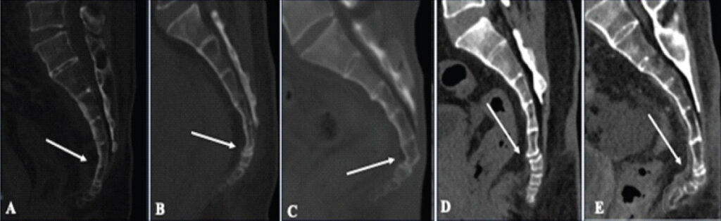
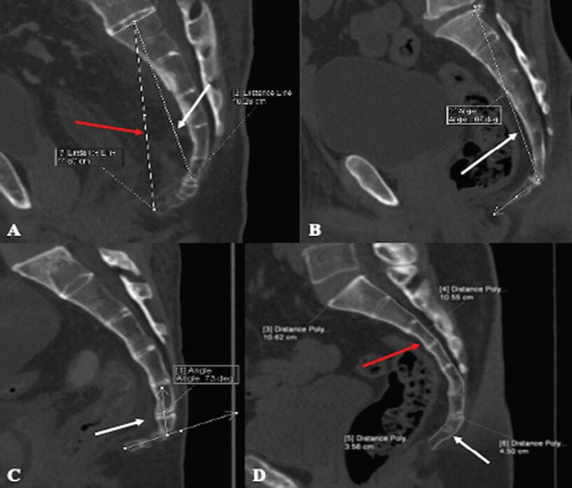
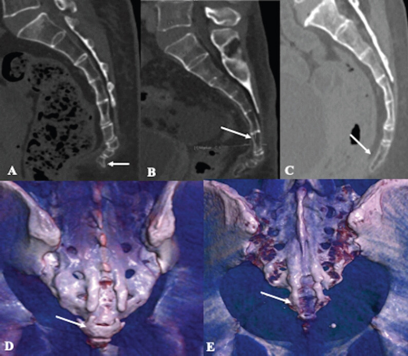
The data were entered in an Excel sheet. The patient information in these scans was masked carefully to maintain confidentiality. Two observers independently classified cases. Interobserver reliability was assessed using Cohen’s kappa coefficient. Finally, the data were analyzed descriptively using the Statistical Package for the Social Sciences v30.
Results
The study included 109 patients: 56 males and 53 females. Based on Nathan et al.’s modified classification, the most common coccyx type was Type I (41.3%), followed by Type II (19.3%). The coccyx was found to have four segments (58.7%), most commonly, followed by three segments (24.8%). The various dimensions related to the coccyx are described in a tabular manner (Tables 3–5).
Number of coccygeal segments present in the spine and prevalence of each type in our study population
| No. of coccygeal segments | Frequency (%) |
|---|---|
| 2 | 4 (3.7) |
| 3 | 27 (24.8) |
| 4 | 64 (58.7) |
| 5 | 14 (12.8) |
| Total (n) | 109 |
Morphological type of coccyx in our study population
| Coccyx morphological type | Frequency (%) |
|---|---|
| 1 | 45 (41.3) |
| 2 | 21 (19.3) |
| 3 | 13 (11.9) |
| 4 | 20 (18.3) |
| 5 | 6 (5.5) |
| 6 | 4 (3.7) |
| Total(n) | 109 |
Dimensions of sacral and coccygeal curved length, straight length, and sacrococcygeal and intercoccygeal angle
| Measurement | Mean±SD (cm) |
|---|---|
| Sacral curved length | 11.03±0.878 |
| Coccygeal curved length | 3.32±0.724 |
| Sacral straight length | 10.38±0.856 |
| Sacrococcygeal straight length | 11.99±1.16 |
| Sacrococcygeal angle | 116.28±11.81 |
| Intercoccygeal angle | 38.99±19.71 |
Sacralization of the first coccygeal segment was around 12.8%, and coccygeal spicules were seen in 30.3%. Sacrococcygeal and intercoccygeal joint fusion was seen in 32.1% and 67.9% of the study population, respectively. Intercoccygeal joint fusion occurred in 67.9% of people, with the 2nd–4th joints most common at 18.4%, and full coccyx fusion in 1.8%. Subluxation of the intercoccygeal segments was most commonly seen at the C1-C2 vertebral level in 15.6% of the study population. In comparison, 74.3% of the population had no intercoccygeal or sacrococcygeal joint subluxation (Table 6).
Describes the frequency of coccygeal spicule, sacralization, sacrococcygeal and intercoccygeal joint fusion, and sacrococcygeal and intercoccygeal joint subluxation in our study population
| Variables | Frequency (%) |
|---|---|
| Coccygeal spicule | 33 (30.3) |
| Sacralization | 14 (12.8) |
| Sacrococcygeal joint fusion | 35 (32.1) |
| Intercoccygeal joint fusion | 74 (67.9) |
| Sacrococcygeal and intercoccygeal joint subluxation | 28 (25.7) |
The calculated Cohen’s kappa value was 0.67, indicating substantial agreement between observers.
Discussion
The coccyx is considered to be a mobile structure rather than a static one and usually contains 3–5 segments [2,10]. Just as the morphology of the skeleton varies between countries, ethnicities, and even small communities, the coccyx also has many variations in its morphology. There are only a handful of data available on the Asian population and less data on the Indian population. Notably, the coccyx is a site for attaching several structures such as ligaments, tendons, and ganglions [11]. The morphological variations in the coccyx may lead to an imbalance in the forces acting on the surrounding structures. They can also lead to pain referred to as coccydynia, which was initially described by Simpson in the year 1859 as severe pain in the coccyx region provoked by sitting or changing the position from sitting to standing.[12] Females are more prone to develop coccydynia than males [13]. The dissimilarity in the female and male pelvis anatomy was proposed by Duncan [14].
Radiographs are the initial modality of investigation for the coccyx, and dynamic acquisitions can be made using sitting and standing X-rays. However, due to poor resolution and overlapping soft tissue, these are suboptimal for evaluation. Cross-sectional imaging such as CT clarifies the bony anatomy of the coccyx, whereas MRI provides detailed views of soft tissues and ligaments.
Postacchini and Massobrio were the pioneers in presenting a classification of coccyx morphology in radiographs [15]. Following this, Nathan et al. presented a revised classification of coccygeal morphology, describing six categories [7].
A study on Korean cohorts and the Chinese population showed Type II to be the most common (51%) [9,16], and an MRI-based study in the Nepalese population by Paudyal et al. also showed Type II (46.3%) as the most common, followed by Type I (40.5%) [17]. However, in our study, the predominant coccyx type was Type I (41.3%), followed by Type II (19.3%). These are similar to the European study that showed Type I being the most common (64%), followed by Type II (16%) [2]. Marwan et al. also found type I (47.5%) in arab population.[8] This finding reflects a notable ethnic variation. However, remarkably, we found a relatively high prevalence of Type IV (18.3%), which may suggest a regional or lifestyle influence. In our study, most subjects had four coccygeal segments (58.7%), followed by three segments (24.8%). Similar findings were noted in both the European (76%) and Korean (64.8%) cohorts, but the Nepalese cohort showed three segments (45.3%) to be more common [2,9,17].
Types III to VI were found to have a significantly higher incidence in patients with coccydynia than Type II, followed by Type I, which is shown to have the lowest incidence. We have a higher incidence of Type I in our cohort. Shams et al. compared the coccygeal features of patients with and without coccydynia using MRI and reported Type II as the most common morphology in their coccydynia cohort [18].
Sacrococcygeal joint fusion was 32.1%, similar to the 34% reported by Yoon et al. but lower than Woon et al.’s 57%. 2.9 intercoccygeal fusion in our study was seen in 67.9% [2]. Paudyal et al. noted similar fusion patterns, suggesting comparable prevalence across South Asian populations [17].
The mean sacrococcygeal angle in our study was 116.3°, slightly higher than the values observed by Woon et al. (106° ± 13°), Paudyal et al. (107.1°), and Yoon et al. (110°) [2,17]. Similarly, our mean intercoccygeal angle was 39°, lower than Korean males (53.9°) and females (44.7°), potentially indicating a straighter coccyx in our Indian population.
Coccygeal spicules were present in 30.3% of our subjects, which is higher than the 23% prevalence reported by Woon et al. and 11.6% in Paudyal et al.’s MRI study [2,17]. This discrepancy may stem from differences in imaging modality sensitivity (CT vs. MRI) or regional bone density patterns. Subluxation was observed only in about 25.7% of cases, with 74.3% not having any subluxation, which is a considerable proportion compared to Woon et al., where it is typically reported as rare (3%) [2]. Authors have reported that the presence of a posterior spicule is a significant factor in coccydynia, especially due to chronic adventitious bursitis, in at least 14% of coccydynia patients [5]. While subluxation does not always correlate with pain, its radiological presence can serve as an anatomical risk factor for coccydynia.
Sacralization was observed in 12.8% of cases, which is consistent with other Asian studies and higher than the rates reported by Woon et al. in European populations [2]. Type I coccyx was the most common in our study, showing higher rates of joint fusion, coccygeal spicule presence, and increased sacrococcygeal angle. Our study included only the static scan performed in the supine position in adults, so dynamic alterations in the coccyx morphology could not be evaluated.
Strengths and limitations
The study is limited by its single-center design and lack of clinical correlation with symptoms such as coccydynia. In addition, morphometric data were derived from supine CT scans, which may not fully represent the weight-bearing configuration of the coccyx. A larger sample would better represent the spectrum of coccygeal morphology. However, our sample exceeded the calculated minimum sample size and provides important baseline morphometric data from an underrepresented eastern Indian population. Ours was a cross-sectional morphometric study, and symptoms/clinical outcomes/coccydynia could not be evaluated. Therefore, we could not compare the morphological differences between those with and without symptoms. Future studies can be longitudinal, incorporating dynamic imaging or correlations with age-stratified analysis, body mass index, occupational and lifestyle factors, obstetric history, and symptomatology. Multicenter studies involving different regions of India would enhance understanding.
Conclusion
In our CT-based cross-sectional study, we analyzed various morphological parameters of the coccyx to provide an understanding of its basic prevalent morphology in the Indian population. This study contributes vital regional anatomical data and aids orthopedic surgeons and pain specialists. The data from the analysis can be used as a baseline for future anthropological studies and comparative ethnicity-based morphometry studies.
Clinical Message
A predominantly Type I, relatively straight coccyx with four segments is the common anatomical pattern in the eastern Indian population, suggesting that deviations from this morphology – such as increased angulation, spicules, or joint subluxation – may have greater clinical relevance in patients presenting with idiopathic coccydynia.
Conflict of Interest:
Nil
Source of Support:
Nil
Consent
The authors confirm that informed consent was obtained from the patient for publication of this article
How to Cite this Article
Nayak MK, Sahoo B, Sneha A, Mayur M, Jain M, Kamboj K. Morphometric Variants of the Coccyx in an Eastern Indian Cohort: Analysis from a Tertiary Care Center. Journal of Orthopaedic Case Reports 2026 October;16(10):314-319.
References
- Yagi F, Yamada Y, Yamada M, Yokoyama Y, Mukai K, Nakahara T. Three-dimensional evaluation of the coccyx movement between supine and standing positions using conventional and upright computed tomography imaging. Sci Rep 2021;11:6886. [Google Scholar] | [PubMed]
- Woon JT, Perumal V, Maigne JY, Stringer MD. CT morphology and morphometry of the normal adult coccyx. Eur Spine J 2013;22:863-70. [Google Scholar] | [PubMed]
- Shalaby SA, Eid EM, Allam OA, Ali AM, Gebba MA. Morphometric study of the normal Egyptian coccyx from (age 1-40 year). Int J Clin Dev Anat 2015;1:32-41. [Google Scholar] | [PubMed]
- Woon JT, Stringer MD. Clinical anatomy of the coccyx: A systematic review. Clin Anat 2012;25:158-67. [Google Scholar] | [PubMed]
- Maigne JY, Rusakiewicz F, Diouf M. Postpartum coccydynia: A Case series study of 57 women. Eur J Phys Rehabil Med 2012;48:387-92. [Google Scholar] | [PubMed]
- Nasiri A, Farajzadeh Vajari F, Sane S, Afsargharehbagh R. Assessment of Ganglion impar block effect on treatment results of coccydynia: A cross-sectional study. Anesthesiol Pain Med 2024;14:e142137. [Google Scholar] | [PubMed]
- Nathan ST, Fisher BE, Roberts CS. Coccydynia: A review of pathoanatomy, aetiology, treatment and outcome. J Bone Joint Surg Br 2010;92-B:1622-7. [Google Scholar] | [PubMed]
- Marwan YA, Al-Saeed OM, Esmaeel AA, Kombar OR, Bendary AM, Azeem ME. Computed tomography-based morphologic and morphometric features of the coccyx among Arab adults. Spine (Phila Pa 976) 2014;39(20):E1210-9. [Google Scholar] | [PubMed]
- Yoon MG, Moon MS, Park BK, Lee H, Kim DH. Analysis of sacrococcygeal morphology in koreans using computed tomography. Clin Orthop Surg 2016;8:412-9. [Google Scholar] | [PubMed]
- Fogel GR, Cunningham PY 3rd, Esses SI. Coccygodynia: Evaluation and management. J Am Acad Orthop Surg 2004;12:49-54. [Google Scholar] | [PubMed]
- Ashby K, Yilmaz E, Mathkour M, Olewnik Ł, Hage D, Iwanaga J. Ligaments stabilizing the sacrum and sacroiliac joint: A comprehensive review. Neurosurg Rev 2022;45:357-64. [Google Scholar] | [PubMed]
- Simpson: On Coccyodynia and the Diseases of the Coccyx. Available from: https://www.coccyx.org/medabs/simpson.htm. [Last accessed on 2025 Jun 07]. [Google Scholar] | [PubMed]
- Karadimas EJ, Trypsiannis G, Giannoudis PV. Surgical treatment of coccygodynia: An analytic review of the literature. Eur Spine J 2011;20:698-705. [Google Scholar] | [PubMed]
- Duncan GA. Painful Coccyx. Arch Surg 1937;34:1088-104. [Google Scholar] | [PubMed]
- Postacchini F, Massobrio M. Idiopathic coccygodynia. Analysis of fifty-one operative cases and a radiographic study of the normal coccyx. J Bone Joint Surg Am 1983;65:1116-24. [Google Scholar] | [PubMed]
- Feng H, Chen X, Cao C, Wan D, Liu H, Zhang E. The susceptibility of coccydynia in morphology: A cross-sectional study. Pain Res Manag 2025;2025:2932223. [Google Scholar] | [PubMed]
- Paudyal S, Karna BP, Joshi BR. Evaluation of anatomical variation of human coccyx on magnetic resonance imaging. Nepal J Radiol 2022;12:2-6. [Google Scholar] | [PubMed]
- Shams A, Gamal O, Mesregah MK. Sacrococcygeal morphologic and morphometric risk factors for idiopathic coccydynia: A magnetic resonance imaging study. Global Spine J 2023;13:140-8. [Google Scholar] | [PubMed]
© 2026 Journal of Orthopaedic Case Reports - Published by Indian Orthopaedic Research Group
About the Authors
-

Manoj K Nayak
-

Biswajit Sahoo
-

Sneha Aribaskar
-

M Mayur
-

Mantu Jain
-

Kulbhushan Kamboj
Related Articles in Journal of Orthopaedic Case Reports
 October 1, 2026 Arthrokinematic and Magnetic Resonance Imaging-Based Outcomes of Customized Lateral Wedge Insoles in Medial Compartment Knee Osteoarthritis: Six-Month Interim Findings of a Prospective Cohort Study
 July 1, 2026 Managing Complications of Total Hip Arthroplasty for Avascular Necrosis Hip Performed Via Anterior Approach
 March 1, 2026 Indigenous Innovation in Orthopedic Robotics: Making Joint Replacement Affordable in India
 December 1, 2025 Influential Factors to Patients When Choosing a Joint Replacement Surgeon in India