


Introduction
Osgood-Schlatter disease (OSD), sometimes referred to as Lannelongue disease, is a form of osteochondrosis affecting adolescents. It develops when repeated pulling forces from the rectus femoris portion of the quadriceps muscle lead to irritation and inflammation at the growth plate, a condition known as traction apophysitis [1]. Although considered a self-limiting condition, it can significantly impair participation in sports and daily activities, with potential for persistent symptoms if not managed appropriately [2].
OSD usually begins between ages 10–15 in boys and 8–13 in girls, with boys affected more often (about 3:1). It occurs in roughly 9.8% of cases and can affect both knees in 20–30% of patients. Despite its relatively common occurrence in active youth, the clinical management of OSD remains inconsistent, particularly in physiotherapy practice. Although several risk factors and activities are linked to its development, the exact cause remains unclear [3]. Frequent quadriceps contraction – especially during activities like running and jumping – can lead to irritation, small avulsions, and eventually osteochondritis at the site, further complicating rehabilitation and delaying recovery in athletic populations [4].
In conditions such as OSD, where pain and altered biomechanics may mask true muscle performance, isokinetic testing enables safe and precise evaluation of quadriceps and hamstring function without excessive joint stress. Incorporating isokinetic parameters in this case report adds novelty by introducing high-resolution, reproducible kinetic data, thereby enhancing the scientific rigor and offering deeper insights into rehabilitation outcomes beyond subjective and conventional clinical measures [5,6].
Current rehabilitation strategies largely include on activity modification, stretching, and progressive strengthening; however, there is no standardized, evidence-based protocol guiding load progression or safe return to sport, especially in symptomatic individuals who cannot tolerate high-load resistance training. This highlights a critical gap in physiotherapy practice [2].
Blood flow restriction therapy (BFRT) is widely used to increase muscle strength and hypertrophy by partially restricting arterial inflow and fully restricting venous outflow in working musculature during exercise [7,8].
BFRT is increasingly used in sports rehabilitation; its application in OSD remains relatively underexplored, with limited case-based or clinical evidence supporting its use in adolescent populations. Hence, this study is needed to explore BFRT as a novel approach that can safely induce comparable muscle adaptations, potentially expanding current rehabilitation strategies.
Case Report
A 15-year-old boy reported to at the physiotherapy department complaining of right knee pain that had been bothering him for the previous 2 months and getting worse when he played sports. He is an active football player who plays as a striker position for a club team and was reportedly asymptomatic before the development of symptoms.
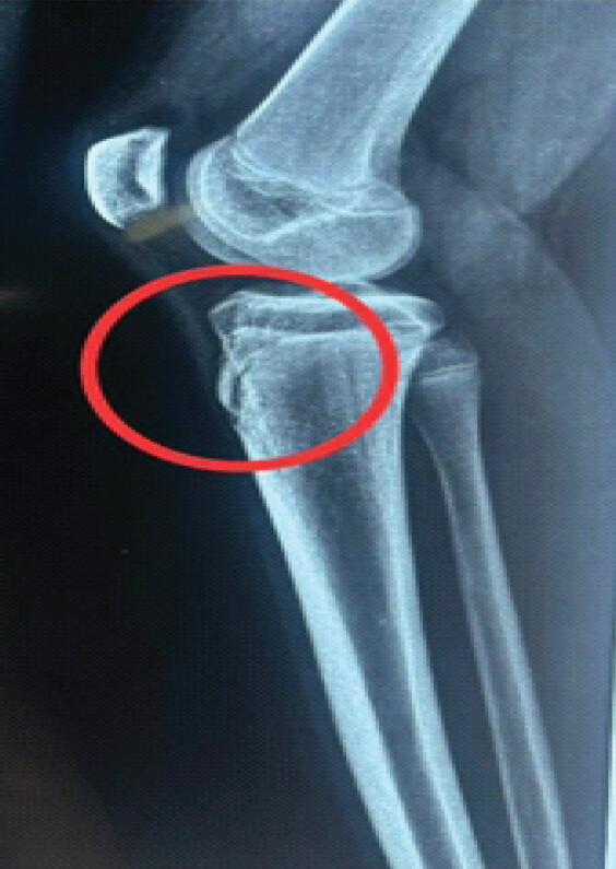
His right knee started to hurt when he kicked the ball during play about 2 months ago. Post-activity icing was used to treat the initial mild 3/10 pain level on the Numeric Rating Scale (NRS). For the next 2 months, he played sports at the same level of intensity despite the pain. He consulted with an orthopedic surgeon when the pain progressively worsened to a 6/10 on the NRS over time. The diagnosis of Osgood-Schlatter Syndrome was verified by radiographic evaluation using an X-ray that showed fragmentation formation at the patellar tendon insertion (Fig. 1). After being diagnosed, the patient was prescribed medications -non steroidal anti inflammatory analgesics and advised to take a 2-month break, with a referred for physical therapy. In addition, he was told to wear a rubber knee sleeve when playing sports. He stated that he was jogging for up to an hour every day without experiencing any pain during the rest interval. He was able to play football at his prior level of intensity while wearing the rubber knee sleeve when his symptoms temporarily subsided. However, the patient’s pain recurred after about 15 days of resuming sports, ranging to 7/10 on the NRS. As a result, he visited the physiotherapy outpatient department for further management.
No traumatic history existed. The patient’s prior surgical and medical history was ordinary. The current state was not influenced by family or psychological history.
Clinical findings
The patient gave their informed permission. When the upper anterior tibia near the tibial tuberosity was palpated, there was grade 1 tenderness (i.e., the patient reported discomfort). There was no noticeable swelling. The HUMAC NORM isokinetic machine was used to measure isokinetic muscle strength during the examination to objectively quantify hip abductor/adductor and quadriceps/hamstring strength (Table 1) [9]. Tape measurements were used to assess the quadriceps and calf muscle girths (Table 2).
Isokinetic values
| Parameters | Pre-intervention | Post-intervention | ||
|---|---|---|---|---|
| Right | Left | Right | Left | |
| PTE 60°/s (Nm) | 100 | 87 | 140 | 142 |
| PTF 60°/s (Nm) | 53 | 66 | 85 | 115 |
| H/Q ratio 60°/s (Nm) | 53 | 77 | 83 | 81 |
| PTE 180°/s (Nm) | 81 | 68 | 100 | 89 |
| PTF 180°/s (Nm) | 56 | 54 | 76 | 79 |
| H/Q ratio 180°/s (Nm) | 68 | 80 | 76 | 83 |
| Hip abductors 60°/s (Nm) | 42 | 35 | 52 | 46 |
| Hip adductors 60°/s (Nm) | 77 | 69 | 84 | 98 |
PTE: Peak torque of the knee extensors, PTF: Peak torque of the knee flexors, H/Q: Hamstring-to-quadriceps
Girth measurement (in cm)
| Girth measurement | Pre-intervention | Post-intervention | ||
|---|---|---|---|---|
| Right | Left | Right | Left | |
| Quadriceps | 44 | 43 | 49 | 50 |
| Calf | 35 | 35 | 38 | 38 |
Isokinetic examination
He warmed up for 5 min at a moderate speed before the examination. Testing was done in concentric force at angular speeds of 60°/s and 180°/s for the hamstring/quadriceps and hip adductor/abductor. When he was ready, five maximal warm-ups were repeated after three submaximal warm-ups at 60°/180°/s of angular speed, followed by a 20-s cool-down. The test was completed after 10 min of cooling down and tensioning activities. Table 1 provides isokinetic parameters before and after the intervention.
Girth measurement
Girth measurement of the quadriceps and calf muscles is performed using a flexible, non-elastic measuring tape. Pre and post-intervention girth measurements are provided in Table 2.
Physiotherapy intervention
The physiotherapy rehabilitation program was divided into three phases and was planned to be implemented for 12 weeks, 3 days a week, starting on day 1 (the day of admission for physiotherapy).
During the initial visit, patient education focused on avoiding aggravating activities such as prolonged sitting, running, and jumping. The patient received instruction on self-management techniques, such as applying ice, wearing a knee brace while practicing, and being aware of the risk factors linked to OSD. A general lower extremity strengthening program was then started. At the end of the session, McConnell taping was applied to the knee for additional support and symptom relief and the participant was asked to keep the tape for 2 days and not to remove it forcefully.
Blood flow restriction (BFR) was commenced at during the second visit. The participant was informed about the treatment plans and procedures before the procedure, and written informed consent was obtained. The BFR cuff (12 × 51 cm) was placed on the proximal thigh and inflated to occlude 60% of the limb occlusion pressure (LOP) in accordance with the clinical practice guidelines [10]. The pressure of total arterial flow blockage is known as LOP. The cuff was deflated only after 75 repetitions for a rest period of 120 s (Fig. 2, 3, 4) [8].
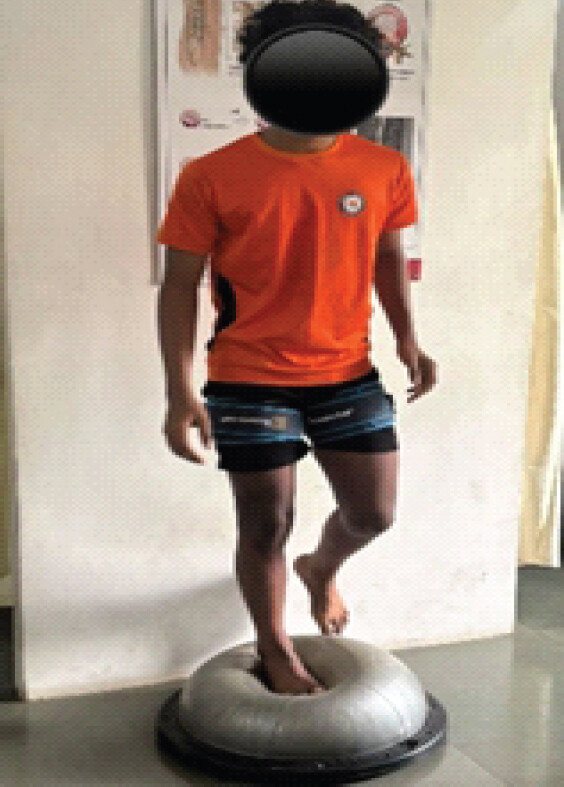
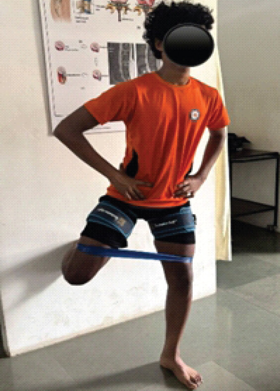
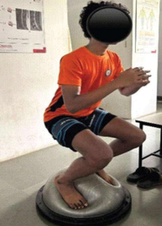
Every session began with a normal warm-up. To restore strength, neuromuscular control, and cardiovascular endurance for a return to sport, the rehabilitation program was gradually advanced based on the patient’s tolerance.
Before initiating this phase 3 that is return to sport, an isokinetic evaluation was carried out (Table 1) to objectively assess limb symmetry and the recovery of quadriceps and hamstring strength before moving on to sport-specific training. Despite the lack of widely recognized return-to-sport guidelines for OSD, objective strength evaluation was included to aid in clinical judgment. In particular, it was discovered that a quadriceps limb symmetry index higher than 87% considerably raised the probability of a successful return to play. Thus, the rehabilitation program was associated with improved lower-limb strength symmetry, restoration of the H/Q ratio, and pain-free execution of sport-specific activities. [11].
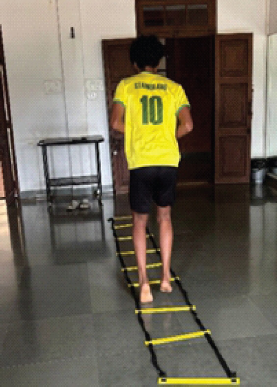
This phase emphasized plyometric and agility training without the use of BFRT (Fig. 5).
Functional outcome measure
Clinical outcome measures were used to assess pre and post-rehabilitation as shown in Table 3. Knee Injury and Osteoarthritis Outcome Score is a self-reported, validated questionnaire used to assess knee-related symptoms and functional status. Vertical jump height is a performance-based outcome measure used to evaluate lower limb power and functional capacity [12]. For 15–16-year-old males, performance can be interpreted as follows: >65 cm = excellent, 56–65 cm = above average, 50–55 cm = average, 40–49 cm = below average, and <40 cm = poor [13].
Rehabilitation protocol
| Training | Exercise | Dosage | Rationale |
|---|---|---|---|
| Phase 1 (0–4 weeks) | |||
| Strength training | Static squats, multiple-angle isometrics at 30, 60° angles, bridging, standing clamshell, standing hip abduction, ball press with knee flexion, wall squat at 90° with calf raises | 30, 15, 15, and 15 with 30 s of rest between the sets | Facilitate early quadriceps activation while minimizing tendon strain, preventing muscle inhibition and atrophy. Improve proximal stability to reduce stress on the knee joint and enhance quadriceps-hamstring co-contraction, thereby improving joint stability and reducing shear forces at the knee. |
| Proprioception and balance training | Ball toss on stability trainer, single-leg standing on stability trainer | ||
| Cardiovascular endurance training | Cycling with low resistance | 5 min | Reducing the risk of fatigue-related biomechanical compensations and improving aerobic fitness |
| Phase 2 (4–8 weeks) | |||
| Strength training | Hamstring isometrics with Swiss ball, bridging on Swiss ball, lateral band walks, lunges, lateral step down | 30, 15, 15, and 15 with 30 s of rest between the sets | Required for full restoration of muscle strength and performance capacity. Once pain subsides, higher loads improve tendon capacity and muscular resilience. |
| Proprioception and balance training | Ball toss on BOSU ball, single-leg standing on BOSU ball, squats on BOSU ball | ||
| Cardiovascular endurance training | Cycling with resistance, elliptical | 5 min each | |
| Phase 3: Return to sport (6–12 weeks) | |||
| Agility training | Box jumps, single-leg hops, reactive jump and sprint | 15 reps each 2 sets | Restoring speed, coordination, and reaction time. |
| Plyometric training | Cone walking drills, ladder drills, T drill, ball dribbling with direction change | 2 rounds each | Reintroduces controlled tendon loading and improves power, elasticity, and neuromuscular efficiency. |
outcome measure
| Outcome measure | Pre- intervention | Post- intervention |
|---|---|---|
| Knee injury and osteoarthritis outcome score | 61/100 | 100/100 |
| Vertical jump height | 50 cm | 56 cm |
The rehabilitation program was well tolerated, with no reports of excessive pain or discomfort during or after therapy sessions.
As a result of the rehabilitation process, he managed to regain his confidence when it came to getting back into playing football again. Initially, there were difficulties with his performance, especially in running and kicking, as well as changing directions quickly. This gradually changed with progressive physical training. Through physical training, significant progress was evident. Not only did the performance on field improve, his confidence in himself improved too. There was also less fear of re injury.
Discussion
In this case study, a 15-year-old male football player with OSD showed significant improvements in isokinetic strength, functional outcomes, and symptom resolution following a 12-week BFRT rehabilitation program.
The most noteworthy outcome was a 40% increase in quadriceps peak torque on the affected right side, along with a 63% rise on the left (unaffected) side at 60°/s and a 60% and 74% increase in hamstring peak torque on the right and left, respectively. Research demonstrates that by increasing metabolic stress, cellular swelling, and the recruitment of fast-twitch muscle fibers, BFRT in conjunction with low-load resistance training (20–30% 1RM) results in hypertrophy and strength increases equivalent to high-load resistance training (70% 1RM) [14, 15].
As a result, the H/Q ratio increased to 81% on the left and 83% on the right. The H/Q ratio of 60–80% is considered optimal for knee joint stability and injury prevention in athletic populations. Following the intervention, the H/Q ratio returned to normal at 83%, suggesting improved muscle balance and a decreased risk of further injuries. A successful return to sport has been shown to depend on quadriceps strength symmetry [11,16].
Increased hip abductor and adductor strength in both extremities improves frontal plane knee control during dynamic exercises while reducing valgus collapse and patellofemoral joint stress [17].
This case study offers initial proof that BFRT could be a helpful supplement in the treatment of teenage athletes with OSD. While the H/Q ratio assisted in identifying muscular imbalances not apparent on manual testing, isokinetic testing provided objective data to inform decisions about return to sport [18]. The use of BFRT was inspired by symptoms that persisted despite activity moderation and traditional strengthening, as well as the difficulty of handling high-load resistance exercise [8]. However, its single-case approach hinders causal inference and restricts generalizability.
To the best of the authors’ knowledge, this is one of the first studies to report BFR-assisted phased rehabilitation in a teenage football player with OSD using isokinetic dynamometry, H/Q ratio analysis, and return-to-sport results. The study provides unbiased evidence supporting BFRT as a low-load strategy for treating strength impairments in individuals with symptomatic OSD.
Conclusion
Improvements were observed in isokinetic strength, H/Q ratio, and functional performance following the intervention. While these findings suggest a potential role for BFRT as a low-load training strategy in this population, conclusions are limited by the single-case design, and further research is required to establish its efficacy, safety, and optimal application.
Clinical Message
BFRT-assisted rehabilitation may restore quadriceps-hamstring balance, enhance functional performance, facilitate safe return to sport, and reduce the risk of symptom recurrence in adolescents with persistent OSD. Isokinetic assessment provides objective evaluation of muscle recruitment, strength and limb symmetry, supporting clinical decision-making for rehabilitation progression and return-to-sport readiness.
Conflict of Interest:
Nil
Source of Support:
Nil
Consent
The authors confirm that informed consent was obtained from the patient for publication of this article
How to Cite this Article
Gaikwad S, Mehendale P, Desai V, Raman. Blood Flow Restriction-Assisted Rehabilitation for Persistent Osgood-Schlatter Disease in an Adolescent Footballer: A Case Report with Isokinetic Evaluation. Journal of Orthopaedic Case Reports 2026 October;16(10): 173-179.
References
- Corbi F, Matas S, Álvarez-Herms J, Sitko S, Baiget E, Reverter-Masia J. Osgood-Schlatter disease: Appearance, diagnosis and treatment: A narrative review. Healthcare (Basel) 2022;10:1011. [Google Scholar] | [PubMed]
- Neuhaus C, Appenzeller-Herzog C, Faude O. A systematic review on conservative treatment options for Osgood-Schlatter disease. Phys Ther Sport 2021;49:178-87. [Google Scholar] | [PubMed]
- Lucenti L, Sapienza M, Caldaci A, Cristo C, Testa G, Pavone V. The etiology and risk factors of Osgood-Schlatter disease: A systematic review. Children (Basel) 2022;9:826. [Google Scholar] | [PubMed]
- Seyfettinoğlu F, Köse Ö, Oğur HU, Tuhanioğlu Ü, Çiçek H, Acar B. Is there a relationship between patellofemoral alignment and Osgood-Schlatter disease? A case-control study. J Knee Surg 2020;33:67-72. [Google Scholar] | [PubMed]
- American College of Sports Medicine. ACSM's Guidelines for Exercise Testing and Prescription. 10th ed. Netherlands: Wolters Kluwer; 2017. [Google Scholar] | [PubMed]
- Dvir Z. Isokinetics: Muscle Testing, Interpretation and Clinical Applications. 2nd ed. United Kingdom: Churchill Livingstone; 2004. [Google Scholar] | [PubMed]
- Lorenz DS, Bailey L, Wilk KE, Mangine RE, Head P, Grindstaff TL. Blood flow restriction training. J Athl Train 2021;56:937-44. [Google Scholar] | [PubMed]
- Patterson SD, Hughes L, Warmington S, Burr J, Scott BR, Owens J. Blood flow restriction exercise: Considerations of methodology, application, and safety. Front Physiol 2019;10:533. [Google Scholar] | [PubMed]
- Aydın CG, Kargın D, Bayhan İA, Ercan S, Örsçelik A. Isokinetic evaluation of adolescent athletes with os good schlatter disease. Acta Med Alanya 2018;2:3-8. [Google Scholar] | [PubMed]
- Nascimento DC, Rolnick N, Neto IV, Severin R, Beal FL. A useful blood flow restriction training risk stratification for exercise and rehabilitation. Front Physiol 2002;13:808622. [Google Scholar] | [PubMed]
- Burland JP, Kostyun RO, Kostyun KJ, Solomito M, Nissen C, Milewski MD. Clinical outcome measures and return-to-sport timing in adolescent athletes after anterior cruciate ligament reconstruction. J Athl Train 2018;53:442-51. [Google Scholar] | [PubMed]
- Rathleff MS, Winiarski L, Krommes K, Graven-Nielsen T, Hölmich P, Olesen JL. Activity modification and knee strengthening for Osgood-Schlatter disease: A prospective cohort study. Orthop J Sports Med 2020;8:2325967120911106. [Google Scholar] | [PubMed]
- Beashel P, Taylor J. The World of Sport Examined. Croatia: Thomas Nelson and Sons; 1997. 57. [Google Scholar] | [PubMed]
- Loenneke JP, Wilson JM, Marín PJ, Zourdos MC, Bemben MG. Low intensity blood flow restriction training: A meta-analysis. Eur J Appl Physiol 2012;112:1849-59. [Google Scholar] | [PubMed]
- Pearson SJ, Hussain SR. A review on the mechanisms of blood-flow restriction resistance training-induced muscle hypertrophy. Sports Med 2015;45:187-200. [Google Scholar] | [PubMed]
- Aagaard P, Simonsen EB, Magnusson SP, Larsson B, Dyhre-Poulsen P. A new concept for isokinetic hamstring: Quadriceps muscle strength ratio. Am J Sports Med 1998;26:231-7. [Google Scholar] | [PubMed]
- Ferber R, Bolgla L, Earl-Boehm JE, Emery C, Hamstra-Wright K. Strengthening of the hip and core versus knee muscles for the treatment of patellofemoral pain: A multicenter randomized controlled trial. J Athl Train 2015;50:366-77. [Google Scholar] | [PubMed]
- Croisier JL, Ganteaume S, Binet J, Genty M, Ferret JM. Strength imbalances and prevention of hamstring injury in professional soccer players: A prospective study. Am J Sports Med 2008;36:1469-75. [Google Scholar] | [PubMed]
Related Articles in Journal of Orthopaedic Case Reports
 October 1, 2026 Outcomes of Blood Flow Restriction Therapy as an Adjunct to Standard Rehabilitation Following Anterior Cruciate Ligament Reconstruction: A Prospective Observational Cohort Study
 October 1, 2026 Combined Endoscopic SpeedBridge™ Reinsertion and Proximal Medial Gastrocnemius Lengthening for Chronic Insertional Achilles Tendinopathy in an Elite Sprinter: A Case Report
 September 1, 2026 Extensive Post-Traumatic Heterotopic Ossification with Multi-joint Involvement: A Case Report
 September 1, 2026 Opportunistic Biological Augmentation during Acute Achilles Tendon Repair: A Novel Surgical Concept With Three-Year Follow-Up—A Case Report