


Introduction
Acute Achilles tendon rupture is one of the most common sports-related tendon injuries, with an increasing incidence over recent decades, particularly among middle-aged men participating in recreational sports such as football [1]. Surgical repair is widely favored in active individuals due to lower re-rupture rates compared with non-operative management, while achieving comparable functional outcomes in most studies [2, 3].
The Krackow locking-loop technique is commonly used for primary repair due to its strong tendon purchase and biomechanical reliability. However, in cases where tendon ends are severely frayed or macerated, suture holding capacity may be compromised, increasing the risk of gap formation. Augmentation strategies, including suture reinforcement and autologous tissue augmentation, have been described to improve mechanical strength and reduce failure risk [4, 5]. The use of plantaris tendon augmentation combined with the Krackow technique has been demonstrated in acute repairs [4], and more recent evidence continues to support the role of local autologous tissue augmentation in selected cases [6].
Despite this, to date, little attention has been given to the preservation of viable traumatically displaced local soft-tissue encountered during acute Achilles tendon repair. These tissues are often discarded without consideration of their biological potential. This report describes a case of acute Achilles tendon rupture with severely compromised “mop-end” tissue treated with Krackow repair, circumferential suture reinforcement, and preservation of viable local soft-tissue sutured over the repaired tendon as a biological onlay augmentation layer, with favorable 3-year outcome.
Case Report
Clinical presentation
A 43-year-old male presented 2 days after sustaining a right Achilles tendon injury during recreational football. He reported sudden posterior ankle pain with inability to plantarflex or push off. He had no significant past medical history.
Clinical findings
- Positive Thompson test [7]
- Palpable gap over the Achilles tendon
- Loss of active plantarflexion strength.
Imaging
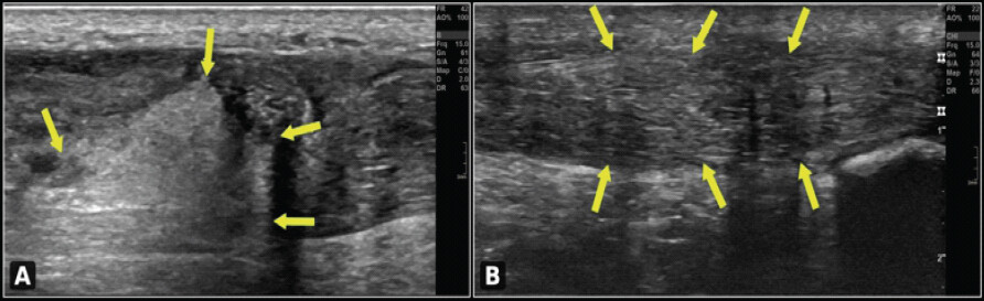
Ultrasound confirmed a complete Achilles tendon rupture with loss of continuity and a hypoechoic defect at the rupture site. At 3-year follow-up, ultrasound demonstrated restored tendon continuity with no evidence of re-rupture (Fig. 1a and b).
Operative findings and technique
Open repair was performed 6 days post-injury under general anesthesia with the patient in the prone position. A posterior longitudinal incision (~11 cm) was used with careful protection of the sural nerve.
Intraoperative findings
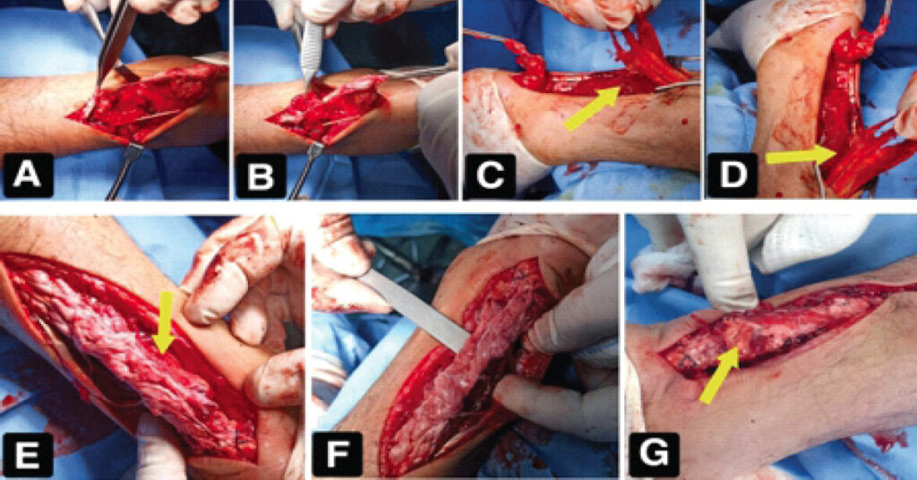
The rupture margins demonstrated (Fig. 2a, b, c, d):
- Severely frayed and macerated tendon ends
- Poorly defined margins consistent with “mop-end” appearance
- High-risk tissue quality for standard repair.
Primary repair
The Krackow locking-loop technique was performed using non-absorbable polypropylene sutures. Tendon approximation was achieved with appropriate tension balanced against the contralateral side. A circumferential running polypropylene suture was added to reinforce the repair construct and reduce gap formation (Fig. 2e and f).
Biological augmentation
A substantial viable local soft tissue was identified interposed between the repaired tendon and the posterior tibial surface. Its macroscopic appearance – including mixed pink-red coloration with white fibrous streaking, punctate bleeding on gentle manipulation, supple musculotendinous consistency, and anatomical location – was presumed to represent traumatically avulsed tissue arising from the distal gastrosoleus musculotendinous complex (yellow arrows, Fig. 2c and d). Histological confirmation was not obtained, as biopsy was not clinically indicated; the intraoperative decision was based on macroscopic assessment of viability, and histological analysis would not have altered the surgical plan regardless of the tissue type identified. Rather than discarding it, the tissue was preserved and sutured directly over the repaired tendon, where it served as a biological onlay augmentation layer (yellow arrows, Fig. 2e and g). The wound was closed in layers (Fig. 2g) and the ankle immobilized in plantar flexion using a below-knee cast.
Post-operative management
Post-operative management followed a conservative, staged protocol as documented in the medical record:
- Weeks 0–5: Below-knee cast in plantar flexion
- Weeks 5–7: Posterior slab in neutral (plantigrade) position
- Week 7: Removal of immobilization – wound healed and tendon strength clinically accepted; the patient was encouraged to begin gentle self-directed ankle mobilization
- Week 8 (~2 months): Formal supervised physiotherapy initiated; partial weight-bearing (PWB) commenced; range-of-motion (ROM) exercises continued
- Week 11: PWB with single crutch; ROM exercises continued
- Week 13 (~3 months): Full weight-bearing (FWB) achieved; occasional ankle swelling noted, managed with limb elevation and continuation of ROM exercises.
Following the 13-week clinic visit, the patient did not attend further scheduled appointments. All subsequent rehabilitation milestones were reported by the patient at the 3-year follow-up assessment and are based on patient recall.
Return-to-sport timeline (Table 1)
| Activity | Time postoperatively |
|---|---|
| Below-knee cast in plantar flexion | 0–5 weeks |
| Posterior slab in neutral (plantigrade) position | 5–7 weeks |
| Removal of immobilization; wound healed; initiation of gentle self- directed ankle mobilization | 7 weeks |
| Supervised physiotherapy initiated; partial weight-bearing (PWB) commenced | 8 weeks (~2 months) |
| PWB with single crutch + range-of- motion exercises | 11 weeks |
| Full weight-bearing; occasional ankle swelling noted | 13 weeks (~3 months) |
| Initiation of jogging (sand surface)* | 5 months |
| Return to team training* | 9 months |
| First competitive match* | 12 months |
| Full return to pre-injury level* | 16 months |
*
Milestones marked with an asterisk are based on patient recall at the 3-year follow-up visit (subject to recall bias), as scheduled clinic follow-up was discontinued after 13 weeks
Outcome at 3 years
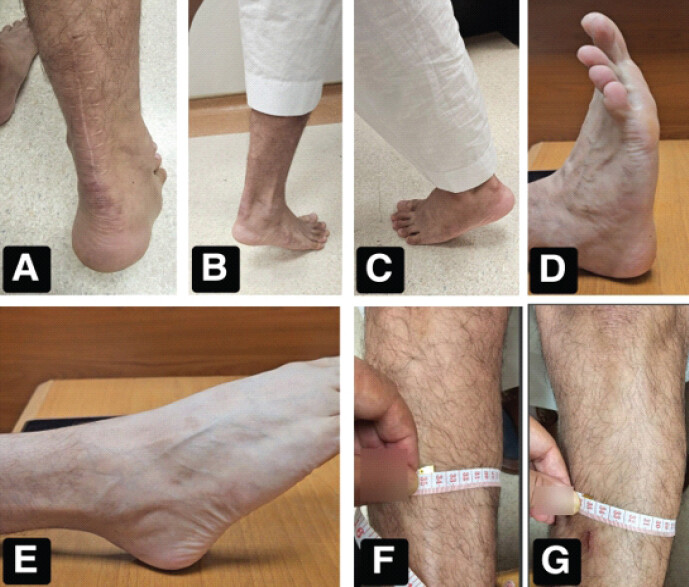
At final follow-up, the patient demonstrated (Fig. 3):
- Full return to competitive football
- No pain or functional limitation
- Full symmetric ankle range of motion
- Successful single-leg heel-rise
- No significant calf atrophy
- Negative Thompson test.
Although numerical goniometric values were not formally recorded, clinical examination and supplementary photographic documentation (Fig. 3d and e) confirmed symmetrical full ankle motion comparable to the contralateral side. Video documentation of the post-operative full range of motion is available as supplementary material, comprising three video clips: Bilateral heel-rise (Video 1), single-leg heel-rise on the operated side (Video 2), and ankle range of motion on the operated side (Video 3). The Achilles Tendon Total Rupture Score (ATRS) was 100/100, with maximal scores across all domains including pain, stiffness, muscle strength, and daily activities [8].
Discussion
This case highlights the management challenges of acute Achilles tendon rupture with severely compromised “mop-end” tissue quality. Despite poor tissue integrity, excellent long-term outcomes were achieved using Krackow repair with circumferential reinforcement and opportunistic biological augmentation, resulting in a 100/100 ATRS score and full return to competitive football at 3 years.
Surgical considerations
The Krackow technique provides strong tendon fixation and is widely accepted in Achilles tendon repair. Biomechanical studies demonstrate high failure loads, especially when combined with augmentation techniques [9]. Alternative techniques such as the Giftbox or triple Kessler methods offer comparable strength profiles, but the Krackow technique remains preferred by many surgeons due to its familiarity and reproducibility [10].
In cases with severely compromised tissue quality, primary repair alone may be insufficient, and additional mechanical reinforcement is often required. Circumferential suture reinforcement offers additional mechanical stability and may reduce early gap formation, particularly in poor-quality tendon tissue [5]. This approach is simple, cost-effective, and applicable in any surgical setting.
Opportunistic biological augmentation
This case proposes the concept of “opportunistic biological augmentation” – where viable local soft tissue is preserved and utilized rather than excised. The rationale for preservation was based on four considerations:
- Viability: Confirmed by punctate bleeding on gentle manipulation, contrasting sharply with the avascular macerated tendon ends.
- Biological contribution: Vascularized tissue may contribute to a favorable biological environment that supports tendon healing through mechanisms including cellular activity, neovascularization, collagen deposition, and tissue remodeling.
- Mechanical support: The augmentation layer may provide an additional protective covering over the repair construct, potentially helping to shield it during the early healing phase.
- No donor-site morbidity: Tissue already present within the operative field is utilized [4].
The central concept of this report is not the introduction of a new repair technique, but rather a change in intraoperative decision-making: The recognition and preservation of viable local soft tissue that would otherwise be discarded.
This concept aligns with previous descriptions of local tissue augmentation in Achilles reconstruction [11,12], but extends its application to the acute setting using tissue that would conventionally be discarded. Hallock described the “Achilles wraparound flap,” using local soleus muscle to enhance healing [11], and Maffulli and Ajis advocated local muscle flaps in chronic ruptures [12]. This report extends these biological principles to the acute setting.
To the best of our knowledge, no previous publication has formally described the preservation of traumatically displaced viable local tissue as an opportunistic biological augmentation strategy during acute Achilles tendon repair. We therefore propose this as a novel surgical concept warranting further study.
Importantly, the augmentation in this case was not pre-planned. It resulted from an intraoperative judgment when unexpectedly viable local tissue, which would otherwise have been discarded, was encountered during routine exploration of the rupture site. This opportunistic decision represents the essence of the proposed concept: not a new suture technique or a new tissue flap, but a new way of thinking – transforming tissue that would conventionally be discarded into a potentially valuable biological resource without additional planning, dissection, or morbidity.
Unlike conventional augmentation techniques – which require pre-operative planning, tendon harvest, additional dissection, or formal reconstruction procedures – opportunistic biological augmentation is an intraoperative decision based solely on the preservation of traumatically displaced but viable local tissue that would otherwise have been discarded. (Table 2)
| Feature | Conventional augmentation | Opportunistic biological augmentation |
|---|---|---|
| Pre-operative planning | Required | Not required |
| Tissue source | Harvested from remote or local donor site | Traumatically displaced local tissue within injury zone |
| Additional dissection | Required | None |
| Additional operative time | Significant | None formally recorded; tissue preservation incorporated into standard repair without a separate operative stage |
| Donor-site morbidity | Present (e.g., plantaris harvest, muscle flap) | None |
| Decision timing | Pre-operative or planned intraoperative | Opportunistic intraoperative |
| Equipment required | May require additional instruments | None beyond standard repair set |
| Histological certainty of tissue | Generally known | Not routinely available; macroscopic viability guides decision-making |
| Biological rationale | Planned biological augmentation | Utilization of unexpectedly encountered viable tissue within the operative field |
| Applicability | Selected cases with planned augmentation | Any case where viable displaced tissue is encountered |
This approach requires no additional equipment beyond the standard Achilles tendon repair set, no additional tissue harvest, and no donor-site morbidity. This maneuver did not require appreciable additional operative time or special equipment, as tissue preservation was incorporated into the standard repair steps without a separate operative stage.
Regarding the precise identity of the augmentation tissue: Its macroscopic characteristics – including a mixed pink-red coloration with white fibrous streaking, punctate bleeding on gentle manipulation, supple musculotendinous consistency, and anatomical location between the repaired tendon and the posterior tibial surface – were collectively consistent with traumatically avulsed viable tissue from the distal gastrosoleus musculotendinous complex – though this could not be confirmed histologically. Importantly, the mixed coloration and texture observed intraoperatively (Fig. 2e and f) preclude definitive identification as purely muscular tissue; the white fibrous component suggests a tendinous or fascial contribution alongside the muscular element. Formal histological confirmation was not performed, as biopsy was not clinically indicated in this acute setting. The tissue is therefore described in descriptive anatomical terms rather than as a definitively identified structure. Regardless of its precise histological identity, its macroscopic viability made it a valuable biological augmentation substrate, and its utilization is justified on functional and biological grounds rather than histological certainty.
Functional outcome
The patient achieved a 100/100 ATRS and full return to competitive sport at 3 years, with outcomes appearing comparable to published return-to-sport data following Achilles tendon repair [13,14]. Surgical management is also associated with lower re-rupture rates compared with non-operative treatment [3,15]. A recent systematic review and meta-analysis reported that 65–100% of patients returned to sport between 3 and 13.4 months post-surgery [13,14], while among professional athletes the mean time to return to competitive play is approximately 11 months with a return rate of 76% [16]. A scoping review of elite athletes confirmed return rates exceeding 70% following surgical repair [17], and a review of return-to-sport outcomes further supports surgery in active patients with high functional demands [18]. The 12-month milestone to first competitive match in this case falls within the published range, and the extended timeline to full pre-injury fitness at 16 months is consistent with a cautious approach appropriate for cases with severely compromised tissue quality.
The post-operative rehabilitation milestones documented up to 13 weeks fall within the accepted range for complex Achilles tendon repairs, reflecting an appropriately cautious approach given the poor intraoperative tissue quality. While more accelerated early mobilization protocols have been described [19], the graduated progression to FWB at 13 weeks is consistent with published protocols for cases with compromised tendon integrity.
It is important to acknowledge that the favorable outcome reported in this case cannot be attributed solely to the biological augmentation. The meticulous primary repair, circumferential suture reinforcement, and structured early rehabilitation each contributed to the overall result. The present report does not seek to establish causality between the augmentation and the outcome. Rather, it demonstrates the technical feasibility of preserving viable local soft tissue and introduces this maneuver as a potentially valuable biological adjunct that warrants prospective investigation.
Limitations
This is a single case report, limiting generalisability. Histological confirmation of the augmented tissue was not performed, as discussed in Section 3.2; the decision was based on macroscopic assessment of viability, and histological analysis would not have altered the surgical plan. Furthermore, the exact time interval between identification of the viable tissue and its incorporation into the repair was not formally recorded, as the decision to preserve and utilize the tissue was made immediately upon encounter during the operative procedure, without a defined waiting period. Post-operative follow-up was documented in the medical record up to 13 weeks. Beyond this point, the patient did not attend scheduled clinic appointments, and all subsequent milestones – including return to jogging, team training, and competitive match play – were based entirely on patient recall at the 3-year follow-up visit. This represents a limitation in the objectivity of the return-to-sport timeline. Furthermore, the rehabilitation protocol was not formally standardised. Despite these limitations, the robust 3-year functional outcomes – including objective measures such as single-leg heel-rise testing, bilateral calf circumference measurement, and a 100/100 ATRS score – provide meaningful evidence supporting the feasibility and potential value of this approach. Furthermore, because this is a single case report, it is not possible to determine the relative contribution of the circumferential suture reinforcement versus the biological augmentation to the excellent clinical outcome. Because only a single patient is presented, reproducibility of this approach across different rupture patterns and tissue presentations cannot be established. Future prospective comparative studies are required to determine whether opportunistic biological augmentation provides measurable benefits beyond meticulous primary repair alone. Advanced post-operative imaging such as magnetic resonance imaging or ultrasound elastography may also help clarify the structural contribution of preserved augmentation tissue in future studies.
Conclusion
Krackow repair with circumferential suture reinforcement combined with opportunistic preservation of viable local soft tissue was associated with an excellent long-term functional outcome in this patient, with a 100/100 ATRS score and full return to competitive football at 16 months.
This case proposes the concept of opportunistic biological augmentation – a simple, cost-free adjunct that may provide additional biological support for healing in selected cases of poor tendon tissue quality. While further studies are required to establish its broader applicability, this approach may be considered when viable local soft tissue is encountered intraoperatively. The underlying principle may extend to other acute tendon repair settings – such as rotator cuff, patellar tendon, or quadriceps tendon repair – wherever traumatically displaced viable soft tissue is encountered. This concept requires prospective validation in larger patient cohorts before routine clinical adoption can be recommended.
Clinical Message
In acute Achilles tendon repair, surgeons may consider inspecting the rupture site for viable displaced soft tissue before debridement. When such tissue is encountered – identifiable by punctate bleeding on gentle manipulation and macroscopic viability – preservation and utilization as an onlay biological augmentation layer is a simple, cost-free, and low-risk maneuver that may provide additional biological support for healing in cases with poor tendon tissue quality.
Conflict of Interest:
Nil
Source of Support:
Nil
Consent:
The authors confirm that informed consent was obtained from the patient for publication of this article
Acknowledgement: The author wishes to express sincere gratitude to Dr. Fath Allah Fathy Awad, MD, Radiology Consultant, Radiology Department, Khafji General Hospital, for his expertise and cooperation in performing and reporting the pre- and postoperative ultrasound studies that contributed to this case report.
References
- Lantto I, Heikkinen J, Flinkkilä T, Ohtonen P, Leppilahti J. Epidemiology of Achilles tendon ruptures: Increasing incidence over a 33-year period. Scand J Med Sci Sports 2015;25:e133-8. [Google Scholar] | [PubMed]
- Khan RJ, Fick D, Keogh A, Crawford J, Brammar T, Parker M. Treatment of acute Achilles tendon ruptures. A meta-analysis of randomized, controlled trials. J Bone Joint Surg Am 2005;87:2202-10. [Google Scholar] | [PubMed]
- Fan L, Hu Y, Zhou L, Fu W. Surgical vs. nonoperative treatment for acute Achilles' tendon rupture: a meta-analysis of randomized controlled trials. Front Surg 2024;11:1483584. [Google Scholar] | [PubMed]
- Akgün U, Erol B, Karahan M. Primary surgical repair with the Krackow technique combined with plantaris tendon augmentation in the treatment of acute Achilles tendon ruptures. Acta Orthop Traumatol Turc 2006;40:228-33. [Google Scholar] | [PubMed]
- Zhang YJ, Zhang C, Wang Q, Lin XJ. Augmented versus nonaugmented repair of acute Achilles tendon rupture: a systematic review and meta-analysis. Am J Sports Med 2018;46(7):1767-1772. [Google Scholar] | [PubMed]
- Prejbeanu R, Vermesan D, Balanescu A, Mioc ML, Haragus H. Repair of acute Achilles tears with plantaris augmentation. Int Orthop 2021;45:2285-2290. PMID: 34350472. [Google Scholar] | [PubMed]
- Thompson TC, Doherty JH. Spontaneous rupture of tendon of Achilles: A new clinical diagnostic test. J Trauma 1962;2:126-9. [Google Scholar] | [PubMed]
- Nilsson-Helander K, Thomée R, Silbernagel KG, Thomée P, Faxén E, Eriksson BI. The Achilles tendon total rupture score (ATRS): Development and validation. Am J Sports Med 2007;35:421-6. [Google Scholar] | [PubMed]
- Diniz P, Pacheco J, Fernandes RM, Pereira H, Castelo Ferreira F, Kerkhoffs GMMJ. Modified triple Kessler with least risk of elongation among Achilles tendon repair techniques: a systematic review and network meta-analysis of human cadaveric studies. Knee Surg Sports Traumatol Arthrosc 2023;31 (5):1644-1657. [Google Scholar] | [PubMed]
- Clanton TO, Haytmanek CT, Williams BT, Civitarese DM, Turnbull TL, Massey MB. et al. A biomechanical comparison of an open repair and 3 minimally invasive percutaneous Achilles tendon repair techniques during a simulated, progressive rehabilitation protocol. Am J Sports Med 2015;43:1957-1964. PMID: 26063402. [Google Scholar] | [PubMed]
- Hallock GG. Multiple, synchronous ipsilateral soleus muscle flaps, including the Achilles “wraparound” flap. Ann Plast Surg 1998;41(1):46-51. [Google Scholar] | [PubMed]
- Maffulli N, Ajis A. Management of chronic ruptures of the Achilles tendon. J Bone Joint Surg Am 2008;90:1348-60. [Google Scholar] | [PubMed]
- Bak BM, Seow D, Teo YZE, Hasan MY, Pearce CJ. Return to play and functional outcomes following treatment of acute Achilles tendon ruptures: A systematic review and meta-analysis. J Foot Ankle Surg 2024;63:420-9. [Google Scholar] | [PubMed]
- Zellers JA, Carmont MR, Grävare Silbernagel K. Return to play post-Achilles tendon rupture: A systematic review and meta-analysis of rate and measures of return to play. Br J Sports Med 2016;50:1325-32. [Google Scholar] | [PubMed]
- Ochen Y, Beks RB, Van Heijl M, Hietbrink F, Leenen LP, Van Der Velde D. Operative treatment versus nonoperative treatment of Achilles tendon ruptures: Systematic review and meta-analysis. BMJ 2019;364:k5120. [Google Scholar] | [PubMed]
- Johns W, Walley KC, Seedat R, Thordarson DB, Jackson B, Gonzalez T. Career outlook and performance of professional athletes after Achilles tendon rupture: a systematic review. Foot Ankle Int 2021;42(4):495-509. [Google Scholar] | [PubMed]
- LaPrade CM, Chona DV, Cinque ME, Freehill MT, McAdams TR, Abrams GD. Return-to-play and performance after operative treatment of Achilles tendon rupture in elite male athletes: A scoping review. Br J Sports Med 2022;56:515-20. [Google Scholar] | [PubMed]
- Vaidya SR, Sharma SC, Al-Jabri T, Kayani B. Return to sport after surgical repair of the Achilles tendon. Br J Hosp Med 2023;84:1-14. [Google Scholar] | [PubMed]
- McCormack R, Bovard J. Early functional rehabilitation or cast immobilisation for the postoperative management of acute Achilles tendon rupture? A systematic review and meta-analysis of randomised controlled trials. Br J Sports Med 2015;49:1329-35. [Google Scholar] | [PubMed]
© 2026 Journal of Orthopaedic Case Reports - Published by Indian Orthopaedic Research Group
About the Authors
-
Ahmed Alsaedawy A. Albeshlawy
Related Articles in Journal of Orthopaedic Case Reports
 October 1, 2026 Combined Endoscopic SpeedBridge™ Reinsertion and Proximal Medial Gastrocnemius Lengthening for Chronic Insertional Achilles Tendinopathy in an Elite Sprinter: A Case Report
 October 1, 2026 Blood Flow Restriction-Assisted Rehabilitation for Persistent Osgood-Schlatter Disease in an Adolescent Footballer: A Case Report with Isokinetic Evaluation
 May 1, 2026 Surgical Treatment of Anchor-induced Heel Pain after Minimally Invasive Repair of Acute Achilles Tendon Rupture Using Percutaneous Achilles Repair System Achilles Midsubstance SpeedBridge™ Repair: A Case Report
 February 1, 2026 Early Return to Competitive Sport Following a Surgically Managed Mandibular Fracture: A Multimodal Ultrasound-Guided Rehabilitation Case Report