


Introduction
Fibular hemimelia (FH), also known as longitudinal fibular deficiency, is a congenital condition marked by partial or complete absence of the fibula and associated abnormalities of the lower limb, including the tibia, knee, and ankle [1]. Several classification systems have been proposed to describe the spectrum of FH [1,2,3]. These musculoskeletal alterations may include tibial deformity, ligamentous instability, and limb-length discrepancy, which affect lower-limb biomechanics and complicate surgical management [1,4,5,6]. In particular, the absence of the fibular neck – the standard anatomical landmark for common peroneal nerve (CPN) localization during the lateral approach – and the dysplastic proximal tibial geometry directly influence surgical exposure and implant selection. The coexistence of complete FH and an intra-articular tibial plateau fracture has not been previously reported; a related report described a tibial plateau fracture in a patient with an isolated dysplastic fibular head, a distinct and more localized congenital anomaly [7]. We report the management of a comminuted Schatzker V (AO/OTA 41-C3) tibial plateau fracture, highlighting the anatomical and technical challenges encountered during surgical treatment.
Case Report
A 37-year-old Portuguese man presented with severe right knee pain, swelling, and inability to bear weight after a fall from an electric scooter. He had congenital right-sided FH, classified as Achterman and Kalamchi type II, indicating a complete absence of the fibula. This condition is associated with limb-length discrepancy, knee instability, and altered bone morphology. His surgical history before presentation was incompletely characterized: Records indicated a femur fracture approximately 15 years earlier and a possible prior intervention on the right leg, but further detail was unobtainable as this care had occurred abroad. Clinical examination revealed a swollen, tender knee with restricted range of motion, preserved distal pulses, and sensation. A lateral femoral scar was noted. The limb demonstrated malalignment and anisomelia consistent with his underlying congenital condition, confirmed on pre-operative standing radiographs. The patient was an active smoker (1 pack per day) and reported regular alcohol consumption (1 unit/day).
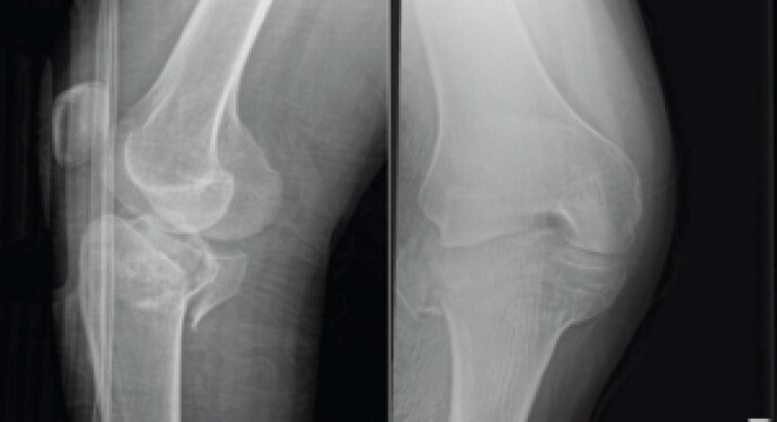
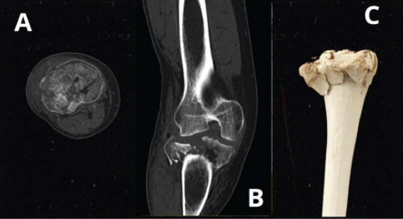
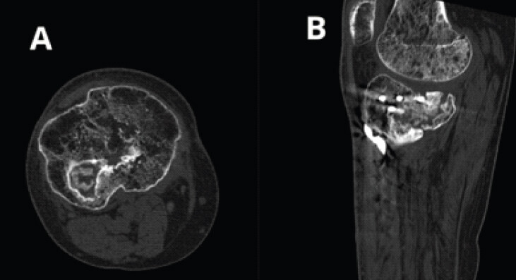
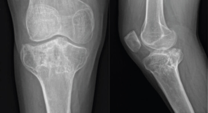
Radiographs demonstrated a comminuted bicondylar (AO/OTA 41-C3) tibial plateau fracture with intra-articular extension and posterior fragment displacement, associated with the absence of the ipsilateral fibula (Fig. 1). Computed tomography (CT) confirmed multifragmentary articular depression and metaphyseal comminution (Fig. 2).
Given the high-energy mechanism and soft-tissue swelling, a staged approach was used. Initial stabilization involved a bridging external fixator with Schanz pins in the distal femur and proximal tibia under fluoroscopy.
Definitive fixation was performed 10 days after injury once soft tissues had improved. During the lateral approach, the CPN was deliberately identified and protected, as the fibular neck – its standard anatomical landmark – was absent due to FH, making its course uncertain. This step required more careful dissection than is typical in standard tibial plateau surgery. The fracture was highly comminuted, with lateral and posterior articular depression and metaphyseal bone loss requiring elevation and cancellous bone grafting.
After reduction and temporary Kirschner wire fixation, a lateral plate was applied. This sequence decreases the lateral articular surface and stabilizes severe comminution and unusual shape before fixing the posteromedial fragment.
Intraoperative plate adjustment was needed; the right-sided Stryker AXSOS plate did not fit; a contralateral left-sided plate was required.
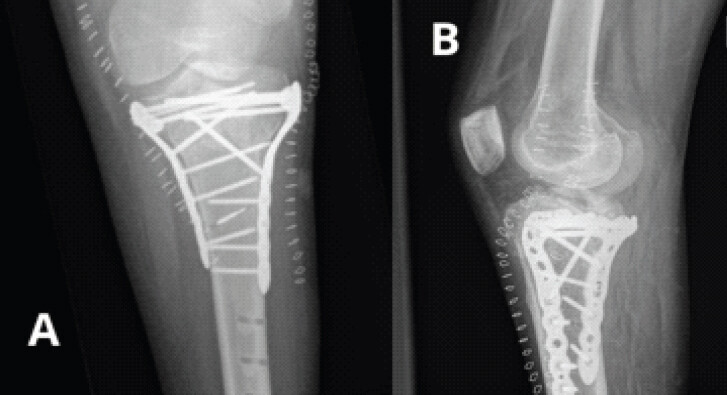
A posteromedial approach was then performed with identification and protection of the pes anserinus. Fixation was completed, and hardware positioning was confirmed on post-operative radiographs (Fig. 3). A fragment of a broken drill bit remained intraosseous without immediate risk.
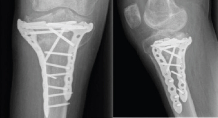
The patient was non-weight-bearing for 12 weeks, with early passive range-of-motion exercises. At 5 months, he reported persistent pain and limping, with knee flexion limited to 100°. Imaging showed maintained alignment but delayed consolidation of the posterior fragment (Fig. 4).
Further evaluation with CT confirmed delayed union of the posteromedial fragment (Fig. 5). Hardware removal was performed approximately 11 months after definitive fixation (Fig. 6), during which the retained drill-bit fragment was retrieved; however, symptoms persisted, and complex regional pain syndrome (CRPS) was diagnosed clinically according to the Budapest criteria during follow-up. Magnetic resonance imaging (MRI) subsequently demonstrated a complete rupture of the anterior cruciate ligament (ACL) with a positive anterior drawer, degenerative meniscal changes, and moderate joint effusion; arthroscopy confirmed the ACL rupture, with the intercondylar notch filled with fibrous tissue, and identified a hypoplastic anterior horn of the medial meniscus. The origin of the ACL injury – congenital, related to a prior undocumented trauma, or attributable to the current fracture – could not be determined.
At 4 years, the patient had persistent knee stiffness with flexion limited to 100°, calf muscle amyotrophy, chronic pain, and needed a single crutch. A 5 cm limb-length discrepancy, measured radiographically, and pelvic obliquity contributed to secondary low back pain. Early post-traumatic gonarthrosis, predominant laterally, was noted on subsequent imaging, and the patient was no longer able to work; referral for medicolegal evaluation and consideration of a limb-lengthening intramedullary nail were undertaken.
Discussion
FH is the most common congenital longitudinal deficiency of the lower limb and involves partial or complete fibular absence with associated femoral, tibial, knee, ankle, and foot deformities [1,8,9]. First described by Gollier in 1698 [8], FH presents with limb-length discrepancy, valgus alignment, cruciate ligament deficiency, and abnormal foot morphology [1,4,5,6,10]. The absence of the fibula alters surgical landmarks, making identification of structures such as the CPN more challenging [1,5]. As demonstrated in this case, dysplastic proximal tibial morphology in FH may render standard ipsilateral anatomic plates non-conforming, necessitating alternative implant strategies such as contralateral plating. Surgeons may wish to anticipate potential implant fit challenges and consider the availability of contralateral implants when operating on patients with FH, pending confirmation of this observation in larger series. Bicondylar tibial plateau fractures represent complex injuries with variable three-dimensional morphology, requiring careful pre-operative planning and adaptation of surgical approach and fixation strategy [11]. Functional outcomes of these injuries remain variable even with appropriate surgical management, reflecting the inherent difficulty of achieving stable fixation and anatomical reconstruction in high-energy patterns [12]. Therefore, it is important to distinguish between complications related to fracture severity and those potentially influenced by underlying anatomical abnormalities.
Intraoperative altered anatomy complicated dissection, reduction, and implant placement. Anatomical plates are designed for normal bone and may not fit a dysplastic tibia in FH. The use of a contralateral plate, as required in this case, aligns with reports of implant fit challenges in FH reconstruction [1,2,5]. During the lateral approach, identification of the CPN was more demanding than usual, as the absent fibular neck eliminated the standard anatomical landmark for nerve localization, requiring deliberate systematic dissection. In addition, metaphyseal bone loss and articular depression required elevation and bone grafting, which are known factors that may increase the risk of delayed healing in complex plateau fractures. While altered bone anatomy has been described in FH, bone-healing complications have mainly been reported in the context of limb reconstruction procedures rather than intrinsic impairment [13].
Management of FH is individualized, often involving limb reconstruction techniques such as external fixation and limb lengthening [4,14]. Staged management with external fixation followed by open reduction and internal fixation is common for high-energy plateau fractures but has not been reported for FH, making this case unique. Congenital deformity complicated both steps, especially in restoring alignment, articular reconstruction, and implant placement.
The fixation sequence – lateral stabilization followed by posteromedial support – was guided by both the fracture pattern and intraoperative judgment. On pre-operative CT, the lateral fragment was predominantly posterior, likely due to the absence of the fibular head, which buttresses the lateral aspect, requiring primary lateral reduction. During the lateral approach, the CPN was deliberately identified and protected, as the absent fibular neck eliminated its standard anatomical landmark. These factors, combined with intraoperative assessment, favored a lateral-first approach. The posteromedial approach then proceeded through the standard interval; the CPN was not at risk during this phase. While medial-first fixation is often recommended, this case illustrates the need to balance anatomical reconstruction with protection of potentially atypical neurovascular structures.
The delayed union of the posteromedial fragment most likely resulted from multiple, overlapping factors rather than the congenital condition alone. High-energy comminution, metaphyseal bone loss, and the mechanical challenges of achieving stable fixation in this fracture pattern are recognized risk factors for delayed healing independent of any underlying congenital anomaly. Smoking (1 pack/day) and regular alcohol use (1 unit/day), both well-documented inhibitors of fracture healing, were present throughout follow-up and cannot be excluded as contributors. FH-related tibial dysplasia and chronic limb asymmetry may have additionally altered biomechanical loading, but given these confounders, causation cannot be attributed to the congenital deformity specifically.
Post-operative recovery in FH is more challenging due to limb-length discrepancy, altered alignment, and joint instability, which cause abnormal loading and may lead to stiffness, pain, and degeneration [4,15,16,17]. Our patient’s prolonged stiffness, persistent pain, anisomelia, and pelvic tilt highlight biomechanical challenges.
The ACL injury in this patient illustrates the difficulty of establishing causation in congenital limb deficiency. Aplasia or hypoplasia of the ACL is reported in up to 95% of knees in FH, independent of trauma [16], and the hypoplastic anterior horn of the medial meniscus identified at arthroscopy supports a congenital predisposition to intra-articular anomaly in this patient. However, both MRI and arthroscopy described a complete rupture with a positive anterior drawer and a notch filled with fibrous tissue, findings also consistent with a longstanding traumatic tear, potentially related to the undocumented injury 15 years earlier or to the current fracture. The precise origin could therefore not be determined, and clinicians should be aware that ligamentous findings in these patients may reflect congenital, remote traumatic, or acute etiologies without a clear means of distinguishing between them retrospectively.
CRPS type II, although not specific to FH, further delays functional recovery.
This report has several limitations. As a single retrospective observation, it lacks a comparator group of tibial plateau fractures without FH, and neither the anatomical findings nor the suggestion to keep contralateral implants available should be generalized without confirmation in larger series. The patient’s pre-injury knee function could not be established. No validated knee-specific outcome instrument was used; functional status was instead tracked through range of motion, pain, and ambulation over 4 years of follow-up. Ligamentous laxity was assessed clinically (Lachman test, anterior drawer) but not with instrumented measurement. A review found no prior reports of tibial plateau fracture in FH, underscoring the case’s novelty. It highlights that, while complications are not solely due to the congenital condition, FH can affect surgical planning, implant choice, and intraoperative decisions. Individualized planning, meticulous technique, and close follow-up are essential, and patients should be counseled about the risk of delayed recovery and complications.
Informed consent for publication of this case report and accompanying images was obtained from the patient.
Conclusion
FH is a rare congenital condition associated with anatomical and biomechanical alterations that can complicate fracture management. In this case, fibular absence and atypical proximal tibial morphology altered surgical landmarks and implant fit, but the guarded 4-year outcome – persistent stiffness, chronic pain, and early gonarthrosis – cannot be attributed to the congenital deformity alone, given the confounding effects of fracture severity, comminution, smoking, and alcohol use. Surgeons managing tibial plateau fractures in patients with FH should anticipate distorted anatomic landmarks and be prepared to adapt implant strategy, while counseling patients that outcomes remain guarded and that these single-case observations do not constitute a validated management protocol.
Clinical Message
Surgeons encountering tibial plateau fractures in patients with congenital FH should anticipate distorted anatomic landmarks – particularly CPN localization and standard implant fit – and plan accordingly, including the availability of contralateral hardware. Ligamentous and meniscal anomalies may coexist and should be assessed intraoperatively. Given the guarded functional outcome observed at 4 years despite adapted surgical technique, complications in such cases should not be presumed attributable to the congenital deformity alone, as fracture severity, comminution, and modifiable factors such as smoking and alcohol use may contribute independently. Individualized planning, transparent counseling on prognosis, and long-term multidisciplinary follow-up remain essential.
Conflict of Interest:
Nil
Source of Support:
Nil
Consent:
The authors confirm that informed consent was obtained from the patient for publication of this article
How to Cite this Article
Warnau L, Koulischer S, Bobu V, Maatou SY. Tibial Plateau Fracture in a Patient with Fibular Hemimelia: A Case Report. Journal of Orthopaedic Case Reports 2026 October;16(10): 180-185.
References
- Zhang Z, Yi D, Xie R, Hamilton J, Kang QL, Chen D. Postaxial limb hypoplasia (PALH): The classification, clinical features, and related developmental biology. Ann N Y Acad Sci 2017;1409:67-78. [Google Scholar] | [PubMed]
- Achterman C, Kalamchi A. Congenital deficiency of the fibula. J Bone Joint Surg Br 1979;61-B:133-7. [Google Scholar] | [PubMed]
- Shikhare S, See PL, Dubey N. I stand corrected. BJR Case Rep 2015;1:20150237. [Google Scholar] | [PubMed]
- Paley D. Surgical reconstruction for fibular hemimelia. J Child Orthop 2016;10:557-83. [Google Scholar] | [PubMed]
- Fordham LA, Applegate KE, Wilkes DC, Chung CJ. Fibular hemimelia: more than just an absent bone. Semin Musculoskelet Radiol 1999;3:227-37. [Google Scholar] | [PubMed]
- Hamdy RC, Makhdom AM, Saran N, Birch JG. Congenital fibular deficiency. J Am Acad Orthop Surg 2014;22:246-55. [Google Scholar] | [PubMed]
- Wang B, Zhu A, Chen H, Xu B. Tibial plateau fracture in a patient with dysplastic fibular head. Asian J Surg 2025;48:1550-1. [Google Scholar] | [PubMed]
- Coventry MB, Johnson EW. Congenital absence of the fibula. J Bone Joint Surg Br 1952;34:941-55. [Google Scholar] | [PubMed]
- Bedoya MA, Chauvin NA, Jaramillo D, Davidson R, Horn D, Ho-Fung V. Common patterns of congenital lower extremity shortening: Diagnosis, classification, and follow-up. Radiographics 2015;35:1191-207. [Google Scholar] | [PubMed]
- Hootnick DR, Vargesson N. The syndrome of proximal femur, fibula, and midline metatarsal long bone deficiencies. Birth Defects Res 2018;110:1188-93. [Google Scholar] | [PubMed]
- Kfuri M, Schatzker J. Revisiting the Schatzker classification of tibial plateau fractures. Injury 2018;49:2252-63. [Google Scholar] | [PubMed]
- Barei DP, Nork SE, Mills WJ, Coles CP, Henley MB, Benirschke SK. Functional outcomes of severe bicondylar tibial plateau fractures treated with dual incisions and medial and lateral plates. J Bone Joint Surg Am 2006;88:1713-21. [Google Scholar] | [PubMed]
- Mishima K, Kitoh H, Iwata K, Matsushita M, Nishida Y, Hattori T. Clinical results and complications of lower limb lengthening for fibular hemimelia. Medicine (Baltimore) 2016;95:e3787. [Google Scholar] | [PubMed]
- Ibrahima F, Motah M, Ngo Nonga B, Ngandeu Singwe M, Bahebeck J, Sosso M, Eimo Malonga E. Type II fibular hemimelia: A case treated with the Ilizarov technique and literature review [Article in French. Health Sci Dis 2011;12:2. doi:10.5281/hsd.v12i2.92. [Google Scholar] | [PubMed] | [CrossRef]
- Pate JW, Hancock MJ, Tofts L, Epps A, Baldwin JN, McKay MJ. Longitudinal fibular deficiency: A cross-sectional study comparing lower limb function of children and young people with that of unaffected peers. Children (Basel) 2019;6:45. [Google Scholar] | [PubMed]
- Roux MO, Carlioz H. Clinical examination and investigation of the cruciate ligaments in children with fibular hemimelia. J Pediatr Orthop 1999;19:247-51. [Google Scholar] | [PubMed]
- Orphanet. Fibular Hemimelia. Available from: https://www.orpha.net. [Last accessed on 2022 Aug 20]. [Google Scholar] | [PubMed]
© 2026 Journal of Orthopaedic Case Reports - Published by Indian Orthopaedic Research Group
About the Authors
-

L Warnau
-

S Koulischer
-

V Bobu
-

S Y Maatou
Related Articles in Journal of Orthopaedic Case Reports
 May 1, 2026 Complications of Limb Salvage in Concordant Bilateral Fibular Hemimelia and Tetramelic Ectrodactyly: A Case Report
 October 1, 2026 Congenital Asymmetrical Multiple Limb Deficiency in 1-Year-Old Male Child: A Case Report
 September 1, 2026 Fixed Flexion Deformity due to Displaced Hoffa Fracture: A Case Report
 July 1, 2026 Three-column Plating for Complex Tibial Plateau Fractures: Multivariate Predictors of Superior Functional Recovery