


Introduction
The radial artery has emerged as the preferred second arterial conduit for coronary artery bypass grafting (CABG), with superior long-term patency and clinical outcomes compared to saphenous vein grafts [1]. A landmark patient-level meta-analysis of six randomized trials demonstrated that radial artery grafts were associated with a significantly lower risk of the composite outcome of death, myocardial infarction, or repeat revascularization at 5 years (hazard ratio 0.67, 95% confidence interval 0.49–0.90, P = 0.01) [2]. Current guidelines from the American College of Cardiology/American Heart Association and the Society of Thoracic Surgeons recommend the radial artery as the preferred second arterial conduit after the left internal mammary artery for patients undergoing multivessel CABG [3]. Radial artery utilization has been increasing, with recent data showing growth from 3.34% in 2017 to 14.24% in 2022 [4].
Endoscopic radial artery harvest (ERAH) was introduced in the early 2000s as a minimally invasive alternative to traditional open harvest, offering superior cosmetic outcomes and reduced wound complications [5]. In a recent population-level analysis of 6,840 patients undergoing CABG with radial artery use between 2015 and 2022, 57.6% underwent endoscopic harvest compared to 42.4% open harvest [6]. Meta-analyses have demonstrated no difference in graft patency or 5-year major adverse cardiac events between techniques [6,7,8,9].
However, concerns persist regarding neurological sequelae following radial artery harvest. The lateral antebrachial cutaneous nerve (LABC) is a purely sensory nerve that provides cutaneous innervation to the dorsoradial aspect of the forearm and base of the wrist. Its anatomical course creates zones of vulnerability during harvest. The LABC was found to be lying directly on the radial artery in 63.4% of cadaveric specimens and was in close proximity in the remainder [10].
Society of Thoracic Surgeons guidelines state that mild pain, paresthesia, weakness, and other neurologic symptoms occur in up to one-third of patients in the early post-operative period, though these are described as usually transient and self-limiting [1]. However, patient-reported outcome studies suggest higher rates of persistent symptoms. Denton et al. found neurologic complications in 30.1% of patients at a mean 14.5-month follow-up, with 12.1% reporting symptoms without any improvement [11]. The comparative incidence between ERAH and open harvest presents a complex picture. While a 2025 randomized controlled trial demonstrated that overall neurologic deficits occurred in 21.2% of ERAH patients versus 55.0% of open harvest patients, Bleiziffer et al. found that sensory branch of the radial nerve (SBRN) lesions specifically occurred in 45% of ERAH patients compared to 23% of open harvest patients (P = 0.014) [12,13]. Regarding the LABC, Kim et al. and Bleiziffer et al. found LABC injury occurred in 18% and 23% of patients in the open group, respectively [13,14]. However, both found injuries to the LABC occurred exclusively in the open group, without incidence of sensory abnormalities of the LABC distribution in the ERAH group. It is to be noted that the sensory territories of the SBRN and LABC demonstrate considerable anatomical overlap in the distal forearm and radial hand, and precise clinical localization based on symptom distribution alone may be challenging. The prevailing assumption that LABC injury is exclusive to open harvest may therefore warrant reconsideration, as some injuries attributed to the SBRN in the ERAH group could involve the LABC.
Although LABC sensory disturbances have been documented following radial artery harvest, detailed characterization of the clinical presentation, natural history, and functional impact of these injuries remains limited in the literature. Most published studies report aggregate complication rates without comprehensive description of individual patient experiences. We present a case of LABC injury following ERAH for CABG, detailing the clinical presentation, sensory examination findings, and patient-reported functional outcomes to contribute to the understanding of this complication and inform pre-operative counseling.
The patient was informed that data concerning the case would be submitted for publication, for which she provided consent.
Case Report
A 68-year-old female presented with chronic left radial and dorsal forearm pain with onset a few days after heart surgery. She had previously undergone a 3-vessel CABG with ERAH. The pain radiated from the dorsal aspect of her thumb and index finger to just distal to her elbow on the lateral aspect of her forearm. The pain was described as a constant burning pain with occasional searing “zingers” or “lightning down her arm” with unpredictable onset. She also experienced numbness and tingling, which was more pronounced on the back of her hand than her palm, although the thenar eminence also experienced significant pain. She had weakness with twisting motions and lifting, impacting her daily activities of living, such as cooking and lifting household objects. Conservative management with Tylenol, lidocaine, ice, and heat provided minimal relief. She was prescribed Cymbalta, which had provided her with some relief, but had not eliminated her pain.
Before she visited with orthopedic surgery, she presented to Physical Medicine and Rehabilitation. An electromyogram was obtained showing electrophysiological evidence of a left LABC sensory mononeuropathy.
Diagnostic ultrasound was performed to evaluate for possible nerve injury to explain patient’s symptoms that showed normal appearance of the superficial branch of the radial nerve, with hypoechoic thickening of the LABC nerve with a hyperechoic metallic clip sitting just superficial and distal to the area of nerve enlargement consistent with the area of the patient’s pain and positive Tinel’s in the area of suspected entrapment. This demonstrated that the nerve was intact and followed down to the distal forearm. Hydrodissection was performed to try to dissect the nerve from the overlapping clip, and while the surround soft tissue was able to be dissected away from the nerve, the metallic clip was unable to be separated, concerning for clipping of the nerve. The procedure provided temporary relief on the day of the injection, but she noted recurrence shortly after the anesthetic period (Fig. 1).
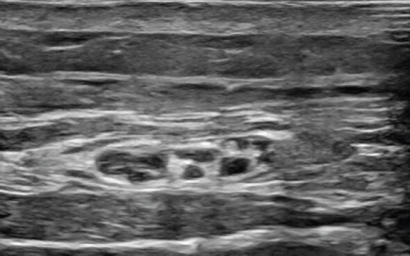
On examination, there was a well-healed scar; the skin appeared clean, dry, and intact, with adequate perfusion to the fingers. There were no open wounds, ulcerations, erythema, or signs of infection. There was intact motor function of the biceps, triceps, extensor pollicis longus, flexor pollicis longus, and intrinsic muscles of the hand. There was a positive Tinel’s sign with tapping the forearm.
She subsequently obtained a radiograph which demonstrated 2 surgical clips in the superficial soft tissues of the volar and radial aspect of the proximal forearm with multiple additional surgical clips in the soft tissues at the volar and radial aspect of the distal forearm at the level of the radial metaphysis (Fig. 2).
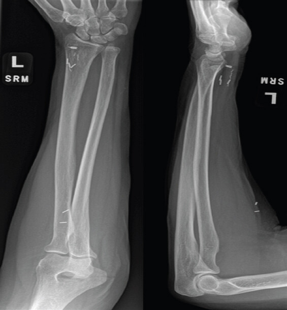
After a discussion about different options, and because she failed non-surgical measures, the patient engaged in shared decision-making and elected to proceed with surgical exploration of the left forearm, removal of foreign bodies, and left antebrachial cutaneous nerve reconstruction with allograft. On the day of the surgery, the regional anesthesia team utilized ultrasound to identify the course of the LABC nerve and position of the foreign bodies to minimize surgical exposure. These locations were marked with an indelible marker (Fig. 3). A pre-operative single injection supraclavicular block with 20 mL of 0.5% ropivacaine was then performed for surgical anesthesia.
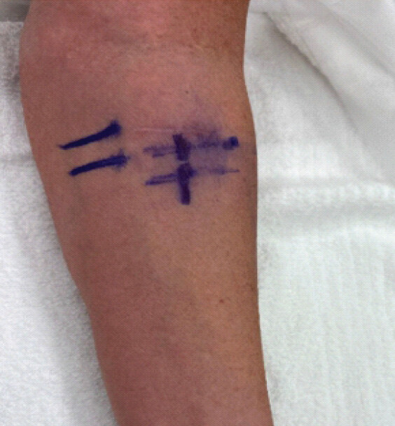
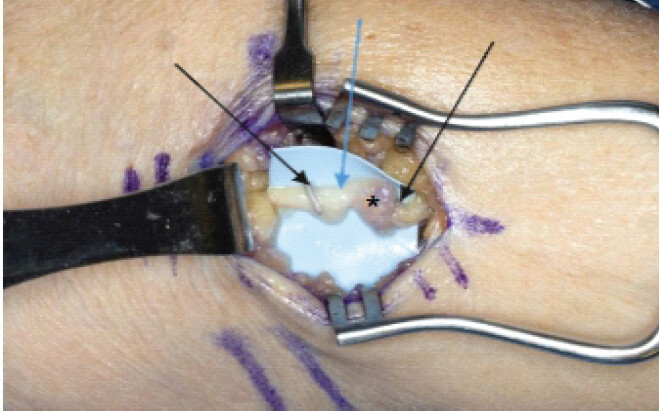
A 3 cm longitudinal incision was made on the ulnar border of the brachioradialis, in line with the course of the nerve confirmed by ultrasound (Fig. 4).
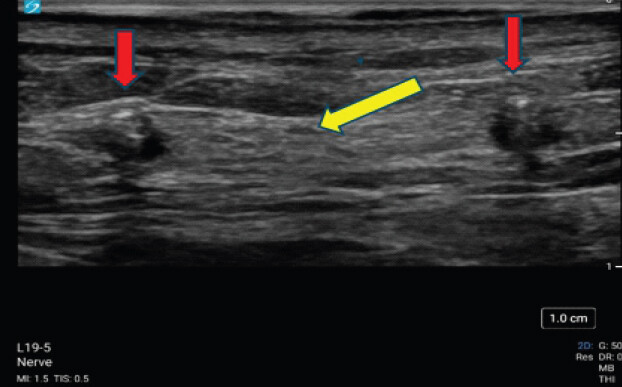
Blunt dissection was carried through the subcutaneous tissue, sparing any subcutaneous nerves and veins. The vessel clips were identified, as well as the LABC nerve. The clips were on the LABC nerve along with apparent neural swelling proximal to the more proximal clip (Fig. 5). Excision and reconstruction with allograft were determined to be the most appropriate next step. The nerve was cut just proximal and distal to the damaged nerve segment and clips, and a 2–3 mm diameter allograft by 50 mm in length was used. Three epineural 8-0 nylon sutures were used to secure the allograft on the proximal end; the allograft was cut to the appropriate length, and three additional epineural 8-0 nylon sutures secured the distal end of the allograft. The neurorrhaphy was augmented with Tisseel glue, and the resected nerve segment was sent for pathology.
At her 6-week follow-up, she endorsed improvement in her nerve pain but persistent numbness, which is expected in the early post-operative period following nerve reconstruction. She noted normal ROM with mild weakness, but improving use in her affected extremity.
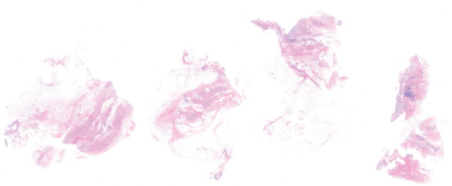
The pathology report described the gross specimen as tan-gray soft tissue. The biopsy was interpreted as a benign peripheral nerve with focal perineural fibrosis and chronic inflammation (Fig. 6).
Discussion
ERAH has become a common technique for obtaining a second arterial conduit for CABG, driven by the known long-term advantages of radial artery grafting and the minimally invasive benefits of endoscopic harvest [7, 15]. However, neurologic sequelae in the donor arm remain an important source of morbidity. Post-operative pain, paresthesia, weakness, and other neurologic symptoms may occur in up to one-third of patients early after harvest and are often characterized as transient, yet patient-reported outcome data demonstrate that a meaningful subset of patients experience persistent symptoms without improvement [11].
This case adds to the limited body of literature describing the functional impact of a focal sensory mononeuropathy after ERAH, demonstrating that the LABC is a vulnerable structure in the harvest corridor [10]. The anatomic proximity of the LABC to the radial artery places it at risk during dissection, clip placement, and instrumentation. In our patient, symptoms were neuropathic in quality and started in the immediate post-operative period, and objective testing supported a focal sensory mononeuropathy. Ultrasound localization further suggested a structural etiology with a metallic clip positioned immediately superficial to an enlarged superficial sensory nerve at the point of maximal tenderness.
This case suggests that LABC injury may be under-recognized in prior literature. Many published studies report neurologic complications in aggregate and do not consistently characterize injuries by discrete nerve distribution or functional impact. Given the demonstrated anatomic vulnerability of the LABC in relation to the radial artery harvest corridor, and the potential for overlapping/adjacent sensory territories among superficial radial-sided cutaneous nerves, it is plausible that some previously reported “radial sensory symptoms” after harvest were not attributed specifically to the LABC because sensory mapping was not performed or because outcomes were not reported by individual nerve territory [16, 17]. More granular post-operative assessment and standardized reporting (e.g., LABC vs. other radial-sided sensory distributions) would improve the diagnosis and strengthen pre-operative counseling regarding the expected area of paresthesia.
Conclusion
This case report illustrates a rare cause of chronic lateral forearm pain secondary to ERAH using surgical clips that manifested following the procedure without improvement of symptoms. Surgical clips placed in close proximity to nerves can cause direct mechanical compression or entrapment, leading to nerve dysfunction manifested as sensory deficits, motor impairment, or neuropathic pain. The compressive force from clips can impair nerve conduction and, if sustained, may result in irreversible electrophysiologic changes and structural nerve damage [18]. When conservative therapies fail, and diagnostic findings suggest focal entrapment or clip-associated injury, surgical exploration with foreign body removal and targeted nerve treatment (including reconstruction when indicated) should be considered. In this case, operative exploration confirmed clip-associated nerve pathology, and pathology showed chronic perineural changes, underscoring the importance of recognizing what is a treatable cause for persistent donor-arm neuropathic pain after ERAH.
Clinical Message
This article highlights detailed characterization of the clinical presentation, natural history, and functional impact of the neurological sequelae of lesion to the left antebrachial cutaneous nerve following endoscopic radial artery harvest. In addition, it demonstrates detailed therapies and surgical management to improve the symptoms of neuropathic pain.
Conflict of Interest:
Nil
Source of Support:
Nil
Consent:
The authors confirm that informed consent was obtained from the patient for publication of this article
How to Cite this Article
Tzoucalis SA, Troyer WD, Warfield DJ, Clendenen SR, Aziz KT. Chronic Neuropathy of the Lateral Antebrachial Cutaneous Nerve after Endoscopic Radial Artery Harvest: A Case Report. Journal of Orthopaedic Case Reports 2026 October;16(10): 23-28.
References
- Aldea GS, Bakaeen FG, Pal J, Fremes S, Head SJ, Sabik J. The society of thoracic surgeons clinical practice guidelines on arterial conduits for coronary artery bypass grafting. Ann Thorac Surg 2016;101:801-9. [Google Scholar] | [PubMed]
- Gaudino M, Benedetto U, Fremes S, Biondi-Zoccai G, Sedrakyan A, Puskas JD. Radial-artery or saphenous-vein grafts in coronary-artery bypass surgery. N Engl J Med 2018;378:2069-77. [Google Scholar] | [PubMed]
- Lawton JS, Tamis-Holland JE, Bangalore S, Bates ER, Beckie TM, Bischoff JM. 2021 ACC/AHA/SCAI guideline for coronary artery revascularization: A report of the american college of cardiology/American heart association joint committee on clinical practice guidelines. J Am Coll Cardiol 2022;79:e21-129. [Google Scholar] | [PubMed]
- Noona SW, Young SD, Young AM, Strobel RJ, Weber MP, Ajadi E. Predictors of radial artery use in coronary bypass grafting: A multi-institutional cohort study. J Surg Res 2025;308:193-201. [Google Scholar] | [PubMed]
- Connolly MW, Torrillo LD, Stauder MJ, Patel NU, McCabe JC, Loulmet DF. Endoscopic radial artery harvesting: Results of first 300 patients. Ann Thorac Surg 2002;74:502-5. [Google Scholar] | [PubMed]
- Razavi AA, Lim B, Sallam A, Perez C, Sharew B, Krishnan A. Endoscopic versus open radial artery harvesting: A population-level analysis. Ann Thorac Surg 2026;121:1385-93. [Google Scholar] | [PubMed]
- Huang TY, Huang TS, Cheng YT, Wang YC, Chen TP, Yin SY. Radial artery harvesting in coronary artery bypass grafting surgery-endoscopic or open method? A meta-analysis. PLoS One 2020;15:e0236499. [Google Scholar] | [PubMed]
- Rahouma M, Kamel M, Benedetto U, Ohmes LB, Di Franco A, Lau C. Endoscopic versus open radial artery harvesting: A meta-analysis of randomized controlled and propensity matched studies. J Card Surg 2017;32:334-41. [Google Scholar] | [PubMed]
- Lei JJ, Ravendren A, Snosi M, Harky A. In patients undergoing coronary artery bypass grafting, is endoscopic harvesting superior to open radial artery harvesting? Interact Cardiovasc Thorac Surg. 2021;32:447-51. [Google Scholar] | [PubMed]
- Khadanovich A, Benes M, Kaiser R, Herma T, Kachlik D. Clinical anatomy of the lateral antebrachial cutaneous nerve: Is there any safe zone for interventional approach?. Ann Anat 2024;252:152202. [Google Scholar] | [PubMed]
- Denton TA, Trento L, Cohen M, Kass RM, Blanche C, Raissi S. Radial artery harvesting for coronary bypass operations: Neurologic complications and their potential mechanisms. J Thorac Cardiovasc Surg 2001;121:951-6. [Google Scholar] | [PubMed]
- Carranza CL, Petersen JJ, Ballegaard M, Werner MU, Hasbak P, Kjaer A. Endoscopic or open radial artery harvest in coronary artery bypass surgery. NEJM Evid 2026;5:EVIDoa2500199. [Google Scholar] | [PubMed]
- Bleiziffer S, Hettich I, Eisenhauer B, Ruzicka D, Voss B, Bauernschmitt R. Neurologic sequelae of the donor arm after endoscopic versus conventional radial artery harvesting. J Thorac Cardiovasc Surg 2008;136:681-7. [Google Scholar] | [PubMed]
- Kim G, Jeong Y, Cho Y, Lee J, Cho J. Endoscopic radial artery harvesting may be the procedure of choice for coronary artery bypass grafting. Circ J 2007;71:1511-5. [Google Scholar] | [PubMed]
- Gaudino M, Burzotta F, Bakaeen F, Bertrand O, Crea F, Di Franco A. The radial artery for percutaneous coronary procedures or surgery?. J Am Coll Cardiol 2018;71:1167-75. [Google Scholar] | [PubMed]
- Mackinnon SE, Dellon AL. The overlap pattern of the lateral antebrachial cutaneous nerve and the superficial branch of the radial nerve. J Hand Surg Am 1985;10:522-6. [Google Scholar] | [PubMed]
- Poublon AR, Walbeehm ET, Duraku LS, Eilers PH, Kerver AL, Kleinrensink GJ. The anatomical relationship of the superficial radial nerve and the lateral antebrachial cutaneous nerve: A possible factor in persistent neuropathic pain. J Plast Reconstr Aesthet Surg 2015;68:237-42. [Google Scholar] | [PubMed]
- Hubbard RD, Winkelstein BA. Transient cervical nerve root compression in the rat induces bilateral forepaw allodynia and spinal glial activation: Mechanical factors in painful neck injuries. Spine (Phila Pa 1976) 2005;30:1924-32. [Google Scholar] | [PubMed]
© 2026 Journal of Orthopaedic Case Reports - Published by Indian Orthopaedic Research Group
About the Authors
-

Sean A Tzoucalis
-

Wesley D Troyer
-

Dennis J Warfield
-

Steven R Clendenen
-

Keith T Aziz
Related Articles in Journal of Orthopaedic Case Reports
 October 1, 2026 Intrapelvic Neck Screw Migration after Trochanteric Fixation Nail Advanced Fixation of a Trochanteric Femoral Fracture: A Case Report and Proposed Treatment Algorithm
 October 1, 2025 Functional and Radiological Outcomes of Anterior Clavicle Plating in the Treatment of Midshaft Clavicle Fractures – Allman Group 1 Type
 September 1, 2025 Chronic Saphenous Nerve Entrapment Neuropathy after Closed Blunt Trauma: A Case Report
 July 10, 2020 Αbsorbable Plates for Isolated Ulnar Diaphyseal Fractures in Adults – A Case Series Study