


Introduction
Flexion deformity of the knee is the inability to fully extend the knee; it could be due to poor muscle power of the quadriceps muscle or failure of the hamstring muscle to lengthen [1]. In delayed presentations after septic arthritis, bony ankylosis could be the cause of stiffness. Marginal joint distraction with gradual joint narrowing can also cause fibrous ankylosis in tuberculosis arthritis [2].
Progressive knee flexion deformity is common in spastic type cerebral palsy primarily because of soft tissue contracture and shortening [3]. Arthrogryposis which usually manifests after birth and involves multiple joints could be the cause of severe knee contracture [4].
Disabling knee contracture can be caused by popliteal pterygium syndrome, juvenile rheumatoid arthritis, hemophilic arthropathy, trauma, prolonged immobilization, and burns [5,6,7,8].
Diagnosis of bony ankylosis can be made using X-rays or computed tomography scans, and this imaging shows the extent of bony growth around the affected joint [9].
Management of knee contracture is a challenging procedure and it ranges from stretching physical therapy to complex bony and soft tissue procedures [10,11,12].
Case Report
A 14-year-old male patient presented to our hospital with inability to extend his left knee and difficulty walking for the past 8 years. Since he could not afford walking aids, he had to crawl whenever he needed to move (Fig. 1). On inspection, there was an old, healed longitudinal surgical scar over the posterolateral aspect of the distal thigh. The knee was locked in 140° flexion, with no further flexion or extension possible (Fig. 2). The hip and ankle ranges of motion were normal. Plain radiographs revealed disappearance of the joint space, with the distal femur slightly deformed in a fixed flexion position (Fig. 3).
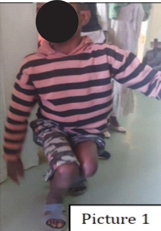
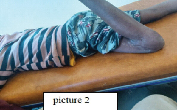
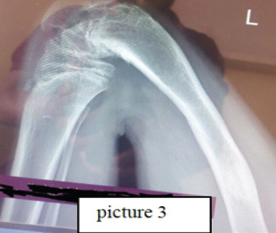
The past medical history revealed that the patient had knee swelling, pain, and fever 8 years earlier, for which he underwent surgery at a local health facility. He was discharged after his symptoms subsided but was not advised to undergo physical rehabilitation. Since then, he has had progressive difficulty walking and increasing stiffness of the knee.
Laboratory findings
-
WBC: 7,800/mm³
-
Hemoglobin: 13 g/dL
-
ESR: 15 mm/h
-
CRP: < 5 ng/L
Surgical technique
Informed consent was obtained from the patient’s family. One gram of intravenous ceftriaxone was administered preoperatively. Under spinal anesthesia, the patient was placed in the right lateral position, and the surgical site was prepared and draped in a sterile manner.
A longitudinal incision was made along the posterolateral aspect of the distal femur. After careful dissection of the scar tissue, the peroneal nerve was identified and found entrapped within fibrous tissue. The nerve was gently freed by meticulous soft tissue dissection. The distal femur was then exposed, and a wedge osteotomy was performed. Soft tissue tension was assessed, and further femoral shortening was carried out to relieve tension on the peroneal nerve. The bone was reduced, and a uniplanar dynamic external fixator was applied, achieving compression at the osteotomy site.
During extension, a skin contracture was noted over the popliteal area. The scarred skin was excised, and a rotational flap was performed to cover the defect. The patient remained stable postoperatively.
The osteotomy ends were kept compressed for 10 days, after which distraction was started at a rate of 1 mm/day for 10 days, achieving an extra 1 cm of lengthening compared to the contralateral limb. Considering the patient’s age of 14 years and his proximity to skeletal maturity, this additional lengthening was carefully planned to account for the remaining growth potential of the contralateral side, ensuring no significant limb length discrepancy would remain at skeletal maturity. Once the limb length discrepancy was corrected, the external fixator was maintained for an additional 2 months for consolidation (Fig. 4a–c).
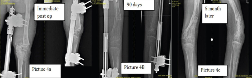
Outcome
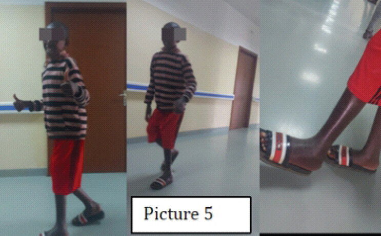
At the 5-month follow-up, the patient achieved a straight, fused leg with improved alignment. He was able to walk upright, without pain and without the need for walking aids (Fig. 5).
Discussion
Severe knee flexion contractures combined with joint ankylosis present a complex surgical challenge. Conventional soft-tissue release is often inadequate in these cases, particularly when contractures exceed 90° [4,5]. Our patient had a 140° flexion contracture with an ankylosed knee joint, making standard correction techniques insufficient.
Femoral extension osteotomy is a recognized method for correcting fixed flexion deformities, particularly in post-traumatic, post-poliomyelitis, or ankylosed knees [12]. In extreme deformities, acute correction carries risks of neurovascular compromise, soft-tissue injury, and instability [3, 7].
Distraction osteogenesis using circular external fixators allows gradual correction while minimizing tension on soft tissues and neurovascular structures [1,5,6,8,11,13]. This technique has been successfully applied in hemophilic knee contractures [5,10], arthrogryposis [4], post-traumatic or post-burn flexion deformities [7], and juvenile-onset ankylosing spondylitis [8]. Kim et al. reported using the Ilizarov frame for popliteal pterygium, highlighting the safety and effectiveness of gradual angular correction [6].
In our patient, we employed a hybrid approach: Femoral osteotomy with acute shortening followed by gradual distraction. Osteotomy provided mechanical realignment, shortening protected neurovascular structures, and gradual distraction facilitated soft-tissue adaptation [11,12]. However, due to the severity of the deformity and underlying ankylosis, the final outcome was a straight-leg fused knee, which restored functional alignment and stability.
This case emphasizes that in extreme flexion contractures with ankylosed joints, the surgical goal may shift from achieving normal range of motion to obtaining a stable, functional limb. Careful planning, staged correction, and gradual soft-tissue adaptation are essential to minimize complications and optimize functional outcome [1,4,5,8,11,12]. However, extended follow-up is ongoing to monitor long-term skeletal remodeling and functional outcomes in this growing patient.
Conclusion
For severe knee flexion contractures with ankylosis, a combined approach of femoral osteotomy, acute shortening, and distraction osteogenesis using uniplanar dynamic external fixation in a resource-limited setting can safely restore functional limb alignment. In cases where full motion cannot be achieved, a fused straight-leg outcome provides a stable, functional extremity, consistent with principles of limb reconstruction and gradual deformity correction.
Clinical Message
In managing severe knee flexion contractures with ankylosis in resource-limited settings, achieving joint extension can expose a popliteal skin contracture defect that requires excision of scarred tissue and coverage with a rotational flap. Integrating this soft-tissue reconstruction with distal femoral wedge osteotomy, acute shortening, peroneal nerve decompression, and gradual distraction osteogenesis via a uniplanar dynamic external fixator safely restores functional limb alignment and achieves a stable, straight fused extremity when full joint mobility is unachievable.
Conflict of Interest:
Nil
Consent:
The authors confirm that informed consent was obtained from the patient for publication of this article
How to Cite this Article
Mulugeta B, Zenebe T, Peltzer J, Babst R, Andualem T. Management of Severe Knee Flexion Contracture of 140° with Fibrous Ankylosis in a Resource-Limited Setting: A Case Report. Journal of Orthopaedic Case Reports 2026 October;16(10): 29-33.
References
- Kwan MK, Penafort R, Saw A. Treatment for flexion contracture of the knee during Ilizarov reconstruction of tibia with passive knee extension splint. Med J Malaysia 2004;59 Suppl F:39-41. [Google Scholar] | [PubMed]
- Korim M, Patel R, Allen P, Mangwani J. Foot and ankle tuberculosis: Case series and literature review. Foot (Edinb) 2014;24:176-9. [Google Scholar] | [PubMed]
- İnan M, Sarikaya İA, Yildirim E, Güven MF. Neurological complications after supracondylar femoral osteotomy in cerebral palsy. J Pediatr Orthop 2015;35:290-5. [Google Scholar] | [PubMed]
- Pontén E. Management of the knees in arthrogryposis. J Child Orthop 2015;9:465-72. [Google Scholar] | [PubMed]
- Balci HI, Kocaoglu M, Eralp L, Bilen FE. Knee flexion contracture in haemophilia: Treatment with circular external fixator. Haemophilia 2014;20:879-83. [Google Scholar] | [PubMed]
- Kim HM, Park IJ, Jeong C. Treatment of popliteal pterygium using an Ilizarov external fixator. Clin Orthop Surg 2009;1:236-9. [Google Scholar] | [PubMed]
- Grishkevich VM, Vishnevsky AV. Postburn knee flexion contractures: Anatomy and methods of their treatment. Trop Med Surg 2013;1:147. [Google Scholar] | [PubMed]
- Xia LW, Xu C, Huang JH. Use of Ilizarov technique for bilateral knees flexion contracture in juvenile-onset ankylosing spondylitis: A case report. World J Clin Cases 2023;11:7179-86. [Google Scholar] | [PubMed]
- Chouinard AF, Kaban LB, Peacock ZS. Acquired abnormalities of the temporomandibular joint. Oral Maxillofac Surg Clin North Am 2018;30:83-96. [Google Scholar] | [PubMed]
- Tepeneu NF, Tepeneu P, Serban M, Petrescu C, Pop L, Popescu B. Orthopaedic non-surgical treatment in chronic haemophilic joint contractures of the knee. Timisoara Med J 2010;60:215-22. [Google Scholar] | [PubMed]
- Hosny GA, Fadel M. Managing flexion knee deformity using a circular frame. Clin Orthop Relat Res 2008;466:2995-3002. [Google Scholar] | [PubMed]
- Zouari O, Gargouri A, Jenzri M, Hadinane R, Slimane N. Supracondylar femoral extension osteotomy for knee flexion contracture correction in poliomyelitic conditions. Rev Chir Orthop Reparatrice Appar Mot 2001;87:361-6. [Google Scholar] | [PubMed]
- Conway JD, Mont MA, Bezwada HP. Arthrodesis of the knee. J Bone Joint Surg Am 2004;86:835-48. [Google Scholar] | [PubMed]
© 2026 Journal of Orthopaedic Case Reports - Published by Indian Orthopaedic Research Group
About the Authors
-

Mulugeta Bekele Geneti
-

Zenebe Teklu Gebrehana
-

Jorge Peltzer
-

Reto Babst
-

Andualem Tesfaye Alemayehu
Related Articles in Journal of Orthopaedic Case Reports
 October 1, 2026 Soft-Tissue Closure through Acute Deformation and Gradual Correction Using Taylor Spatial Frame in a Gustilo-Anderson Type IIIB Open Tibial Pilon Fracture: A Case Report
 August 1, 2026 Understanding Diagnostic Challenges in Ewing Sarcoma – A Report of Two Cases
 July 1, 2026 Step-cut and Butterfly Femoral Osteotomies Following Intraoperative Fracture Propagation during Distraction Osteogenesis: Report of Two Cases
 July 1, 2026 Outcomes of Surgical Treatment for Masada Type lVa Forearm Deformity in Children with Multiple Hereditary Exostosis: A Case Series