


Introduction
Open (compound) fractures are injuries in which a soft-tissue wound communicates the fracture site with the external environment, exposing bone and/or fracture hematoma to the outside [1]. They pose a major reconstructive challenge due to the attendant risk of infection, soft-tissue loss, and delayed union. The annual incidence of open long-bone fractures is estimated at around 11.5/100,000 population, with open tibial fractures accounting for a substantial proportion of these injuries; infection rates rise sharply with increasing injury severity, from under 2% in low-grade injuries to approximately 50% in the most severe grade [2].
Historically, staged management – external fixation followed by delayed internal fixation once soft-tissue swelling had settled – was preferred, chiefly to reduce infection risk. Advances in debridement technique, antibiotic prophylaxis, and soft-tissue reconstruction have since supported primary (early) definitive internal fixation, generally within 72 h of injury, as a viable alternative that may reduce hospital stay and accelerate functional recovery without a proportionate rise in infection.
Selecting patients appropriately for early fixation, however, depends on an accurate assessment of injury severity. The Gustilo-Anderson classification, first described in an analysis of over 670 open fractures [3], remains widely used but has recognized limitations – imprecise definitions, interobserver reliability of only about 60%, inability to grade the extent of skeletal and soft-tissue damage within its broad Type IIIB category, and no guidance on limb salvage [4,5]. The Orthopedic Trauma Association classification refined this approach by grading skin, vascular, muscle, contamination, and bone loss separately [6], while the Ganga Hospital Open Injury Score (GHOIS) incorporates skin, bone, and functional-tissue components to guide both salvage decisions and the timing of soft-tissue reconstruction; a total score of ≤9 identifies lower-violence injuries amenable to early definitive treatment [7,8].
Against this background, we undertook a prospective cohort study to evaluate the outcomes of primary definitive internal fixation performed within 72 h of injury in patients with compound fractures selected using GHOIS (≤9), with the objectives of determining the pattern of injury on the basis of GHOIS, treating patients with the early fixation under a standard antibiotic protocol, and following them for fracture union, soft-tissue healing, functional outcome, and complications over 1 year.
Materials and Methods
This prospective cohort study was conducted in a tertiary-care orthopedic center in India over 21 months (March 2023 to November 2024), after obtaining Institutional Ethics Committee approval (147/IEC AMC/MAY 2024) and written informed consent from all participants. This study was conducted in accordance with the World Medical Association Declaration of Helsinki.
All consecutive patients presenting to the casualty department of the hospital with open tibial fractures and a GHOIS ≤9 who fulfilled the predefined eligibility criteria during the study period (March 2023 to November 2024) were considered for inclusion. A total of 140 patients met the inclusion criteria and were enrolled in the study.
Inclusion criteria were as follows: Patients who gave informed consent, with at least 1 year of follow-up, who underwent definitive fixation and wound closure within 72 h of injury, and who had a GHOIS ≤9. Exclusion criteria were as follows: Loss of limb vascularity or Gustilo-Anderson Type IIIC injury; severe soft-tissue injury warranting initial external fixation; refusal of consent; incorrect antibiotic protocol; loss to follow-up; fixation and/or closure beyond 72 h; polytrauma requiring prior intervention from other specialties; associated head injury; and a GHOIS skin or bone sub-score of 5.
Data were collected using a predesigned, semi-structured questionnaire covering demographic details (age, gender, occupation, and tobacco/alcohol use), mechanism, time and place of injury, associated injuries, and GHOIS scoring. At presentation, all wounds were irrigated with 3–5 L of sterile saline and covered with a sterile dressing. Patients then underwent thorough surgical debridement followed by early definitive internal fixation (intramedullary nailing, open reduction and internal fixation with plating, or screw fixation, as indicated) within 72 h of injury. All patients received a standardized antibiotic protocol of injectable piperacillin-tazobactam 4.5 g 3 times daily, metronidazole 500 mg 3 times daily, and amikacin 500 mg twice daily for 5 days, followed by 10–12 days of oral antibiotics. Soft-tissue cover, where required, was individualized (primary split-skin grafting or flap cover) and, when needed, undertaken jointly with the plastic surgery department within the 72-h window.
Postoperatively, patients underwent isometric quadriceps/hamstring strengthening and knee range-of-motion exercises (lower limb) or isometric elbow flexion-extension exercises (upper limb); tibial intramedullary nail dynamization was performed at 16–20 weeks in selected cases at the treating surgeon’s discretion. Patients were followed for 1 year and assessed for time to soft-tissue healing, time to radiological union, hospital stay, complications, and functional outcome, the last graded using the modified criteria of Ketenjian and Sheldon as excellent, good, fair, or poor [9]. Data were summarized as means for continuous variables and as frequencies and percentages for categorical variables.
Results
A total of 140 patients with compound fractures meeting the inclusion criteria were enrolled and followed for 1 year.
Demographic and injury profile (Table 1)
Demographic and injury profile of patients (n=140)
| Variable | n | % |
|---|---|---|
| Age group (years) | ||
| 20–30 | 39 | 27.85 |
| 31–40 | 49 | 35 |
| 41–50 | 38 | 27.15 |
| 51–60 | 14 | 10 |
| Mean age | 37.08 years | |
| Gender | ||
| Male | 85 | 60.71 |
| Female | 55 | 39.29 |
| Side involved | ||
| Right | 74 | 52.85 |
| Left | 66 | 47.15 |
| Mechanism of injury | ||
| Road traffic accident | 114 | 81.42 |
| Self-fall | 15 | 10.71 |
| Fall from height | 11 | 7.87 |
| Total | 140 | 100 |
The mean age of patients was 37.08 years; the largest proportion was aged 31–40 years (35%), followed by 20–30 years (27.85%), 41–50 years (27.15%), and 51–60 years (10%). Males predominated (60.71%, n = 85) over females (39.29%, n = 55). The right limb was affected in 74 patients (52.85%) and the left in 66 (47.15%). Road traffic accidents were the leading mechanism of injury (81.42%, n = 114), followed by self-fall (10.71%, n = 15) and fall from height (7.87%, n = 11).
Fracture severity (Table 2)
Fracture severity (Gustilo-Anderson classification) and mean GHOIS
| Gustilo-Anderson type | n | % |
|---|---|---|
| Type I | 26 | 18.57 |
| Type II | 20 | 14.28 |
| Type IIIA | 56 | 40 |
| Type IIIB | 38 | 27.13 |
| Total | 140 | 100 |
| Mean GHOIS | 7.25 | — |
GHOIS: Ganga hospital open injury score
By Gustilo-Anderson grading, Type IIIA fractures were the most frequent (56 cases, 40%), followed by Type IIIB (38, 27.13%), Type I (26, 18.57%), and Type II (20, 14.28%); higher-grade (Type III) injuries accounted for 67.13% of the cohort. The mean GHOIS was 7.25.
Treatment timelines, fixation, and soft-tissue management (Table 3)
Treatment timelines, fixation method and soft-tissue management (n=140)
| Parameter | n /Mean | % |
|---|---|---|
| Mean time since injury to presentation (hours) | 3.82 | — |
| Mean time to definitive fixation (hours) | 45.8 | — |
| Type of fixation | ||
| Intramedullary nailing | 76 | 54.28 |
| Open reduction and internal fixation with plating | 48 | 34.28 |
| Screw fixation | 16 | 11.44 |
| Soft-tissue coverage | ||
| No coverage required | 66 | 47.14 |
| Primary SSG | 50 | 35.71 |
| Fasciocutaneous flap+primary SSG | 18 | 12.85 |
| Gastrocnemius flap+SSG | 6 | 4.28 |
SSG: Split-skin grafting
The mean time from injury to hospital presentation was 3.82 h, and the mean time to definitive fixation was 45.8 h, both within the intended 72-h window. Intramedullary nailing was the most frequently used fixation method (76 cases, 54.28%), followed by open reduction and internal fixation with plating (48, 34.28%) and screw fixation (16, 11.44%). No additional soft-tissue coverage was required in 66 cases (47.14%); primary split-skin grafting was performed in 50 (35.71%), fasciocutaneous flap with primary split-skin grafting in 18 (12.85%), and gastrocnemius flap with split-skin grafting in 6 (4.28%).
Functional and healing outcomes (Table 4)
Functional outcome and healing parameters (n=140)
| Parameter | n /Mean | % |
|---|---|---|
| Functional outcome (modified Ketenjian and Sheldon) | ||
| Excellent | 26 | 18.57 |
| Good | 64 | 45.71 |
| Fair | 36 | 25.72 |
| Poor | 14 | 10 |
| Mean time to soft-tissue healing (weeks) | 3.75 | — |
| Mean time to bone union (weeks) | 28.49 | — |
| Mean hospital stay (days) | 11.59 | — |
Functional outcome, assessed using the modified Ketenjian and Sheldon criteria, was excellent in 26 patients (18.57%) and good in 64 (45.71%), together comprising 64.28% of the cohort; a fair outcome was recorded in 36 patients (25.72%) and a poor outcome in 14 (10%). The mean time to soft-tissue healing was 3.75 weeks, the mean time to radiological union was 28.49 weeks, and the mean hospital stay was 11.59 days.
Complications (Table 5)
Complications (n=140)
| Complication | n | % |
|---|---|---|
| Infection (8 antibiotic-responsive; 8 required re-debridement) | 16 | 11.42 |
| Stiffness | 6 | 4.28 |
| Non-union | 6 | 4.28 |
| Delayed union | 1 | 0.71 |
Infection was the most common complication (16 cases, 11.42%); of these, eight resolved with antibiotics alone while the remaining eight required re-debridement. Stiffness occurred in 6 patients (4.28%) and improved with physiotherapy. Non-union occurred in 6 patients (4.28%) and required revision surgery, while delayed union was seen in a single patient (0.71%). No patient in this series required amputation or died as a result of the injury or its treatment.
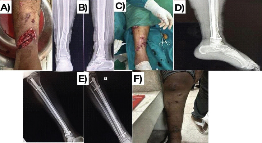
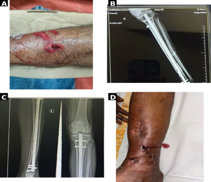
Representative case illustrations demonstrating the treatment pathway and outcomes are shown in Fig. 1 and 2.
Discussion
The management of open fractures continues to evolve, balancing the historic caution around infection with growing evidence favoring early definitive stabilization when injury severity is objectively assessed [10,11]. Contemporary guidance recommends antibiotic administration within 60 min of arrival, debridement within 24 h, and soft-tissue coverage within 7 days, with definitive fixation ideally performed within 24–72 h when feasible [10]. The mean time to fixation of 45.8 h in our cohort, following a mean presentation time of 3.82 h after injury, was consistent with this window and reflects timely triage and surgical decision-making.
The demographic profile in this study – predominantly young to middle-aged males injured in road traffic accidents – mirrors most reported open-fracture cohorts, though the reported mean age varies between series: 41.5 years in the cohort of Cullen et al. [12], 49 years in Nishida et al. [13], and 43.8 years in Prabhakaran et al. [14], compared with 37.08 years in the present study, likely reflecting differences in trauma demographics and case mix across populations. The male predominance (60.71%) and road traffic accidents as the leading mechanism (81.42%) in this study are similarly consistent with the pattern reported by Prabhakaran et al. and Azam et al., underscoring high-energy trauma as the principal driver of compound fractures in this age group. [14,15] (Table 6).
Comparison of key parameters with previous studies
| Study | Mean age (years) | Male (%) | Predominant mechanism | Mean time to fixation (h) |
|---|---|---|---|---|
| Nishida et al. | 49 | 90 | RTA/industrial | — |
| Prabhakaran et al. | 43.8 | — | RTA | 11.97 |
| Azam et al. | — | 73 | RTA (55.55%) | 4.70* |
| Cullen et al. | 41.5 | — | — | — |
| Jaidev et al. | 32.19 | — | — | — |
| Present study | 37.08 | 60.71 | RTA (81.42%) | 45.8 |
*
Refers to mean time to debridement rather than definitive fixation in the study by Azam et al. RTA: Road traffic accident
The predominance of Gustilo-Anderson Type IIIA and IIIB injuries (67.13% of the cohort) reflects the referral pattern of a tertiary trauma center and is broadly comparable to the case mix reported by Nishida et al., in whom 116 of 117 fractures were Grade IIIA or IIIB [13]. The mean GHOIS of 7.25 in our cohort falls well within the ≤9 threshold that Rajasekaran and Sabapathy identified as denoting lower-violence injury amenable to early soft-tissue reconstruction and definitive fixation, suggesting that the cohort was consistent with the lower-violence injury group described in previous GHOIS literature [7,8].
Intramedullary nailing was the most frequently used fixation method in our series (54.28%), consistent with its established role as a load-sharing, minimally invasive option that preserves periosteal blood supply and permits early mobilization [16,17]. Comparable series report broadly similar fixation choices tailored to the fracture pattern and bone involved [15].
Functional outcomes in our series were favorable, with 64.28% of patients achieving an excellent or good result by the modified Ketenjian and Sheldon criteria [9] – comparable to the 83% (25 of 30) satisfactory-or-better outcomes reported by Prabhakaran et al., in whom outcomes were similarly better for lower-grade injuries and less favorable when the fracture was close to a joint [14]. The mean time to union (28.49 weeks) and infection rate (11.42%) in our cohort were comparable to outcomes reported in previous studies of open tibial fractures treated with internal fixation or external fixation [18]. These findings suggest that, in appropriately selected open fractures, early definitive fixation was associated with acceptable union and infection outcomes. Azam et al. similarly emphasized that meticulous, repeated debridement together with judicious, staged soft-tissue cover and internal fixation shortens hospital stay and improves anatomical and functional recovery without impeding soft-tissue reconstruction – an approach broadly mirrored in our antibiotic and soft-tissue-cover protocol [15]. Jaidev et al. also reported favorable outcomes following primary internal fixation of ballistic open fractures, although their cohort involved a distinct mechanism of injury and substantially earlier fixation than the present study [19]. Infection remained the most frequent complication in our series (11.42%), a rate consistent with the recognized infection risk associated with open fractures [5]. Antibiotic therapy is an established component of open-fracture management [20]. In our cohort, infection was managed successfully with antibiotics or re-debridement in all cases without limb loss. Non-union (4.28%) and stiffness (4.28%) were infrequent and amenable to revision surgery or physiotherapy respectively, while delayed union occurred in only one patient.
Limitations
This study has several limitations. The absence of a concurrent staged-fixation comparator and the single-center, non-randomized cohort design preclude direct comparison with conventional staged fixation and introduce the possibility of selection bias and residual confounding. Although predefined clinical and injury-severity criteria, including GHOIS, were used to select patients for early definitive fixation, treatment suitability was also influenced by injury characteristics and surgeon judgment. The study population was therefore highly selected, comprising open tibial fractures with GHOIS ≤9 and excluding patients with severe soft-tissue injury requiring initial external fixation, vascular compromise or Gustilo-Anderson Type IIIC injury, a GHOIS skin or bone sub-score of 5, polytrauma requiring prior intervention, and delayed fixation or closure; consequently, these findings should not be extrapolated to severe or physiologically unstable open fractures. Although all fractures were tibial, the cohort included different Gustilo-Anderson grades and fracture patterns, and outcomes were not stratified according to fracture pattern or injury severity beyond the overall Gustilo-Anderson and GHOIS distributions. Interobserver reliability of GHOIS was also not assessed. Furthermore, fixation methods and soft-tissue management were heterogeneous and individualized according to fracture characteristics, soft-tissue condition, and surgeon judgment, making it difficult to isolate the independent contribution of fixation timing from the effects of fixation technique and soft-tissue reconstruction. The relatively broad and prolonged antibiotic regimen used in this cohort may also limit generalizability to centers with different antimicrobial protocols, resistance patterns, and stewardship practices. Infection rates were not formally compared across individual Gustilo-Anderson grades or GHOIS categories. Functional outcome was assessed using the modified Ketenjian and Sheldon criteria without a validated patient-reported outcome measure. Statistical analysis was primarily descriptive, without confidence intervals, effect-size estimation, multivariable analysis, or formal subgroup/predictor analysis; therefore, the independent influence of GHOIS, Gustilo-Anderson grade, time to fixation, fixation method, soft-tissue coverage, smoking status, or fracture pattern on infection, union, or functional outcome could not be established. Finally, the requirement for 1-year follow-up may have introduced attrition or survivorship bias if patients with poorer outcomes were less likely to complete long-term follow-up. Given the observational design and multiple co-interventions, the favorable outcomes observed cannot be attributed causally to early definitive fixation alone. Accordingly, the findings should be interpreted as evidence of feasibility and association in a carefully selected cohort rather than comparative effectiveness or proof of superiority, and future multicenter comparative studies are required to determine whether early definitive fixation offers advantages over staged fixation strategies.
Conclusion
Primary definitive internal fixation of open tibial fractures within 72 h of injury, in appropriately selected patients with GHOIS ≤9, was associated with fracture union and good-to-excellent functional outcomes in the majority of patients, with acceptable healing times, hospital stay, and manageable complication rates. These findings support the feasibility of early definitive fixation in selected open tibial fractures but do not establish superiority or relative safety compared with staged fixation. Multicenter prospective comparative studies are required to determine whether early definitive fixation offers advantages over staged fixation strategies.
Clinical Message
Objective severity scoring using GHOIS may help identify appropriately selected open tibial fractures suitable for primary definitive internal fixation within 72 h. In this selected cohort, early definitive fixation was associated with favorable functional recovery and an acceptable complication profile. However, the absence of a staged-fixation comparator precludes conclusions regarding superiority or relative safety, and multicenter comparative studies are required.
Conflict of Interest:
Nil
Source of Support:
Nil
Consent:
The authors confirm that informed consent was obtained from the patient for publication of this article
How to Cite this Article
Ghosh S, Choudhary PC, Mudunuri RM. Outcomes of Primary Definitive Internal Fixation of Compound Fractures within 72 h of Injury: A Prospective Cohort Study. Journal of Orthopaedic Case Reports 2026 October;16(10): 361-367.
References
- Sop JL, Sop A. Open Fracture Management; 2023. In: StatPearls. Treasure Island, FL: StatPearls Publishing; 2025. [Google Scholar] | [PubMed]
- Weber CD, Hildebrand F, Kobbe P, Lefering R, Sellei RM, Pape HC. Epidemiology of open tibia fractures in a population-based database: Update on current risk factors and clinical implications. Eur J Trauma Emerg Surg 2019;45:445-53. [Google Scholar] | [PubMed]
- Gustilo RB. Management of open fractures. An analysis of 673 cases. Minn Med 1971;54:185-9. [Google Scholar] | [PubMed]
- Gustilo RB, Mendoza RM, Williams DN. Problems in the management of type III (severe) open fractures: A new classification of type III open fractures. J Trauma 1984;24:742-6. [Google Scholar] | [PubMed]
- Kim PH, Leopold SS. In brief: Gustilo-Anderson classification. [Corrected]. Clin Orthop Relat Res 2012;470:3270-4. [Google Scholar] | [PubMed]
- Agel J, Rockwood T, Barber R, Marsh JL. Potential predictive ability of the orthopaedic trauma association open fracture classification. J Orthop Trauma 2014;28:300-6. [Google Scholar] | [PubMed]
- Rajasekaran S, Sabapathy SR. A philosophy of care of open injuries based on the Ganga hospital score. Injury 2007;38:137-46. [Google Scholar] | [PubMed]
- Rajasekaran S, Naresh Babu J, Dheenadhayalan J, Shetty AP, Sundararajan SR, Kumar M. A score for predicting salvage and outcome in Gustilo type-IIIA and type-IIIB open tibial fractures. J Bone Joint Surg Br 2006;88:1351-60. [Google Scholar] | [PubMed]
- Ketenjian AY, Shelton ML. Primary internal fixation of open fractures: A retrospective study of the use of metallic internal fixation in fresh open fractures. J Trauma 1972;12:756-63. [Google Scholar] | [PubMed]
- American College of Surgeons Trauma Quality Improvement Program. Best Practices in the Management of Orthopaedic Trauma; 2015. 1-38. [Google Scholar] | [PubMed]
- Halawi MJ, Morwood MP. Acute management of open fractures: An evidence-based review. Orthopedics 2015;38:e1025-33. [Google Scholar] | [PubMed]
- Cullen S, Flaherty D, Fitzpatrick N, Ali A, Elkhidir I, Pillai A. Outcomes following surgical fixation of Gustilo-Anderson IIIb open tibial fractures. Acta Orthop Belg 2024;90:83-9. [Google Scholar] | [PubMed]
- Nishida M, Kamekura S, Nakada I, Kiriyama M, Maeda C, Ozone E. Definitive internal fracture fixation followed by staged free flap coverage (“fix followed by flap” protocol) for open Gustilo type IIIB fractures. J Orthop Sci 2025;30:142-6. [Google Scholar] | [PubMed]
- Prabhakaran AK, Kumar AJ, Nizar AM, Sundar RA, Subash Y. Functional outcome of management of open fractures of both bones of the leg with primary intramedullary interlocking nailing. Int J Orthop Sci 2023;9:285-91. [Google Scholar] | [PubMed]
- Azam Q, Sherwani M, Abbas M, Gupta R, Asif N, Sabir A. Internal fixation in compound type III fractures presenting after golden period. Indian J Orthop 2007;41:204-8. [Google Scholar] | [PubMed]
- Yokoyama K, Shindo M, Itoman M, Yamamoto M, Sasomoto N. Immediate internal fixation for open fractures of the long bones of the upper and lower extremities. J Trauma 1994;37:230-6. [Google Scholar] | [PubMed]
- Whittle AP, Russel TA, Taylor JC, Lavelle DG. Treatment of open fractures of the tibial shaft with the use of interlocking nailing without reaming. J Bone Joint Surg Am 1992;74:1162-71. [Google Scholar] | [PubMed]
- Court-Brown CM, McQueen MM, Quaba AA, Christie J. Locked intramedullary nailing of open tibial fractures. J Bone Joint Surg Br 1991;73:959-64. [Google Scholar] | [PubMed]
- Jaidev KP, Bhaskarwar A, Ghai A. Outcomes of fast-track primary internal fixation of open fractures in ballistic injuries - a single-centre experience. J Orthop Traumatol Rehabil 2021;13:138-43. [Google Scholar] | [PubMed]
- Patzakis MJ, Harvey JP Jr, Ivler D. The role of antibiotics in the management of open fractures. J Bone Joint Surg Am 1974;56:532-41. [Google Scholar] | [PubMed]
© 2026 Journal of Orthopaedic Case Reports - Published by Indian Orthopaedic Research Group
About the Authors
-

Sayam Ghosh
-

Pranay C Choudhary
-

Ramaraju M P Mudunuri
Related Articles in Journal of Orthopaedic Case Reports
 October 1, 2026 Functional Outcome of Operative Titanium Elastic Nailing System Versus Non-Operative Figure-of-Eight Bandage in Displaced Mid-Shaft Clavicle Fractures: A Randomized Controlled Study
 October 1, 2026 Clinical and Functional Outcomes of Closed Intramedullary Interlocking Nailing in Extra-articular Distal Tibial Fractures: A Prospective Observational Study
 October 1, 2026 A Functional Outcome of Closed Intra-articular Fracture of Proximal Interphalangeal Joint of Hand Managed By Suzuki Frame – A Longitudinal Study
 October 1, 2026 Functional Outcome of Lucerne Cast in Patients with Metacarpal Fracture with or without Proximal Phalanx Fracture: An Observational Prospective Study