


Introduction
Total knee arthroplasty (TKA) is one of the most successful orthopedic procedures for treating end-stage knee osteoarthritis, providing substantial pain relief, correction of deformity, and restoration of joint function [1]. The global prevalence of knee osteoarthritis has increased due to aging populations, obesity, and sedentary lifestyles, leading to a significant rise in the demand for TKA. Although modern implants demonstrate excellent long-term survivorship, a considerable proportion of patients remain dissatisfied because of persistent pain, stiffness, instability, or functional limitations. Consequently, recent research has shifted from focusing solely on implant longevity to restoring normal knee kinematics and improving patient-reported outcomes [2].
Mechanical alignment (MA) has long been considered the standard approach in TKA, aiming to achieve a neutral mechanical axis by positioning implants perpendicular to the femoral and tibial mechanical axes [3]. This technique has shown predictable implant survival and reliable clinical outcomes. However, restoring every patient to a neutral alignment may not reproduce individual native anatomy, potentially altering ligament balance, joint line orientation, and patellofemoral biomechanics, thereby contributing to post-operative dissatisfaction [4].
Unrestricted KA has emerged as an individualized alternative that seeks to restore the patient’s pre-arthritic anatomy while preserving constitutional limb alignment and native ligament tension [5]. This personalized approach aims to recreate physiological knee motion, minimize soft tissue releases, and improve functional recovery. Several studies have reported improved patient satisfaction and functional outcomes with KA, although concerns regarding implant positioning and long-term durability remain under investigation [6].
Restoration of normal patellofemoral mechanics is an essential determinant of successful TKA. Abnormal patellar tracking and excessive patellar tilt are major causes of anterior knee pain, instability, reduced range of motion (ROM), and implant wear. Proper positioning of the femoral and tibial components directly influences patellar movement during knee flexion and extension, making accurate assessment of patellofemoral biomechanics clinically important [7]. Conventional post-operative assessment using plain radiographs and static computed tomography (CT) evaluates implant orientation but cannot assess dynamic patellar motion [8]. Dynamic CT has emerged as an advanced imaging modality that provides real-time evaluation of patellar tracking, patellar tilt, translation, and femoropatellar congruence during active knee movement. This technique offers a more comprehensive assessment of post-operative patellofemoral biomechanics than static imaging [9].
Recent comparative studies have suggested that unrestricted KA may better restore native patellofemoral mechanics than conventional MA by preserving trochlear orientation and improving rotational alignment [10]. While some studies have demonstrated reduced patellar tilt, improved knee function, and higher patient satisfaction, others have reported minimal differences between the two alignment strategies, highlighting the need for further prospective comparative research using objective imaging methods [6,7,8]. Therefore, the present study compared post-operative patellar tracking and clinical outcomes following unrestricted KA and conventional MA using dynamic CT.
Materials and Methods
Study design
This prospective, comparative, non-randomized observational study was conducted in the Department of Orthopaedics in collaboration with the Department of Radiodiagnosis at a tertiary care teaching hospital over a period of 18 months. The study aimed to compare post-operative patellar tracking, patellar tilt, and functional outcomes following unrestricted KA and standard MA in primary TKA using dynamic CT. The study protocol was approved by the Institutional Ethics Committee, and written informed consent was obtained from all participants before enrollment in the study.
Participants
Inclusion criteria
-
Patients aged 50–80 years
-
Patients diagnosed with primary end-stage knee osteoarthritis requiring primary TKA
-
Patients willing to undergo post-operative dynamic CT evaluation
-
Patients who provided written informed consent
-
Patients who were able to comply with post-operative follow-up.
Exclusion criteria
-
Revision TKA
-
Previous fracture or surgery around the affected knee
-
Severe extra-articular deformity requiring corrective osteotomy
-
Inflammatory arthritis (rheumatoid arthritis, psoriatic arthritis, etc.)
-
Neuromuscular disorders affecting gait or patellar tracking
-
Active infection around the knee
-
Patients with contraindications to CT imaging
-
Patients unwilling to participate or lost to follow-up.
Study sampling
A consecutive sampling technique was employed. All eligible patients presenting to the orthopedic outpatient department and fulfilling the inclusion criteria during the study period were screened for participation. Patients meeting the eligibility criteria were recruited consecutively until the required sample size was attained. This method minimized selection bias while ensuring representative enrollment of patients undergoing primary TKA.
Study sample size
A total of 50 patients undergoing primary TKA were included in the study. No formal a priori sample size calculation was performed. The sample size was determined based on the expected availability of eligible patients during the 18-month study period and the feasibility of performing post-operative dynamic CT evaluation. The enrolled patients were equally distributed between the two comparison groups, with 25 patients undergoing unrestricted KA and 25 patients undergoing standard MA.
Study groups
The study population was divided into two groups according to the alignment technique used during surgery. Allocation was based on the planned surgical alignment philosophy and was not randomized. Therefore, the study was conducted as a prospective comparative observational study rather than a randomized controlled trial.
Group A (n = 25): Patients who underwent primary TKA using the unrestricted KA technique.
Group B (n = 25): Patients who underwent primary TKA using the standard MA technique.
Both groups received identical perioperative care, rehabilitation protocols, post-operative analgesia, thromboprophylaxis, and physiotherapy to minimize potential confounding.
Study parameters
The study evaluated a comprehensive set of baseline, clinical, radiological, and functional parameters. Baseline variables included age, gender, body mass index (BMI), side of surgery, duration of symptoms, Kellgren–Lawrence grade of osteoarthritis, pre-operative knee ROM, and pre-operative Knee Society Score (KSS). The primary outcome measure was post-operative patellar tracking assessed using dynamic CT. Secondary outcome measures included patellar tilt angle, patellar translation, femoral and tibial component rotational alignment, post-operative KSS, functional KSS (Functional KSS), post-operative ROM, incidence of anterior knee pain, and post-operative complications.
Study procedure
Following informed consent, demographic details, clinical history, physical examination findings, and pre-operative radiographic assessments were recorded. Routine laboratory investigations and anesthetic fitness evaluations were completed before surgery.
All patients underwent primary cemented TKA using the same implant system. Depending upon the planned surgical philosophy, unrestricted KA or standard MA was performed. Femoral and tibial bone resections, component positioning, rotational alignment, ligament balancing, and patellar management were performed according to the respective alignment philosophy. For unrestricted KA, component positioning was individualized to restore the patient’s constitutional anatomy while preserving native joint-line orientation and ligament tension. For MA, the femoral and tibial components were positioned to achieve the planned neutral MA. Rotational alignment and ligament balancing were assessed intraoperatively according to the respective surgical protocol, and patellar management was performed using the same standardized approach in both groups.
Postoperatively, all patients received standardized pain management, antibiotic prophylaxis, thromboprophylaxis, and supervised physiotherapy. Early mobilization and weight-bearing were encouraged according to institutional rehabilitation guidelines. Dynamic CT evaluation of the operated knee was performed after completion of the initial rehabilitation period during active knee flexion and extension. As dynamic CT was performed specifically for research-related assessment rather than routine post-operative care, the potential benefits, radiation exposure, and associated financial implications were explained to all participants before enrollment. Dynamic CT was performed only after obtaining specific informed consent. The examination was conducted using a low-dose CT protocol appropriate for the required dynamic assessment, with radiation exposure minimized according to institutional radiological safety protocols. The study-related imaging costs were [borne by the study/institution/participants – specify the actual arrangement]. Patients were informed that participation was voluntary and that refusal to undergo dynamic CT would not affect their clinical treatment.
Study data collection
Data were collected using a predesigned structured case record form. Demographic characteristics, clinical findings, operative details, implant information, intraoperative observations, post-operative rehabilitation progress, functional scores, and dynamic CT measurements were systematically documented. Imaging measurements were performed independently by experienced musculoskeletal radiologists using standardized software to reduce observer variability. All collected data were verified for completeness before statistical analysis.
Data analysis
The collected data were entered into Microsoft Excel and analyzed using the Statistical Package for the Social Sciences version 26.0. Continuous variables were expressed as mean ± standard deviation (SD), whereas categorical variables were presented as frequency and percentage. Independent Student’s t-test was used for comparison of normally distributed continuous variables between the two groups. Mann–Whitney U test was applied for non-normally distributed variables whenever appropriate. Chi-square test or Fisher’s exact test was used for categorical variables. A P < 0.05 was considered statistically significant.
Results
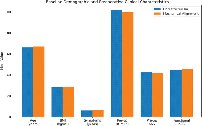
A total of 50 patients undergoing primary TKA were included in the study, with 25 patients each in the unrestricted KA and standard MA groups. Baseline demographic and pre-operative clinical characteristics, including age, sex, BMI, duration of symptoms, pre-operative KSS, functional KSS, ROM, and Kellgren–Lawrence grade, were comparable between the two groups (all P > 0.05), indicating similar baseline characteristics (Table 1 and Fig. 1).
Baseline demographic and pre-operative clinical characteristics
| Variable | KA Group (n=25) | MA Group (n=25) | P-value |
|---|---|---|---|
| Age (years) | 66.4±6.2 | 67.1±5.8 | 0.682 |
| Male, n (%) | 12 (48.0) | 11 (44.0) | 0.777 |
| BMI (kg/m2) | 28.3±2.8 | 28.7±3.0 | 0.641 |
| Duration of symptoms (years) | 6.2±2.4 | 6.5±2.3 | 0.654 |
| Pre-operative ROM (°) | 101.6±12.3 | 99.8±11.6 | 0.593 |
| Pre-operative KSS | 42.5±7.8 | 41.9±8.2 | 0.792 |
| Functional KSS | 44.8±8.4 | 45.3±8.1 | 0.836 |
| Kellgren–Lawrence Grade IV | 17 (68.0) | 16 (64.0) | 0.758 |
BMI: Body mass index, ROM: Range of motion, KSS: Knee society score, KA: Kinematic alignment, MA: Mechanical alignment
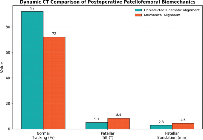
Dynamic CT assessment demonstrated significantly superior post-operative patellofemoral biomechanics in the KA group. Normal patellar tracking was observed in 92.0% of patients treated with KA compared with 72.0% in the MA group (P = 0.043). Furthermore, mean patellar tilt (5.1 ± 2.0° vs. 8.4 ± 2.8°; P < 0.001) and patellar translation (2.8 ± 1.1 mm vs. 4.5 ± 1.6 mm; P < 0.001) were significantly lower following unrestricted KA, indicating improved patellar alignment and tracking (Table 2 and Fig. 2).
Dynamic CT assessment of patellofemoral biomechanics
| Parameter | KA Group | MA Group | P-value |
|---|---|---|---|
| Normal patellar tracking | 23 (92.0%) | 18 (72.0%) | 0.043 |
| Patellar tilt (°) | 5.1±2.0 | 8.4±2.8 | <0.001 |
| Patellar translation (mm) | 2.8±1.1 | 4.5±1.6 | <0.001 |
CT: Computed tomography, KA: Kinematic alignment, MA: Mechanical alignment
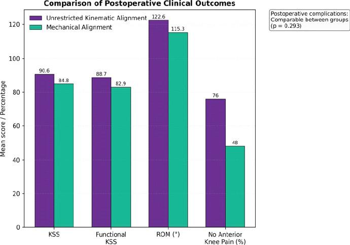
Post-operative functional outcomes were significantly better in patients undergoing unrestricted KA. Mean post-operative KSS (90.6 ± 5.4 vs. 84.8 ± 6.3; P = 0.001), functional KSS (88.7 ± 6.1 vs. 82.9 ± 6.8; P = 0.002), and ROM (122.6 ± 8.5° vs. 115.3 ± 9.6°; P = 0.007) were significantly higher than those observed in the MA group. In addition, anterior knee pain was less frequent following KA, with 76.0% of patients reporting no anterior knee pain compared with 48.0% in the MA group (P = 0.038). Post-operative complications were infrequent and comparable between both groups (P = 0.293), indicating similar short-term safety profiles (Table 3 and Fig. 3).
Post-operative clinical outcomes
| Parameter | KA Group | MA Group | P-value |
|---|---|---|---|
| Knee Society Score | 90.6±5.4 | 84.8±6.3 | 0.001 |
| Functional KSS | 88.7±6.1 | 82.9±6.8 | 0.002 |
| Range of motion (°) | 122.6±8.5 | 115.3±9.6 | 0.007 |
| No anterior knee pain | 19 (76.0%) | 12 (48.0%) | 0.038 |
| Overall complications | Comparable | Comparable | 0.293 |
KA: Kinematic alignment, MA: Mechanical alignment, KSS: Knee Society Score
Discussion
The present prospective comparative observational study evaluated post-operative patellofemoral biomechanics and functional outcomes following unrestricted KA and standard MA in primary TKA using dynamic CT. The principal findings demonstrated that unrestricted KA provided significantly better patellar tracking, lower patellar tilt, reduced patellar translation, higher KSS, improved functional KSS, greater post-operative ROM, and a lower incidence of anterior knee pain compared with conventional MA, while post-operative complication rates were comparable between the two alignment strategies. These findings support the concept that restoration of patient-specific knee anatomy through unrestricted KA results in superior patellofemoral biomechanics without compromising short-term safety. Dynamic CT evaluation provided objective evidence that individualized alignment can improve patellar kinematics, an important determinant of post-operative satisfaction following TKA. The comparable baseline demographic and clinical characteristics between the two groups ensured that post-operative differences were attributable to the alignment philosophy rather than pre-operative confounding factors.
In the present study, normal post-operative patellar tracking was observed in 92.0% of patients undergoing unrestricted KA compared with 72.0% in the MA group (P = 0.043). Furthermore, severe patellar maltracking occurred only in the MA group. These findings indicate that unrestricted KA more effectively restores native trochlear orientation and extensor mechanism biomechanics. Although none of the included previous studies directly evaluated dynamic CT-based patellar tracking, the superior functional performance observed with KA indirectly supports improved patellofemoral mechanics. Koutp et al. [11] reported that patients undergoing KA achieved significantly better WOMAC scores (P = 0.003), higher KSS pain scores (P = 0.024), and superior forgotten joint score-12 (FJS-12) (P = 0.001) compared with MA at 2 years, suggesting improved joint function and patient perception following KA. Similarly, Franceschetti et al. [12] demonstrated significantly higher KSS Part 1 (84.6 ± 15.3 vs. 73.9 ± 18.9; P < 0.001) and FJS (90.5 ± 15.3 vs. 80.4 ± 15.8; P < 0.001) among patients treated with unrestricted KA, supporting the concept that restoration of native knee alignment improves overall post-operative knee function.
One of the most important observations in the present study was the significant reduction in patellar tilt angle following unrestricted KA (5.1 ± 2.0° vs. 8.4 ± 2.8°, P < 0.001). Excessive patellar tilt has been consistently associated with anterior knee pain, abnormal patellar contact pressures, polyethylene wear, and reduced patient satisfaction after TKA. Restoration of native femoral component orientation and preservation of physiological joint line obliquity through unrestricted KA likely contributed to the improved patellar alignment observed in the present study. Although previous comparative clinical studies have not specifically reported post-operative patellar tilt using dynamic CT, the systematic review by Saffarini et al. [13] summarized radiographic alignment after unrestricted KA and MA. They reported mean post-operative lateral distal femoral angles of 88.0° following KA compared with 90.2° after MA, while post-operative medial proximal tibial angles were 87.1° and 89.6°, respectively, demonstrating that unrestricted KA more closely reproduces native anatomy than conventional MA. These radiographic findings provide a plausible explanation for the improved patellofemoral alignment observed in the present study.
The present investigation also demonstrated significantly lower post-operative patellar translation following unrestricted KA (2.8 ± 1.1 mm) compared with MA (4.5 ± 1.6 mm, P < 0.001). Reduced patellar translation reflects improved patellar tracking and femoropatellar congruence during active knee movement, thereby reducing abnormal shear forces across the patellofemoral articulation. Previous studies have emphasized restoration of physiological joint alignment rather than isolated patellar translation; however, the radiographic synthesis by Saffarini et al. [13] showed that unrestricted KA consistently restored hip-knee-ankle alignment closer to constitutional alignment, with post-operative HKA angles of −0.3° compared with −0.9° after MA. Such restoration of physiological limb alignment likely contributes to the improved patellar kinematics demonstrated in the present study.
Functional recovery was significantly superior among patients undergoing unrestricted KA. The post-operative KSS (90.6 ± 5.4 vs. 84.8 ± 6.3; P = 0.001), functional KSS (88.7 ± 6.1 vs. 82.9 ± 6.8; P = 0.002), and ROM (122.6 ± 8.5° vs. 115.3 ± 9.6°; p = 0.007) were all significantly higher in the KA group. These findings are consistent with recent randomized and comparative studies demonstrating improved patient-reported outcomes following individualized alignment strategies. Koutp et al. [11] similarly reported significantly better KSS Pain, WOMAC, and FJS-12 following KA, although ROM did not differ significantly between groups (P = 0.201). Franceschetti et al. [12] observed significantly higher KSS part 1 and FJS among varus knees treated with unrestricted KA compared with MA, particularly in CPAK I phenotypes. The superior functional outcomes observed in the present study may therefore be explained by improved restoration of physiological ligament balance, preservation of constitutional alignment, and enhanced patellofemoral biomechanics.
Unlike the present findings, Waterson et al. [14] conducted one of the earliest randomized controlled trials comparing KA and MA and reported no statistically significant differences in KOOS (P = 0.80), EQ-5D (P = 0.84), ROM (P = 0.99), walking distance (P = 0.58), or timed up-and-go test (P = 0.62) at 1 year following surgery. These differences may be attributed to methodological variations, smaller sample size, shorter follow-up, differences in implant design, and the absence of advanced imaging assessment such as dynamic CT. Moreover, unrestricted KA techniques have evolved considerably over the past decade, with improved understanding of constitutional alignment and individualized component positioning that may explain the superior outcomes demonstrated in more recent investigations.
Anterior knee pain remains one of the leading causes of dissatisfaction following TKA despite technically successful surgery. In the present study, 76.0% of patients in the unrestricted KA group were free from anterior knee pain compared with only 48.0% in the MA group, while moderate pain occurred more frequently following MA (16.0% vs. 4.0%; P = 0.038). The lower incidence of anterior knee pain observed after unrestricted KA is likely related to improved patellar tracking, reduced patellar tilt, and decreased patellar translation demonstrated by dynamic CT evaluation. Although previous studies primarily evaluated overall functional scores rather than isolated anterior knee pain, the significantly higher FJS reported by Koutp et al. (P = 0.001) and Franceschetti et al. (90.5 ± 15.3 vs. 80.4 ± 15.8; P < 0.001) indirectly reflect improved patient comfort and reduced awareness of the operated knee following unrestricted KA [11,12].
The present study demonstrated comparable post-operative complication rates between the two alignment techniques. Although wound infection, stiffness, patellar instability, and deep vein thrombosis occurred numerically more often in the MA group, the overall difference was not statistically significant (P = 0.293). These findings are consistent with the current literature demonstrating that unrestricted KA does not increase early post-operative complications or revision risk. Blackman et al. [15] performed a meta-analysis of 11 randomized controlled trials involving 972 patients and reported no significant differences in all-cause reoperation (RR 1.34; 95% CI 0.71–2.52; P = 0.37), long-term reoperations (RR 1.21; P = 0.59), or component revisions (RR 1.26; P = 0.71) between KA and MA. Similarly, bilateral TKA studies included in their meta-analysis demonstrated that patients were more than twice as likely to prefer their KA knee (RR 2.15; 95% CI 1.36–3.40), despite comparable objective outcomes [15]. These findings support the safety of unrestricted KA while highlighting improved patient perception.
The present study adds important evidence by incorporating dynamic CT assessment of patellofemoral biomechanics, an outcome rarely investigated in previous comparative studies. Most earlier investigations focused predominantly on patient-reported outcome measures, radiographic alignment, or implant survivorship. The systematic review by Saffarini et al. [3] highlighted the heterogeneity among published studies and emphasized that unrestricted KA has often been combined with modified alignment philosophies, limiting direct comparison of true unrestricted KA with MA. By objectively evaluating patellar tracking, patellar tilt, and patellar translation using dynamic CT, the present study provides additional biomechanical evidence supporting the theoretical advantages of unrestricted KA.
Overall, the findings of the present study indicate that unrestricted KA offers superior restoration of patellofemoral biomechanics, improved functional recovery, higher KSSs, and lower anterior knee pain compared with conventional MA without increasing post-operative complications. These findings are consistent with recent comparative investigations demonstrating improved patient satisfaction and functional outcomes following unrestricted KA while remaining compatible with previous randomized trials showing equivalent safety. Larger multicenter randomized trials with long-term follow-up incorporating advanced imaging modalities such as dynamic CT are warranted to determine whether the biomechanical advantages observed in the present study translate into improved implant longevity, reduced revision rates, and sustained patient satisfaction over time.
The strengths of the present study include its prospective design, objective dynamic CT assessment of patellofemoral biomechanics, standardized rehabilitation protocol, and comprehensive evaluation of functional outcomes. However, the study has several limitations. The relatively small sample size and single-center design may limit generalizability. Allocation to alignment strategy was not randomized, introducing the possibility of selection bias. Follow-up was limited to 6 months, precluding assessment of implant survivorship, late complications, and long-term patellofemoral function. Dynamic CT was performed only postoperatively without pre-operative comparison, and cost-effectiveness of unrestricted KA was not evaluated. Larger multicenter randomized studies with longer follow-up are required to validate these findings.
Conclusion
Unrestricted KA demonstrated superior patellofemoral biomechanics, improved functional outcomes, and reduced anterior knee pain compared with MA without increasing short-term complications. Dynamic CT provides an objective method for evaluating post-operative patellar kinematics following TKA.
Clinical Message
Surgeons performing primary TKA should consider unrestricted kinematic alignment to improve patellofemoral biomechanics and post-operative function. Dynamic CT provides objective assessment of patellar tracking and may assist post-operative evaluation.
Conflict of Interest:
Nil
Source of Support:
Nil
Consent:
The authors confirm that informed consent was obtained from the patient for publication of this article
How to Cite this Article
Benjamin E, Dinesh RP, Singh G, Charan D, Ismayil H, Kulkarni T. Dynamic Computed Tomography Evaluation of Patellar Tracking Following Unrestricted Kinematic Versus Mechanical Alignment in Primary Total Knee Arthroplasty: A Prospective Comparative Study. Journal of Orthopaedic Case Reports 2026 October;16(10): 555-562.
References
- Bourne RB, Chesworth BM, Davis AM, Mahomed NN, Charron KD. Patient satisfaction after total knee arthroplasty: Who is satisfied and who is not?. Clin Orthop Relat Res 2010;468:57-63. [Google Scholar] | [PubMed]
- Baker PN, Van Der Meulen JH, Lewsey J, Gregg PJ. National, Joint Registry for England and Wales. The role of pain and function in determining patient satisfaction after total knee replacement. Data from the national joint registry for England and wales. J Bone Joint Surg Br 2007;89:893-900. [Google Scholar] | [PubMed]
- Rivière C, Iranpour F, Auvinet E, Howell S, Vendittoli PA, Cobb J. Alignment options for total knee arthroplasty: A systematic review. Orthop Traumatol Surg Res 2017;103:1047-56. [Google Scholar] | [PubMed]
- Hungerford DS, Krackow KA. Total joint arthroplasty of the knee. Clin Orthop Relat Res 1985;192:23-33. [Google Scholar] | [PubMed]
- Hungerford DS, Kenna RV, Krackow KA. The porous-coated anatomic total knee. Orthop Clin North Am 1982;13:103-22. [Google Scholar] | [PubMed]
- Toksvig-Larsen S, Ryd L, Stentström A, Dansgård F, Jonsson K, Robertson O. The porous-coated anatomic total knee experience. Special emphasis on complications and wear. J Arthroplasty 1996;11:11-7. [Google Scholar] | [PubMed]
- Yeo JH, Seon JK, Lee DH, Song EK. Correction to: No difference in outcomes and gait analysis between mechanical and kinematic knee alignment methods using robotic total knee arthroplasty. Knee Surg Sports Traumatol Arthrosc 2019;27:2385. [Google Scholar] | [PubMed]
- Bargren JH, Freeman MA, Swanson SA, Todd RC. ICLH (Freeman/Swanson) arthroplasty in the treatment of arthritic knee: A 2 to 4-year review. Clin Orthop Relat Res 1976;120:65-75. [Google Scholar] | [PubMed]
- Insall JN, Binazzi R, Soudry M, Mestriner LA. Total knee arthroplasty. Clin Orthop Relat Res 1985;192:13-22. [Google Scholar] | [PubMed]
- Hirschmann MT, Becker R, Tandogan R, Vendittoli PA, Howell S. Alignment in TKA: What has been clear is not anymore! Knee Surg Sports Traumatol Arthrosc 2019;27:2037-9. [Google Scholar] | [PubMed]
- Koutp A, Schroedter R, Leitner L, Vielgut I, Leithner A, Sadoghi P. Unrestricted kinematic alignment offers limited functional benefit over mechanical alignment in medial pivot total knee arthroplasty: A randomized controlled trial using conventional instrumentation. Knee Surg Sports Traumatol Arthrosc 2026;34:140-52. [Google Scholar] | [PubMed]
- Franceschetti E, Giurazza G, Campi S, Hirschmann MT, Samuelsson K, Tanzilli A. Unrestricted kinematic alignment in varus total knee arthroplasty outperforms mechanical alignment in CPAK I phenotype, but yields comparable outcomes in CPAK IV: A retrospective analysis from the FP-UCBM knee study group. Knee Surg Sports Traumatol Arthrosc 2026;34:1759-65. [Google Scholar] | [PubMed]
- Saffarini M, Canetti R, Henry J, Michalewska K, Müller JH, Hirschmann MT. Sparse and inconsistent reporting of pre- and post- operative radiographic angles of total knee arthroplasty using true unrestricted kinematic alignment: An umbrella review and secondary meta-analysis. Knee Surg Sports Traumatol Arthrosc 2025;33:997-1014. [Google Scholar] | [PubMed]
- Waterson HB, Clement ND, Eyres KS, Mandalia VI, Toms AD. The early outcome of kinematic versus mechanical alignment in total knee arthroplasty: A prospective randomised control trial. Bone Joint J 2016;98:1360-8. [Google Scholar] | [PubMed]
- Blackman BW, Burow CA, Macciacchera MP, Hayes EW, Garceau SP. Kinematic and mechanical alignment yield similar outcomes in total knee arthroplasty: A systematic review and a meta-analysis of randomized controlled trials. J Arthroplasty 2026;41:3188-98e1. [Google Scholar] | [PubMed]
© 2026 Journal of Orthopaedic Case Reports - Published by Indian Orthopaedic Research Group
About the Authors
-

Elvis Benjamin
-

R P Dinesh
-

Gagandeep Singh
-

Dilip Charan
-

Hijas Ismayil
-

Tej Kulkarni
Related Articles in Journal of Orthopaedic Case Reports
 May 1, 2026 Coronal Plane Alignment of the Knee Classification and Its Correlation with Post-operative Functional Outcomes in Total Knee Arthroplasty: A Prospective Cohort Study
 February 1, 2026 Assessment of Bone Resection Accuracy in Robotic-Arm-Assisted Total Knee Arthroplasty: A Prospective Observational Study
 March 1, 2025 Does Multiple Intravenous Tranexamic Acid Doses in Patients Undergoing Total Knee Arthroplasty using Kinematic Alignment without Tourniquet Application show any Difference in Blood Loss, Transfusion Requirements and Hospital Stays: A Randomized Controlled Study
 October 1, 2026 Intraoperative Discovery of Incorrect Femoral Component Packaging during Bilateral Total Knee Arthroplasty: A Near-Miss Case Report