


Introduction
The knee joint is one of the most complex and frequently injured joints in the human body, playing a vital role in weight-bearing, stability, and mobility. The menisci are fibrocartilaginous structures located between the femur and tibia that function to distribute load, absorb shock, enhance joint stability, facilitate lubrication, and protect the articular cartilage. Preservation of meniscal integrity is essential for maintaining normal knee biomechanics and preventing premature degenerative changes. Injury to the meniscus, particularly the meniscal root, can significantly impair its biomechanical function and accelerate the progression of osteoarthritis if left untreated [1].
Meniscal root tears (MRTs) are defined as radial tears occurring within 1 cm of the meniscal root attachment or avulsion of the meniscal root from its tibial insertion. These injuries disrupt the circumferential collagen fibers responsible for converting axial loads into hoop stresses, resulting in extrusion of the meniscus and increased contact pressure on the articular cartilage. Consequently, untreated MRTs can lead to rapid cartilage degeneration, joint space narrowing, and early-onset osteoarthritis, making timely diagnosis and appropriate management essential [2].
The clinical presentation of MRTs is often non-specific and may include knee pain, swelling, locking, instability, or reduced range of motion. Because these symptoms overlap with other intra-articular pathologies, clinical examination alone is insufficient for establishing a definitive diagnosis. Conventional physical examination tests have limited sensitivity and specificity for detecting meniscal root injuries, emphasizing the need for advanced imaging techniques to improve diagnostic accuracy before surgical intervention [3].
Magnetic resonance imaging (MRI) has become the imaging modality of choice for evaluating soft tissue injuries of the knee due to its excellent contrast resolution and multiplanar imaging capability. MRI enables detailed visualization of the menisci, ligaments, cartilage, bone marrow, and surrounding soft tissues without exposure to ionizing radiation. It provides comprehensive assessment of meniscal morphology and allows identification of characteristic imaging signs associated with MRTs, thereby assisting clinicians in planning appropriate treatment strategies [4].
Several characteristic MRI signs have been described for diagnosing MRTs. These include the ghost meniscus sign on sagittal images, truncation sign on coronal sections, radial linear defect, meniscal extrusion >3 mm, cleft sign, giraffe neck sign, and associated bone marrow edema. Recognition of these imaging features can improve diagnostic confidence and facilitate early referral for arthroscopic management. However, the sensitivity and specificity of individual MRI signs may vary depending on image quality, tear location, chronicity of injury, and radiologist experience [5].
Despite continuous advancements in MRI technology, certain MRTs remain difficult to identify, particularly partial tears, chronic lesions, and post-operative changes. False-negative findings may delay appropriate treatment, whereas false-positive interpretations may result in unnecessary surgical procedures. Therefore, understanding the diagnostic performance of specific MRI signs remains an important aspect of musculoskeletal imaging and orthopedic practice [6].
Knee arthroscopy is widely regarded as the gold standard for confirming MRTs because it permits direct visualization of the meniscal attachment, assessment of tear morphology, and simultaneous therapeutic intervention. Arthroscopic findings provide the definitive diagnosis against which MRI interpretations are compared when evaluating diagnostic accuracy. Correlating MRI findings with arthroscopic observations helps determine the reliability of different MRI signs and supports evidence-based clinical decision-making [7].
Accurate pre-operative diagnosis of MRTs is particularly important because treatment options vary according to tear type, chronicity, cartilage status, and patient characteristics. Early identification allows timely meniscal root repair, which has been shown to restore knee biomechanics, reduce meniscal extrusion, preserve cartilage, and delay the progression of osteoarthritis. Conversely, delayed or missed diagnosis may result in irreversible joint degeneration and inferior functional outcomes [8].
Evaluation of MRI diagnostic accuracy involves assessment of sensitivity, specificity, positive predictive value (PPV), negative predictive value (NPV), and overall accuracy using arthroscopic findings as the reference standard [9]. These statistical measures provide valuable information regarding the clinical usefulness of MRI signs and help determine which imaging features are most reliable in routine orthopedic practice. Improved diagnostic accuracy enhances surgical planning, optimizes patient selection for repair procedures, and minimizes unnecessary interventions [10].
The present study aims to evaluate the diagnostic accuracy of various MRI signs in predicting MRTs by comparing pre-operative MRI findings with arthroscopic confirmation. Establishing the reliability of these imaging markers may contribute to earlier diagnosis, improved treatment planning, better preservation of knee function, and enhanced long-term clinical outcomes in patients with suspected meniscal root injuries.
Materials and Methods
Study design
This prospective observational diagnostic accuracy study was conducted over a period of 18 months in the Department of Radiodiagnosis in collaboration with the Department of Orthopaedics at a tertiary care teaching hospital. Adult patients presenting with clinical suspicion of MRTs who underwent pre-operative MRI followed by diagnostic or therapeutic knee arthroscopy were consecutively enrolled. Knee arthroscopy served as the reference standard for confirming the diagnosis of MRTs and evaluating the diagnostic performance of MRI. The study protocol was approved by the Institutional Ethics Committee, and written informed consent was obtained from all participants before enrollment.
Inclusion criteria
-
Adult patients aged 18 years and above
-
Patients with clinical suspicion of MRT
-
Patients who underwent pre-operative MRI of the affected knee
-
Patients who subsequently underwent diagnostic or therapeutic knee arthroscopy
-
Patients willing to provide written informed consent.
Exclusion criteria
-
Previous knee surgery on the affected side
-
Acute fractures around the knee
-
Advanced osteoarthritis causing distorted joint anatomy
-
Inflammatory or infective arthritis
-
Poor-quality MRI images unsuitable for interpretation
-
Patients refusing consent or lost to follow-up before arthroscopy.
Study sampling
A consecutive sampling technique was employed throughout the study period. All eligible patients fulfilling the inclusion criteria were recruited consecutively until the desired sample size was attained. Consecutive sampling minimized selection bias by ensuring that every eligible patient presenting during the study period had an equal opportunity to participate. Patient enrollment continued without interruption until the predetermined number of participants was achieved.
Study sample size
The study included a total sample size of 50 patients. The sample size was determined based on the expected number of eligible patients presenting to the study center during the study period and the feasibility of obtaining both MRI and arthroscopic confirmation for each participant. All fifty patients completed the diagnostic evaluation and were included in the final statistical analysis.
Study parameters
The study evaluated various demographic, clinical, radiological, arthroscopic, and diagnostic accuracy parameters. Demographic variables included the patients’ age and gender, while clinical parameters comprised the side of the knee involved, clinical presentation, duration of symptoms, and history of trauma. MRI evaluation included assessment of characteristic imaging findings such as the ghost meniscus sign, truncation sign, radial linear defect, meniscal extrusion, cleft sign, associated ligament injuries, cartilage abnormalities, and bone marrow edema. Arthroscopic examination was performed as the reference standard to determine the presence, type, and location of MRTs. The diagnostic performance of MRI was assessed by calculating sensitivity, specificity, PPV, NPV, and overall diagnostic accuracy through comparison of MRI findings with arthroscopic findings.
Study procedure
Patients attending the orthopedic outpatient department with symptoms suggestive of meniscal injury were initially evaluated through detailed history taking and clinical examination. Those fulfilling the eligibility criteria underwent MRI of the affected knee before arthroscopic intervention. MRI examinations were performed using a standardized knee MRI protocol on a (1.5-T/3-T) system. The protocol included sagittal, coronal, and axial (T1-weighted, proton-density/proton-density fat-suppressed, and T2-weighted/T2-fat-suppressed) sequences with a slice thickness of approximately (X) mm. Images were reviewed for ghost sign, truncation sign, radial linear defect, meniscal extrusion, and associated intra-articular abnormalities. MRI interpretation was performed by experienced radiologists who were blinded to arthroscopic findings. The MRI images were independently interpreted by experienced radiologists who were blinded to the arthroscopic findings. Characteristic MRI signs suggestive of MRTs, including the ghost sign, truncation sign, radial tear, meniscal extrusion, and associated abnormalities, were documented.
Following MRI evaluation, all patients underwent knee arthroscopy performed by experienced orthopedic surgeons. Arthroscopic examination served as the reference standard for confirming the presence or absence of MRTs. The location, type, and extent of the tear were carefully documented. MRI findings were subsequently correlated with arthroscopic findings to determine the diagnostic accuracy of individual MRI signs.
Study data collection
Data were collected using a structured case record form prepared specifically for the study. Demographic information, clinical history, physical examination findings, MRI observations, and arthroscopic findings were recorded prospectively. MRI reports included evaluation of specific imaging signs associated with MRTs. Arthroscopic reports documented the definitive diagnosis and associated intra-articular abnormalities. All collected information was cross-verified for completeness before entry into the study database. Patient confidentiality was maintained by assigning unique identification numbers to each participant.
Data analysis
Diagnostic performance of MRI was assessed by calculating sensitivity, specificity, PPV, NPV, overall diagnostic accuracy, and positive and negative likelihood ratios, with corresponding 95% confidence intervals (Cis). Agreement between MRI and arthroscopic findings was assessed using Cohen’s kappa coefficient. Associations between categorical variables were evaluated using the Chi-square test or Fisher’s exact test, as appropriate. A P < 0.05 was considered statistically significant. Receiver operating characteristic analysis was not performed because MRI findings were assessed as categorical qualitative variables without a continuous diagnostic score.
Results
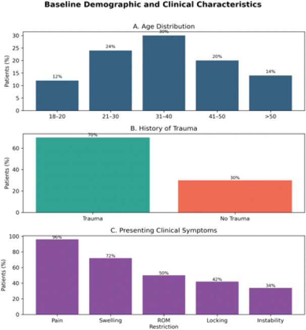
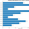
A total of 50 patients with clinically suspected MRTs who underwent pre-operative MRI followed by knee arthroscopy were included in the study. The majority of participants were males (64.0%), with the highest proportion belonging to the 31–40-year age group (30.0%). The right knee was involved in 58.0% of patients, and a history of trauma was present in 70.0%. Knee pain was the most common presenting symptom (96.0%), followed by swelling (72.0%), restricted range of motion (50.0%), locking (42.0%), and instability (34.0%), reflecting the typical clinical presentation of meniscal root injuries (Table 1 and Fig. 1).
Baseline demographic and clinical characteristics of the study population (n=50)
| Variable | Category | Frequency | Percentage |
|---|---|---|---|
| Age group (years) | 18–20 | 6 | 12 |
| 21–30 | 12 | 24 | |
| 31–40 | 15 | 30 | |
| 41–50 | 10 | 20 | |
| >50 | 7 | 14 | |
| Gender | Male | 32 | 64 |
| Female | 18 | 36 | |
| Side involved | Right | 29 | 58 |
| Left | 21 | 42 | |
| History of trauma | Present | 35 | 70 |
| Absent | 15 | 30 | |
| Presenting symptoms | Knee pain | 48 | 96 |
| Swelling | 36 | 72 | |
| Restricted ROM | 25 | 50 | |
| Locking | 21 | 42 | |
| Instability | 17 | 34 |
ROM: Range of motion
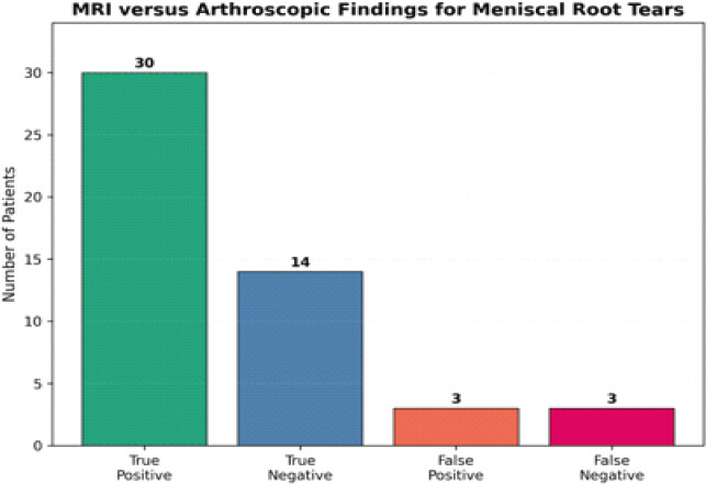
MRI evaluation demonstrated that the ghost sign was the most frequent imaging finding (62.0%), followed by the truncation sign (58.0%), meniscal extrusion (54.0%), radial linear tear (52.0%), and bone marrow edema (36.0%). Arthroscopy, used as the reference standard, confirmed MRTs in 33 (66.0%) patients. MRI correctly identified 30 true-positive, 14 true-negative, 3 false-positive, and 3 false-negative cases, demonstrating excellent agreement with arthroscopic findings (Table 2 and Fig. 2).
MRI findings and arthroscopic correlation in meniscal root tears
| Parameter | Frequency | Percentage |
|---|---|---|
| Ghost sign | 31 | 62 |
| Truncation sign | 29 | 58 |
| Meniscal extrusion | 27 | 54 |
| Radial tear | 26 | 52 |
| Bone marrow edema | 18 | 36 |
| Arthroscopically confirmed root tear | 33 | 66 |
| True positive | 30 | — |
| True negative | 14 | — |
| False positive | 3 | — |
| False negative | 3 | — |
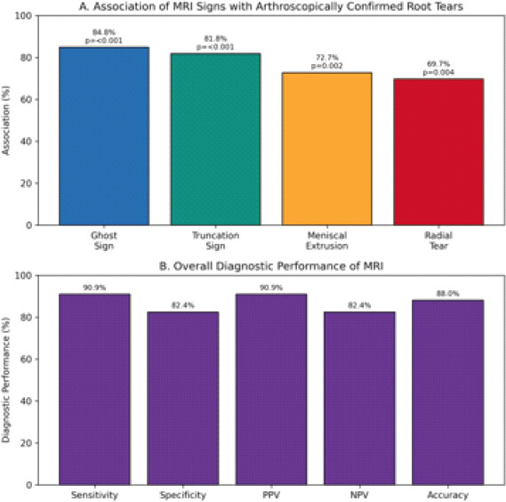
MRI demonstrated a sensitivity of 90.9% (95% CI, 76.4–96.9%), specificity of 82.4% (95% CI, 59.0–93.8%), PPV of 90.9% (95% CI, 76.4–96.9%), NPV of 82.4% (95% CI, 59.0–93.8%), and overall diagnostic accuracy of 88.0% (95% CI, 76.2–94.4%). The positive and negative likelihood ratios were 5.15 and 0.11, respectively. Agreement between MRI and arthroscopy was substantial (Cohen’s κ = 0.73). The association between MRI and arthroscopic findings remained statistically significant (χ2 = 28.64, P < 0.001). Meniscal extrusion (72.7% vs. 17.6%; P = 0.002) and radial tear (69.7% vs. 17.6%; P = 0.004) were also significantly associated with positive arthroscopic findings, confirming their value as reliable MRI indicators of MRTs (Table 3 and Fig. 3).
Diagnostic performance of MRI and association of MRI signs with arthroscopically confirmed meniscal root tears
| Parameter | Value |
|---|---|
| Sensitivity | 90.90% |
| Specificity | 82.40% |
| Positive predictive value | 90.90% |
| Negative predictive value | 82.40% |
| Overall diagnostic accuracy | 88.00% |
| Ghost sign | P<0.001 |
| Truncation sign | P<0.001 |
| Meniscal extrusion | P=0.002 |
| Radial tear | P=0.004 |
MRI: Magnetic resonance imaging
Among the 33 arthroscopically confirmed MRTs, the posterior root of the medial meniscus was the most commonly affected location (72.7%), followed by the posterior root of the lateral meniscus (21.2%) and the anterior root (6.1%). Bone marrow edema was the most frequent associated MRI abnormality (36.0%), followed by anterior cruciate ligament (ACL) injury (24.0%), cartilage defects (20.0%), and medial collateral ligament injury (12.0%), while 30.0% of patients had no associated intra-articular abnormality (Table 4).
Distribution of meniscal root tear location and associated MRI findings
| Parameter | Frequency | Percentage |
|---|---|---|
| Tear location | ||
| Posterior medial root | 24 | 72.7 |
| Posterior lateral root | 7 | 21.2 |
| Anterior root | 2 | 6.1 |
| Associated MRI findings | ||
| Bone marrow edema | 18 | 36 |
| ACL injury | 12 | 24 |
| Cartilage defect | 10 | 20 |
| MCL injury | 6 | 12 |
| No associated abnormality | 15 | 30 |
ACL: Anterior cruciate ligament, MCL: Medial collateral ligament, MRI: Magnetic resonance imaging
Discussion
The present prospective observational diagnostic accuracy study evaluated the ability of MRI to predict MRTs using knee arthroscopy as the gold standard. A total of 50 patients with clinically suspected MRTs who underwent both MRI and arthroscopy were included. MRI demonstrated a sensitivity of 90.9%, specificity of 82.4%, PPV of 90.9%, NPV of 82.4%, and an overall diagnostic accuracy of 88.0%. Furthermore, MRI findings showed a highly significant association with arthroscopic findings (χ2 = 28.64, P < 0.001), indicating that MRI is a reliable non-invasive imaging modality for the pre-operative diagnosis of MRTs.
The demographic profile of the present study showed that the majority of patients belonged to the 31–40-year age group (30.0%), with males constituting 64.0% of the study population. The predominance of young and middle-aged adults observed in our study is consistent with the fact that meniscal injuries commonly occur in physically active individuals and those sustaining sports- or trauma-related knee injuries. Similarly, LaPrade et al. [11] included 287 patients with a mean age of 41.7 years, comprising 156 males and 131 females, thereby demonstrating that MRTs are frequently encountered among middle-aged adults with a slight male predominance, which is comparable to our findings.
Clinically, knee pain was the most common presenting symptom in our study (96.0%), followed by swelling (72.0%), restricted range of motion (50.0%), locking (42.0%), and instability (34.0%). These findings reflect the typical clinical manifestations associated with meniscal root injuries. However, clinical examination alone cannot reliably distinguish MRTs from other intra-articular knee pathologies. Therefore, advanced imaging remains essential for confirming the diagnosis before surgical intervention. This observation is supported by Antinolfi et al., [12], who evaluated 80 patients with suspected meniscal injuries using clinical examination, MRI, and arthroscopy. Although clinical examination performed by an experienced knee surgeon demonstrated excellent diagnostic performance for medial meniscal tears with 91% sensitivity, 87% specificity, 90% accuracy, 94% PPV, and 81% NPV, MRI showed comparatively lower values of 85% sensitivity, 75% specificity, 82% accuracy, 88% PPV, and 71% NPV. Their study concluded that experienced clinical assessment may equal or even surpass MRI for diagnosing meniscal injuries in selected patients. In contrast, the present study specifically evaluated MRTs, where MRI demonstrated considerably higher sensitivity and overall diagnostic accuracy, highlighting its important role in detecting these often subtle lesions.
The present study demonstrated that the ghost sign (62.0%) was the most common MRI finding, followed by the truncation sign (58.0%), meniscal extrusion (54.0%), and radial tear (52.0%). These MRI features are recognized as characteristic indicators of posterior meniscal root injuries. Similar findings were reported by Lee et al., [13] who retrospectively evaluated 39 arthroscopically confirmed medial MRTs. They observed that the ghost sign was present in 100% (36/36) of radial tears, the vertical linear defect on coronal images in 100% (36/36), and the radial linear defect on axial images in 94% (34/36). In addition, they reported a high prevalence of degenerative joint disease (97%), cartilage defects (89%), and meniscal extrusion (67%) associated with medial MRTs. Although the prevalence of individual MRI signs was comparatively lower in our study, the pattern of MRI findings closely resembles those described by Lee et al., [13] confirming that these imaging signs remain highly valuable in identifying MRTs.
Arthroscopy confirmed MRTs in 66.0% of patients in the present study and served as the reference standard for evaluating MRI performance. MRI correctly identified 30 true-positive, 14 true-negative, 3 false-positive, and 3 false-negative cases. Consequently, MRI achieved an overall diagnostic accuracy of 88.0%, supporting its reliability as a pre-operative imaging modality. Comparable findings were reported by Sharifah et al., [14] who prospectively evaluated 65 patients (66 knees) with ACL injuries. Their study reported MRI sensitivity, specificity, and accuracy of 82%, 92%, and 88%, respectively, for medial meniscal tears, while for lateral meniscal tears these values were 83%, 97%, and 92%, respectively. The authors further observed five false-negative medial and four false-negative lateral meniscal tears, with most missed tears involving the peripheral posterior horns. They concluded that MRI possesses high NPV and remains a valuable investigation before arthroscopy, although posterior horn lesions may occasionally be overlooked. These findings closely parallel our results, particularly regarding the high diagnostic accuracy and occasional false-negative MRI examinations.
The diagnostic performance observed in the present study was also comparable with the findings of LaPrade et al., [11] specifically investigated the diagnostic accuracy of 3-Tesla MRI for posterior MRTs using arthroscopy as the gold standard. Their study demonstrated an overall sensitivity of 77.0%, specificity of 72.9%, PPV of 22.0%, and NPV of 97.0%. For medial MRTs, MRI achieved 82.4% sensitivity, 80.0% specificity, and 98.6% NPV, whereas for lateral root tears, sensitivity decreased to 60.0%, although specificity increased to 90.3%. Compared with these findings, the present study demonstrated higher sensitivity (90.9%) and overall accuracy (88.0%). The improved diagnostic performance in our study may be attributed to standardized MRI protocols, careful interpretation of characteristic MRI signs, and direct comparison with arthroscopic findings.
The findings of our study are also supported by the recently published systematic review and meta-analysis by Nguyen et al., [15] which included 75 studies involving 8,507 patients (8,517 knees). Their pooled analysis demonstrated a 91.0% sensitivity for medial meniscal tears and 78.5% sensitivity for lateral tears. In contrast, specificity was higher for lateral tears (94.0%) than medial tears (87.7%). The authors concluded that MRI consistently provides high diagnostic accuracy for detecting meniscal tears irrespective of study design and emphasized that combining meniscal signal intensity with morphologic distortion significantly improves diagnostic performance. The sensitivity of 90.9% observed in our study is remarkably consistent with the pooled sensitivity reported in this meta-analysis, thereby reinforcing the reliability of MRI in diagnosing MRTs.
The statistically significant association between MRI and arthroscopic findings (P < 0.001) observed in our study further supports the clinical usefulness of MRI as an effective screening and pre-operative planning tool. Although arthroscopy remains the definitive diagnostic method, MRI offers the advantages of being non-invasive, widely available, and capable of evaluating associated ligamentous, cartilaginous, and osseous abnormalities simultaneously. The present study therefore supports the routine use of MRI in patients with suspected MRTs while recognizing that equivocal or clinically suspicious cases should ultimately undergo arthroscopic confirmation. The present study demonstrated that MRI possesses excellent sensitivity, good specificity, and high overall diagnostic accuracy for detecting MRTs when compared with arthroscopy. The findings are consistent with previous literature and confirm that recognition of characteristic MRI signs, including the ghost sign, truncation sign, radial tear, and meniscal extrusion, substantially improves diagnostic confidence. Early and accurate MRI diagnosis facilitates appropriate surgical planning, timely meniscal root repair, and may help prevent progressive cartilage degeneration and early osteoarthritis.
This study has several limitations. First, the relatively small sample size of 50 patients may have reduced the precision of diagnostic estimates and limited generalizability. Second, the single-center design may limit applicability to different institutions, MRI protocols, and patient populations. Third, selection bias cannot be excluded because only patients with clinical suspicion of MRTs who subsequently underwent arthroscopy were included, resulting in an enriched disease prevalence of 66%. The exclusion of patients with previous knee surgery, advanced osteoarthritis, fractures, inflammatory or infective arthritis, and poor-quality MRI further limits external validity. Although MRI was interpreted by experienced radiologists blinded to arthroscopic findings, interobserver and intraobserver reliability were not assessed. Subgroup analyses according to medial versus lateral root tears, acute versus chronic presentation, and traumatic versus degenerative etiology were also not performed because of the limited sample size. In addition, the study did not evaluate whether MRI diagnostic findings influenced post-operative functional outcomes, meniscal healing, or osteoarthritis progression. Finally, although the MRI protocol was standardized within the study center, different MRI field strengths, sequences, and technical parameters were not compared. Larger multicenter studies with standardized MRI protocols, multiple blinded observers, appropriate subgroup analyses, and longitudinal clinical follow-up are warranted to validate these findings.
Conclusion
MRI demonstrated high sensitivity, specificity, and overall diagnostic accuracy for detecting MRTs compared with arthroscopy. The ghost sign, truncation sign, meniscal extrusion, and radial tear were significantly associated with arthroscopically confirmed tears. MRI therefore represents a valuable non-invasive pre-operative diagnostic tool, although arthroscopy remains the reference standard in clinically suspicious or inconclusive cases.
Clinical Message
MRI should be considered the primary imaging investigation in patients with suspected MRTs because it provides accurate pre-operative assessment and helps guide surgical planning. However, as some tears may still be missed on MRI, knee arthroscopy remains the gold standard for definitive diagnosis, particularly in patients with persistent clinical suspicion despite inconclusive imaging findings.
Conflict of Interest:
Nil
Source of Support:
Nil
Consent:
The authors confirm that informed consent was obtained from the patient for publication of this article
References
- Aagaard H, Verdonk R. Function of the normal meniscus and consequences of meniscal resection Scand J Med Sci Sports 1999;9:134-40. [Google Scholar] | [PubMed]
- Fox AJ, Wanivenhaus F, Burge AJ, Warren RF, Rodeo SA. The human meniscus: A review of anatomy, function, injury, and advances in treatment Clin Anat 2015;28:269-87. [Google Scholar] | [PubMed]
- Terry GC, Tagert BE, Young MJ. Reliability of the clinical assessment in predicting the cause of internal derangements of the knee Arthroscopy 1995;11:568-76. [Google Scholar] | [PubMed]
- Mohan BR, Gosal HS. Reliability of clinical diagnosis in meniscal tears Int Orthop 2007;31:57-60. [Google Scholar] | [PubMed]
- Nickinson R, Darrah C, Donell S. Accuracy of clinical diagnosis in patients undergoing knee arthroscopy Int Orthop 2010;34:39-44. [Google Scholar] | [PubMed]
- Crawford R, Walley G, Bridgman S, Maffulli N. Magnetic resonance imaging versus arthroscopy in the diagnosis of knee pathology, concentrating on meniscal lesions and ACL tears: A systematic review Br Med Bull 2007;84:5-23. [Google Scholar] | [PubMed]
- Rubin DA, Paletta GA Jr. Current concepts and controversies in meniscal imaging Magn Reson Imaging Clin N Am 2000;8:243-70. [Google Scholar] | [PubMed]
- Esmaili Jah AA, Keyhani S, Zarei R, Moghaddam AK. Accuracy of MRI in comparison with clinical and arthroscopic findings in ligamentous and meniscal injuries of the knee Acta Orthop Belg 2005;71:189-96. [Google Scholar] | [PubMed]
- Scholten RJ, Devillé WL, Opstelten W, Bijl D, Van Der Plas CG, Bouter LM. The accuracy of physical diagnostic tests for assessing meniscal lesions of the knee: A meta-analysis J Fam Pract 2001;50:938-44. [Google Scholar] | [PubMed]
- Hing W, White S, Reid D, Marshall R. Validity of the mcmurray's test and modified versions of the test: A systematic literature review J Manual Manip Ther 2009;17:22-35. [Google Scholar] | [PubMed]
- LaPrade RF, Ho CP, James E, Crespo B, LaPrade CM, Matheny LM. Diagnostic accuracy of 3.0 T magnetic resonance imaging for the detection of meniscus posterior root pathology Knee Surg Sports Traumatol Arthrosc 2015;23:152-7. [Google Scholar] | [PubMed]
- Antinolfi P, Cristiani R, Manfreda F, Brue S, Sarakatsianos V, Placella G, et al. Relationship between clinical, MRI, and arthroscopic findings: A guide to correct diagnosis of meniscal tears Joints 2017;5:164-7. [Google Scholar] | [PubMed]
- Lee YG, Shim JC, Choi YS, Kim JG, Lee GJ, Kim HK. Magnetic resonance imaging findings of surgically proven medial meniscus root tear: Tear configuration and associated knee abnormalities J Comput Assist Tomogr 2008;32:452-7. [Google Scholar] | [PubMed]
- Sharifah MI, Lee CL, Suraya A, Johan A, Syed AF, Tan SP. Accuracy of MRI in the diagnosis of meniscal tears in patients with chronic ACL tears Knee Surg Sports Traumatol Arthrosc 2015;23:826-30. [Google Scholar] | [PubMed]
- Nguyen JC, Yaya-Quezada C, Lerebo WT, Patel VS, Chalian M, Flores DV, et al. MRI diagnosis of meniscus tears in the knee: An updated systematic review and meta-analysis Radiology 2026;319:e252288. [Google Scholar] | [PubMed]
© 2026 Journal of Orthopaedic Case Reports - Published by Indian Orthopaedic Research Group
About the Authors
-

Dr. Aluka Sundeep Kund Reddy
-

Dr. Anmol Kshirsagar
-

Dr. Suhail Ahmad Khanda
-

Dr. Paarth Gautam
-

Dr. Deepankar Satapathy
-

Dr. D M Nikhil Dev
Related Articles in Journal of Orthopaedic Case Reports
 October 1, 2026 Magnetic Resonance Imaging-Arthroscopy Correlation in Traumatic Knee Injuries: A Retrospective Diagnostic Accuracy Study
 October 1, 2026 Incidence, Arthroscopic Detection, and Clinical Outcomes of Ramp Lesions in Patients Undergoing Anterior Cruciate Ligament Reconstruction: A Prospective Observational Study
 October 1, 2026 Acute Cauda Equina Syndrome in Rapid Succession: A Case Series of Eight Consecutive Patients Undergoing Emergency Surgical Decompression Over One Week
 October 1, 2026 Digital Transformation in Orthopedics: Can India Leapfrog with Artificial Intelligence, Augmented Reality, and Robotics?