


Introduction
Total knee arthroplasty (TKA) has been one of the most important orthopedic advances. However, vascular lesions and complications in TKA are well documented with sufficient data. The popliteal artery is more commonly involved [1], but some cases involving an isolated injury to the superficial femoral artery have been reported in a few isolated case reports. Hence, the incidence is unknown.
Calligaro et al. [2] studied an overall incidence of 0.03–0.17% arterial complications among a sample of 13618 (including 1665 revisions) during TKA. There were 24 cases of arterial complications, of which 5 were popliteal artery pseudoaneurysms.
Butt et al. in 2010 explained the mechanisms of arterial complications following TKA [1].
According to He and Yang [3] and Zhao et al. [4], vascular lesions can occur up to 5 months after surgery, ranging from an active hemorrhage to the development of pseudoaneurysms or false popliteal aneurysms. Early diagnosis of such lesions can be life and limb-saving and prove to be the most important factor in prognosis. The mechanism of vascular injury in this case, however, still seems to be unclear.
Case Report
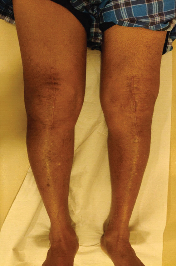
A 72-year-old male with a past medical history of diabetes mellitus was operated on for bilateral total knee replacement. He was not on any anticoagulants preoperatively and he did not have any known preexisting vascular disease. Pre-operative arterial ultrasound Doppler was not done, as it is not routinely done in our institute, and ankle-brachial pressure index was recorded to be 1.0. The pneumatic tourniquet pressure was set at 300 mmHg for a duration of 35 min for each side. In the immediate post-operative period, he was administered with adductor canal blocks for both the limbs on the operating table after completion of the surgery. He was started on 0.6 mL of Clexane (60 mg Enoxaparin) postoperatively, and bilateral deep vein thrombosis pumps were used during the hospital stay of 5 days. He was discharged after an uneventful stay in the hospital. Four weeks later, he presented to the emergency department with right thigh and knee pain and swelling, which, according to him, was increasing in size (Fig. 1). There was no history of fever. After thorough clinical examination, there was a compressible swelling extending into the mid-thigh. There was no redness or local rise in temperature, but there was mild tenderness. Distal neurovascular status was unaffected. On investigation, hemoglobin was found to be 5.7 g/dL with total leukocyte count within normal limits. Radiographic evaluation comprising X-rays, a venous and arterial Doppler, and a computed tomography (CT) angiogram was advised. Doppler showed a hematoma and bleeding from a principal vessel (Fig. 2). CT angiogram revealed a narrow-necked small pseudoaneurysm of size 5 mm × 4 mm arising from a muscular branch of the superficial femoral artery, described as the third branch arising distal to the common femoral artery bifurcation, with opacification of a small pseudoaneurysmal sac. The pseudoaneurysm corresponded to the focus of active extravasation identified on CT angiography and was associated with a large intramuscular hematoma within the vastus intermedius. This hematoma in turn caused a compression of the right superficial femoral vein (Fig. 3).
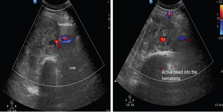
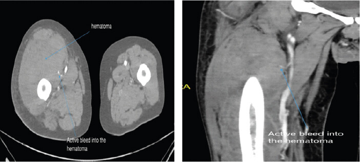
The patient was referred to the vascular intervention team and underwent diagnostic peripheral angiography through a right common femoral artery approach. Angiography confirmed active extravasation from a muscular branch of the superficial femoral artery, corresponding to the third branch distal to the common femoral artery bifurcation and the pseudoaneurysm identified on CT angiography. The lesion was a small, narrow-neck pseudoaneurysm measuring 5 × 4 mm and was associated with a large intramuscular hematoma within the vastus intermedius.
Considering the small size and muscular branch origin of the pseudoaneurysm, selective endovascular embolization was chosen to achieve hemostasis while avoiding intervention on the main superficial femoral artery. Two 3-mm coils were deployed to occlude the culprit branch. Completion angiography demonstrated successful cessation of bleeding from the affected branch. Coil embolization was preferred over open surgical repair because it provided a minimally invasive method of achieving definitive hemostasis in a deep proximal thigh lesion. Ultrasound-guided compression or thrombin injection, although established options for selected femoral pseudoaneurysms, were considered less suitable given the small muscular branch origin and associated deep intramuscular hematoma. Covered stent placement was also not considered appropriate for a small muscular branch because of the need to preserve the parent superficial femoral artery and avoid unnecessary implantation in a branch vessel. 3 units of low-density packed red blood cell components were transfused, and the patient was discharged after a successful recovery.
Discussion
Post-operative pseudoaneurysm in the superficial femoral artery after knee arthroplasty is a very rare occurrence and is an anomaly in the rehabilitative phase of knee arthroplasty.
The SFA is unique in nature, as it runs along the length of the thigh without any important branches and collaterals. Hence, a range of forces is exerted on it that alters the flow in the vessel during flexion, extension, contraction, torsion, and compression [5]. This makes it more susceptible to tethering during manipulation under a tourniquet.
Butt et al. described four mechanisms of arterial complications following TKA [1]. Embolization of an atheromatous plaque, especially with the use of a pneumatic tourniquet, commonly affected the superficial femoral artery [6,7,8]. However, large-scale studies are yet to be published on its incidence.
Excessive manipulation can cause intimal tears of major vessels. When combined with the low-flow state that results from applying a tourniquet, these favor thrombus development and consequent ischemia [6,7,8,9]. Release of severe flexion contractures can cause traction injury and intimal tears of the popliteal artery [6,7,8,9,10]. Extensive releases should be done cautiously. Rubash et al. described, in cadaveric specimens, the vessels at risk during excessive retraction and releases performed during TKA [11].
The last mechanism involves direct injury to major vessels by various instruments used during TKA [8].
Sanchez et al. [12] and Vorobeichik and Abdallah [13] described that not only surgical procedures but also adductor canal blocks given as a method of post-surgical pain management can be linked to inadvertent puncture of the superficial femoral artery during application. Post-operative adductor canal block is routinely practiced in our institute following TKA which can be kept in mind as a cause of the complication. Bomberg et al. [14] demonstrated a 1.1% risk of vascular puncture while performing a femoral block. Cappelleri et al. [15] suggested the possibility of vascular injury even if the adductor canal block was done under ultrasonic guidance.
A pre-operative assessment to identify the risk of arterial injury in patients undergoing TKA is essential to avoid such complications. History of tobacco intake, hypertension, diabetes mellitus, claudication symptoms, and previous history of stroke or cardiovascular surgery are important considerations in history taking before surgery [1,2,16,17]. Skin condition and pulses in comparison to the contralateral limb should be examined by the operating surgeon.
The routine use of ankle-brachial pressure index is an important tool in developing suspicion of an existing arterial disease, but values of >1.3 in cases of diabetes mellitus can be highly misleading. Values <0.5 are suggestive of severe ischemia [16,17]. The presence of vascular calcification on plain radiographs should raise concern for an existing arterial disease [18].
Pre-operative Doppler of arterial and venous systems is recommended in such cases. When intervention is needed or there is a high level of suspicion, CT angiography may be necessary [2,17].
Most complications are diagnosed within 48 h of surgery. Post-operative excessive pain, swelling in the knee, high drain output with fresh blood, and other signs of ischemia are suggestive of intimal tear or laceration of an artery. It is mandatory that there are routine checks in the immediate and early post-operative period to recognize any sign of arterial injury following TKA. A delay in diagnosis can have devastating complications such as irreversible ischemia, compartment syndrome, and surgical amputation [2].
Once clinical suspicion is raised, the opinion of a vascular surgeon is mandatory. Management options include arterial embolization using coils or intraluminal thrombin, endovascular stenting, oversewing of the aneurysm neck, or excision and repair or bypass [19,20,21,22]. Ultrasound-guided compression repair is the gold standard treatment for a proximal femoral artery pseudoaneurysm [23]. Our case was treated with embolization of the affected vessel with 3 mm coils and post-operative blood transfusion.
Mortality and amputations are quite rare. Calligaro et al. [2] reported no deaths, and limbs could be salvaged in all 24 cases with arterial complications. However, other studies reported the need for amputation of the limb after events of an arterial injury, the occurrence being very few. He also reported that a delay in diagnosis and cases where surgery was delayed for more than 24 h from the time of occurrence needed surgical procedures like fasciotomies along with the principal surgery performed by the vascular surgeon.
Only case reports with generally positive results are available for studies on pseudoaneurysms following TKA. Even fewer case reports have mentioned an SFA aneurysm. In most of the case reports, no definite etiology has been mentioned as a reason for the vascular injury. While most cases talk about injury to the popliteal artery or anterior tibial artery, which is in the vicinity of the surgical field, in our case, the SFA pathology is much more proximal to the surgical field. Therefore, the author feels the likely etiology could be due to the application of the tourniquet or due to the adductor canal block in the immediate post-operative period.
Conclusion
Pseudoaneurysm of the superficial femoral artery following TKA is an extremely rare but serious complication that may present in a delayed manner. Although vascular injuries after TKA typically involve the popliteal artery, surgeons should remain vigilant for proximal arterial lesions when patients present with unexplained thigh swelling and pain in the post-operative period.
Prompt recognition with appropriate imaging modalities and early endovascular intervention can lead to favorable outcomes and prevent limb-threatening sequelae. Awareness of various etiologies, including tourniquet-related injury in cases with the aforementioned risk factors and regional anesthesia techniques such as adductor canal block, is essential to ensure a timely diagnosis and management.
Clinical Message
This case emphasizes that superficial femoral artery pseudoaneurysm, although rare, is a potentially limb-threatening delayed complication after TKA that may present with vague symptoms such as thigh swelling and pain. Awareness of these risk factors encourages surgeons and anesthetists to maintain vigilant post-operative vascular monitoring and use early imaging in suspicious cases, especially when tourniquets or adductor canal blocks are employed.
Conflict of Interest:
Nil
Source of Support:
Nil
Consent:
The authors confirm that informed consent was obtained from the patient for publication of this article
How to Cite this Article
Singh C, Ghosh S, Kacker S, Taneja S, Marya SKS, Sahu AK. Superficial Femoral Artery Pseudoaneurysm after Total Knee Arthroplasty: An Uncommon Vascular Complication Journal of Orthopaedic Case Reports 2026 October;16(10): 198-202.
References
- Butt U, Samuel R, Sahu A, Butt IS, Johnson DS, Turner PG. Arterial injury in total knee arthroplasty. J Arthroplasty 2010;25:1311-8. [Google Scholar] | [PubMed]
- Calligaro KD, Dougherty MJ, Ryan S, Booth RE. Acute arterial complications associated with total hip and knee arthroplasty. J Vasc Surg 2003;38:1170-7. Erratum in: J Vasc Surg 2004;39:628. [Google Scholar] | [PubMed]
- He R, Yang L. Acute arterial occlusion in the midpiece of femoral artery following total knee arthroplasty: Report of one case. Chin J Traumatol 2016;19:116-8. [Google Scholar] | [PubMed]
- Zhao R, Li Y, Liu Y, Zhang K, Liu Z. Pseudoaneurysm of a high-division anterior tibial artery following primary TKA. Orthopade 2017;46:275-9. [Google Scholar] | [PubMed]
- Perlstein TS, Krichavsky MZ. Peripheral Artery Disease, Cardiovascular Therapeutics: A Companion to Braunwald's Heart Disease. 4th ed. Ch. 35. Philadelphia, PA: W. B. Saunders; 2013. 539-52. [Google Scholar] | [PubMed]
- Kumar SN, Chapman JA, Rawlins I. Vascular injuries in total knee arthroplasty. A review of the problem with special reference to the possible effects of the tourniquet. J Arthroplasty 1998;13:211. [Google Scholar] | [PubMed]
- McCauley CE, Steed DL, Webster MW. Arterial complications of total knee replacement. Arch Surg 1989;119:260. [Google Scholar] | [PubMed]
- Saleh KJ, Hoeffel DP, Kassim RA, Burstein G. Complications after revision total knee arthroplasty. J Bone Joint Surg 2003;85A:S71-4. [Google Scholar] | [PubMed]
- Ohira T, Fujimoto T, Taniwaki K. Acute artery occlusion after total knee arthroplasty. Arch Orthop Trauma Surg 1997;116:429. [Google Scholar] | [PubMed]
- Robson LJ, Walls CE, Swanson AB. Popliteal artery obstruction following Shiers total knee replacement. A case report. Clin Orthop Relat Res 1975;109:130. [Google Scholar] | [PubMed]
- Rubash HE, Beregr RA, Britton CA, Nettrour WA, Seel MJ. Avoiding neurologic and vascular injuries with screw fixation of the tibial component in total knee arthroplasty. Clin Orthop Rel Res 1993;286:56-63. [Google Scholar] | [PubMed]
- Sanchez CA, Jara AB, Mariño J. Superficial femoral artery pseudoaneurysm, compartment syndrome, and deep vein thrombosis after total knee arthroplasty. Arthroplast Today 2020;6:227-30. Erratum in: Arthroplast Today 2022;15:234. [Google Scholar] | [PubMed]
- Vorobeichik L, Abdallah FW. Femoral artery dissection after adductor canal block. Anesthesiology 2019;130:1037-8. [Google Scholar] | [PubMed]
- Bomberg H, Huth A, Wagenpfeil S, Kessler P, Wulf H, Standl T. Psoas versus femoral blocks: A registry analysis of risks and benefits. Reg Anesth Pain Med 2017;42:719-24. [Google Scholar] | [PubMed]
- Cappelleri G, Molinari P, Stanco A. Iatrogenic pseudoaneurysm after continuous adductor canal block. A A Case Rep 2016;7:200-2. [Google Scholar] | [PubMed]
- Abularrage CJ, Weiswasser JM, Dezee KJ, Slidell MB, Henderson WG, Sidawy AN. Predictors of lower extremity arterial injury after total knee or hip arthroplasty. J Vasc Surg 2008;47:803. [Google Scholar] | [PubMed]
- Smith DE, McGraw RW, Taylor DC, Masri BA. Arterial complications and total knee arthroplasty. J Am Acad Orthop Surg 2001;9:253-7. [Google Scholar] | [PubMed]
- DeLaurentis DA, Levitsky KA, Booth RE, Rothman RH, Calligaro KD, Raviola CA. Arterial and ischemic aspects of total knee arthroplasty. Am J Surg 1992;164:237-40. [Google Scholar] | [PubMed]
- O'Connor JV, Stocks G, Crabtree JD Jr, Galasso P, Wallsh E. Popliteal pseudoaneurysm following total knee arthroplasty. J Arthroplasty 1998;13:830. [Google Scholar] | [PubMed]
- Ibrahim M, Booth RE, Clark TW. Embolization of traumatic pseudoaneurysms after total knee arthroplasty. J Arthroplasty 2004;19:123. [Google Scholar] | [PubMed]
- Plagnol PH, Diard N, Bruneteau P, Roncheau V. Pseudoaneurysm of popliteal artery complicating a total knee replacement: A successful percutaneous endovascular treatment. Eur J Vasc Endovasc Surg 2001;21:81. [Google Scholar] | [PubMed]
- Holmberg A, Milbrink J, Bergqvist D. Arterial complications after knee arthroplasty: 4 cases and review of the literature. Acta Orthop scand 1996;67:75-8. [Google Scholar] | [PubMed]
- Lenartova M, Tak T. Iatrogenic pseudoaneurysm of femoral artery: Case report and literature review. Clin Med Res 2003;1:243-7. [Google Scholar] | [PubMed]
© 2026 Journal of Orthopaedic Case Reports - Published by Indian Orthopaedic Research Group
About the Authors
-

Chandeep Singh
-

Sayam Ghosh
-

Shitij Kacker
-

Shobhit Taneja
-

Sanjiv Kumar Singh Marya
-

Amit Kumar Sahu
Related Articles in Journal of Orthopaedic Case Reports
 August 1, 2025 An Unusual Presentation of Delayed Hemarthrosis Following Total Knee Arthroplasty: A Case Report
 October 1, 2026 Intraoperative Discovery of Incorrect Femoral Component Packaging during Bilateral Total Knee Arthroplasty: A Near-Miss Case Report
 October 1, 2026 Occult Femoral Arteriovenous Fistula and Pseudoaneurysm after a Gunshot-Associated Subtrochanteric Fracture: Transfusion-Refractory Anemia as the Sentinel Sign – A Case Report
 October 1, 2026 Comparative Study of Effect of Periarticular Fentanyl and Dexmedetomidine Injection for Pain Management in Total Knee Arthroplasty a Randomized Control Trial