


Introduction
Acromioclavicular (AC) joint ligament injuries account for a considerable proportion of traumatic shoulder conditions, representing approximately 9% of all shoulder injuries [1]. High-grade acute AC dislocations are characterized by complete disruption of both the AC and coracoclavicular ligament complexes, resulting in significant joint instability and therefore commonly necessitate operative stabilization [2]. To date, no consensus on an optimal surgical technique has been reached, and procedure-related complications have been reported [3].
Clavicular hook plate fixation has demonstrated reliable clinical outcomes in unstable AC joint injuries [4,5]. The overall complication rate associated with hook plate fixation was 15.8%, including subacromial impingement, acromial osteolysis, loss of reduction, AC joint osteoarthritis, and periarticular calcification [6]. Transacromial erosion and associated fractures have been reported as complications of hook plate fixation as well [7,8,9]. In this article, we report a rare stress-related fracture at the base of the acromion following hook plate fixation.
Case Report
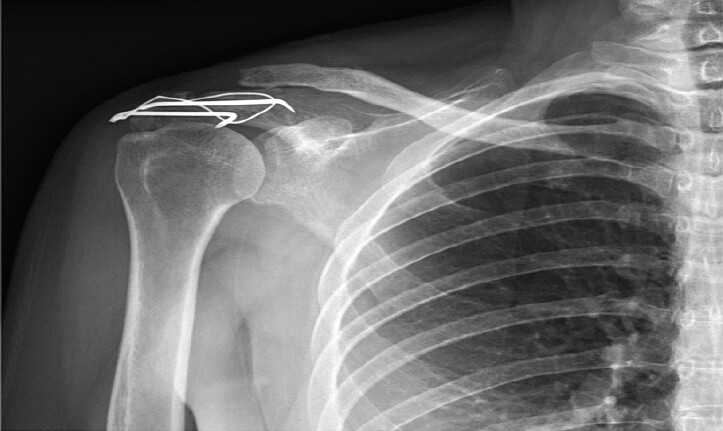
A 48-year-old woman sustained a right shoulder contusion in a traffic accident, resulting in a Rockwood type V AC joint injury (Fig. 1a). Open reduction and internal fixation of the AC joint was performed using a clavicular hook plate, followed by regular outpatient follow-up and a progressive rehabilitation program (Fig. 1b).
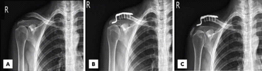
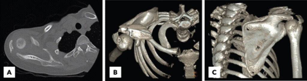
2 months after the index surgery, she presented with painful dysfunction of the right shoulder after a fall onto an outstretched hand. Physical examination revealed localized tenderness over the acromion without evidence of vascular compromise or brachial plexus injury. Plain radiographs demonstrated failure of the hook plate construct associated with a displaced acromial fracture (Fig. 1c). Computed tomography with three-dimensional reconstruction revealed a stress-related fracture originating from the base of the acromion, with a fragment diastasis of 15 mm and narrowing of the subacromial space, consistent with a Kuhn type III acromion fracture (Fig. 2).
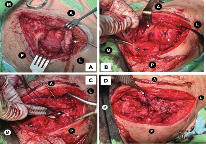
Through a superolateral approach, the hook plate was removed, and osteosynthesis of the acromial fracture was performed using a tension-band wiring technique. The AC joint was stabilized with percutaneous pin fixation, and the coracoclavicular ligament was reconstructed using the coracoacromial ligament augmented with Mersilene tape (Fig. 3). Post-operative radiographs confirmed satisfactory reduction and fixation (Fig. 4).
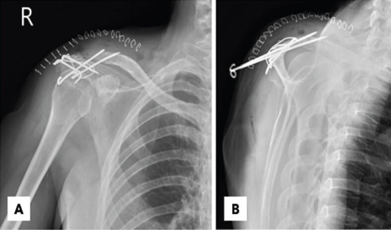
Postoperatively, the shoulder was immobilized in a sling for 2 weeks, followed by passive range-of-motion exercises for 4 weeks. After removal of the percutaneous AC pins at 6 weeks, active range-of-motion exercises were initiated. At 3 months postoperatively, the patient had regained functional shoulder motion for daily activities, and radiographs demonstrated fracture consolidation (Fig. 5).
Discussion
Acromial erosion with subsequent hook plate cut-out is an uncommon but recognized complication following clavicular hook plate fixation [7,8,9]. From a surgical perspective, increased hook depth reduces stress on the clavicle and the force transmitted to the acromion, which may help reduce the risk of acromial osteolysis or peri-implant fracture [10]. In addition, clavicular hook plates with different hook angles may induce varying biomechanical effects on the clavicle and acromion. Therefore, careful selection of an appropriate hook plate based on individual patient anatomy is essential [11].
Beyond these previously described complications, even more rarely, we report a stress-related fracture at the base of the acromion with a fracture pattern distinct from the typical hook plate cut-out mechanism, resulting in disruption of multiple components of the superior shoulder suspensory complex (SSSC). The SSSC, first described by Goss in 1993, comprises a ring of osseous and ligamentous structures essential for maintaining shoulder biomechanics, including the glenoid fossa, coracoid process, coracoclavicular ligaments, distal clavicle, AC joint, and acromion. Goss conceptualized injuries to the SSSC as a ring disruption, proposing that involvement of two or more components leads to shoulder girdle instability and generally warrants surgical stabilization [12]. However, no definitive treatment guidelines currently exist for injuries of the SSSC, and the optimal management of multiple SSSC disruptions remains controversial [13]. Consequently, treatment should be individualized according to patient age, associated injuries, and biomechanical considerations [14].
With respect to acromial fractures, Kuhn et al. proposed a three-stage classification system based on the degree of displacement: Type I, minimal displacement; Type II, displacement without compromise of the subacromial space; and Type III, displacement associated with narrowing of the subacromial space. Type III acromial fractures managed non-operatively have been associated with painful restriction of shoulder motion, indicating that early surgical intervention may be warranted [15]. Recent studies have suggested that surgical treatment is indicated in cases of symptomatic non-union, subacromial impingement, displacement >10 mm, open fractures, or disruption of the SSSC [16,17]. Nevertheless, no widely accepted treatment algorithm or fixation strategy for acromial fractures currently exists. Osteosynthesis may be achieved using tension-band wiring, cannulated screws, or plate fixation, largely based on surgeon preference [16,18]. Each fixation method has distinct advantages and limitations. Cannulated screw fixation is minimally invasive and usually does not require implant removal; however, screw placement can be technically challenging due to the thin cortical bone of the acromion and is less suitable for comminuted lateral fractures. Plate fixation, while more invasive, allows accurate fracture reduction and rigid stabilization but has been associated with a higher risk of infection. The tension-band technique provides both static and dynamic compression of fracture fragments and is less invasive than plate fixation; however, it does not offer the most rigid construct and carries a risk of implant irritation or migration [16,19,20,21]. In the present case, the tension-band technique was selected because the acromial fragment was intact and the fracture pattern was simple.
In conclusion, stress-related fractures of the acromion base represent a rare but important complication following clavicular hook plate fixation and may differ from the typical cut-out–related erosion pattern. Awareness of this injury mechanism is essential for timely diagnosis and appropriate treatment. Surgical management should be individualized based on fracture morphology and stability, and tension-band fixation may be a suitable option in cases with an intact fragment and simple fracture pattern.
Conclusion
Stress-related fractures of the acromion base represent a rare but clinically significant complication following clavicular hook plate fixation for AC joint injuries. This fracture pattern differs from the typical cut-out mechanism and may be associated with disruption of the SSSC, resulting in shoulder girdle instability. Early recognition through appropriate imaging and individualized surgical management based on fracture morphology and stability are essential to achieve favorable clinical and radiographic outcomes.
Clinical Message
Although clavicular hook plate fixation is an effective treatment for high-grade acromioclavicular joint injuries, surgeons should remain vigilant for rare stress-related fractures at the base of the acromion. New-onset or persistent shoulder pain after surgery, even following minor trauma, should prompt thorough imaging evaluation. Treatment should be tailored according to fracture pattern and overall shoulder girdle stability.
Conflict of Interest:
Nil
Source of Support:
Nil
Consent:
The authors confirm that informed consent was obtained from the patient for publication of this article
How to Cite this Article
Chen Y, Chang S, Chiu C, Lu C. Stress-Related Acromion Base Fracture after Clavicular Hook Plate Fixation: A Case Report. Journal of Orthopaedic Case Reports 2026 October;16(10): 292-296.
References
- Mazzocca AD, Arciero RA, Bicos J. Evaluation and treatment of acromioclavicular joint injuries. Am J Sports Med 2007;35:316-29. [Google Scholar] | [PubMed]
- Martetschlager F, Kraus N, Scheibel M, Streich J, Venjakob A, Maier D. The diagnosis and treatment of acute dislocation of the acromioclavicular joint. Dtsch Arztebl Int 2019;116:89-95. [Google Scholar] | [PubMed]
- Takase K, Hata Y, Morisawa Y, Goto M, Tanaka S, Hamada J. Treatment of acromioclavicular joint separations in Japan: A survey. JSES Int 2021;5:51-5. [Google Scholar] | [PubMed]
- Ejam S, Lind T, Falkenberg B. Surgical treatment of acute and chronic acromioclavicular dislocation Tossy type III and V using the Hook plate. Acta Orthop Belg 2008;74:441-5. [Google Scholar] | [PubMed]
- Li G, Liu T, Shao X, Liu Z, Duan J, Akileh R. Fifteen-degree clavicular hook plate achieves better clinical outcomes in the treatment of acromioclavicular joint dislocation. J Int Med Res 2018;46:4547-59. [Google Scholar] | [PubMed]
- Liu G, Hu Y, Ye F, Huang F, Yu T. Clavicular hook plate for acute high-grade acromioclavicular dislocation involving rockwood type V: Clinical and radiological outcomes and complications evaluation. Int Orthop 2022;46:2405-11. [Google Scholar] | [PubMed]
- Koh KH, Shin DJ, Hwang SM. Crossbar technique for the failed clavicular hook plate fixation in an acute acromioclavicular joint dislocation: Salvage for acromial fracture after clavicular hook plate. Clin Shoulder Elb 2019;22:149-53. [Google Scholar] | [PubMed]
- Chiang CL, Yang SW, Tsai MY, Kuen-Huang Chen C. Acromion osteolysis and fracture after hook plate fixation for acromioclavicular joint dislocation: A case report. J Shoulder Elbow Surg 2010;19:e13-5. [Google Scholar] | [PubMed]
- Hoffler CE, Karas SG. Transacromial erosion of a locked subacromial hook plate: Case report and review of literature. J Shoulder Elbow Surg 2010;19:e12-5. [Google Scholar] | [PubMed]
- Lee CH, Shih CM, Huang KC, Chen KH, Hung LK, Su KC. Biomechanical analysis of implanted clavicle hook plates with different implant depths and materials in the acromioclavicular joint: A finite element analysis study. Artif Organs 2016;40:1062-70. [Google Scholar] | [PubMed]
- Hung LK, Su KC, Lu WH, Lee CH. Biomechanical analysis of clavicle hook plate implantation with different hook angles in the acromioclavicular joint. Int Orthop 2017;41:1663-9. [Google Scholar] | [PubMed]
- Goss TP. Double disruptions of the superior shoulder suspensory complex. J Orthop Trauma 1993;7:99-106. [Google Scholar] | [PubMed]
- Nguyen MK, Nguyen VH, Le HG, Cao DB. Multiple disruptions of superior suspensory shoulder complex: A case report. Int J Surg Case Rep 2024;124:110385. [Google Scholar] | [PubMed]
- Wu K, Wu XM, Zha XL, Wang QG. Anatomic restoration of triple disruption of the superior shoulder suspensory complex: A case report and review of the literature. Orthop Surg 2020;12:1526-30. [Google Scholar] | [PubMed]
- Kuhn JE, Blasier RB, Carpenter JE. Fractures of the acromion process: A proposed classification system. J Orthop Trauma 1994;8:6-13. [Google Scholar] | [PubMed]
- Cicekli O, Akar A, Topcu HN. Displaced acromion fracture: A rare injury, case report. Int J Surg Case Rep 2017;39:313-6. [Google Scholar] | [PubMed]
- Hill BW, Anavian J, Jacobson AR, Cole PA. Surgical management of isolated acromion fractures: Technical tricks and clinical experience. J Orthop Trauma 2014;28:e107-13. [Google Scholar] | [PubMed]
- Alawad MO, Alharthi S, Mahmoud J, Alanazi B, Surur S. Open fracture of the acromion: An isolated injury with oblique-type fracture. Case Rep Orthop 2018;2018:2107059. [Google Scholar] | [PubMed]
- Dukan R, Abboud J, Ouchrif Y. Isolated acromial fracture in a cyclist: A case report and review of the literature. JBJS Case Connect 2020;10:e2000079. [Google Scholar] | [PubMed]
- Goss TP. The scapula: Coracoid, acromial, and avulsion fractures. Am J Orthop (Belle Mead NJ) 1996;25:106-15. [Google Scholar] | [PubMed]
- Kurahashi S, Takeda S, Mitsuya S, Makihara K, Yamauchi KI. Plate fixation of acromion fracture using a mesh plate. Trauma Case Rep 2021;33:100470. [Google Scholar] | [PubMed]
© 2026 Journal of Orthopaedic Case Reports - Published by Indian Orthopaedic Research Group
About the Authors
-

Yi-Hsuan Chen
-

Shun-Min Chang
-

Chao-Tse Chiu
-

Cheng-Chang Lu
Related Articles in Journal of Orthopaedic Case Reports
 July 1, 2026 Chronic Acromioclavicular Joint Instability: Outcomes after Stabilization without Tendon Graft Augmentation
 June 1, 2026 Malleolar Stress Fracture as the First Manifestation of Undiagnosed Crohn’s Disease: A Case Report
 April 1, 2026 Migration of Two Broken K-wires into the Cervical Region following Lateral End Clavicle Fixation: A Rare Case Report and Literature Review
 December 1, 2025 Adult-Onset Fanconi Syndrome Presenting as Stress Fractures: A Case Report