

Persistent anterior chest pain and weakness following traumatic anterior shoulder dislocation should prompt evaluation for concomitant pectoralis major rupture, as early recognition and comprehensive surgical management can restore excellent shoulder function.
Dr. Vasileios Kosmas, Orthopedic Clinic OSTEON, Athens Medical Center, Athens, Greece. E-mail: v.d.kosmas@hotmail.com
Abstract
Introduction: Concomitant rupture of the pectoralis major tendon and traumatic anterior shoulder dislocation is an exceptionally rare injury pattern. Although anterior shoulder dislocation is a common orthopaedic injury and pectoralis major rupture is increasingly recognized in athletic populations, the simultaneous occurrence of these injuries has been reported only rarely. This case is important because associated soft tissue injuries may be overlooked during the evaluation of shoulder instability, potentially delaying diagnosis and treatment. The present report contributes to the limited literature describing this combined injury pattern and its successful surgical management.
Case Report: A 47-year-old male recreational water polo athlete sustained a traumatic anterior shoulder dislocation during a water polo match following a forceful abduction and external rotation mechanism while contesting the ball overhead. Clinical examination demonstrated pain, anterior shoulder instability, weakness of adduction and internal rotation, and deformity of the anterior axillary fold. Magnetic resonance imaging revealed an anterior-inferior labral tear, a Hill-Sachs lesion, and complete rupture of the pectoralis major tendon. Surgical treatment consisted of arthroscopic repair of the anterior-inferior labrum, arthroscopic remplissage of the humeral head defect, and open repair of the pectoralis major tendon using suture anchors and non-absorbable sutures. A structured rehabilitation program was followed postoperatively. At final follow-up, the patient demonstrated restoration of shoulder stability, recovery of pectoralis major contour and strength, and return to recreational sports without recurrent instability.
Conclusion: This case highlights the importance of careful assessment for associated tendon injuries in patients presenting with traumatic anterior shoulder dislocation, particularly following high-energy sporting mechanisms involving abduction and external rotation. Recognition of this uncommon injury combination allows timely treatment and may improve functional outcomes. This report adds to the orthopaedic literature by documenting a rare concomitant injury pattern and demonstrating that combined arthroscopic and open surgical management can achieve successful restoration of shoulder stability and function.
Keywords: Shoulder instability, anterior shoulder dislocation, pectoralis major rupture, Bankart lesion, Hill-Sachs lesion.

Anterior shoulder dislocation is the most common major joint dislocation encountered in orthopedic practice and is frequently associated with soft tissue and osseous injuries including Bankart lesions and Hill-Sachs defects. However, concomitant rupture of the pectoralis major tendon in association with traumatic anterior shoulder instability is exceedingly rare. Most pectoralis major ruptures occur during weightlifting activities, particularly the bench press, where eccentric overload is applied to the muscle-tendon unit in extension and external rotation.
Several reports have described isolated pectoralis major rupture and isolated anterior shoulder instability; however, reports describing concomitant traumatic anterior shoulder instability with pectoralis major rupture remain exceedingly rare. Arciero and Cruser first described simultaneous anterior shoulder dislocation and pectoralis major rupture in 1997, while Vaughan et al. later reported a similar combined injury following weightlifting, and Wolfley et al. described a pectoralis major tear associated with anterior labral injury and humeral avulsion of the glenohumeral ligament (HAGL) in an active-duty soldier. Collectively, these reports emphasize the importance of careful physical examination and advanced imaging to identify associated tendon injuries that may otherwise be overlooked [1,2,3].
We present a case of traumatic anterior shoulder dislocation associated with Bankart lesion, Hill-Sachs lesion, and concomitant pectoralis major rupture sustained during a water polo game in a 47-year-old male treated successfully with arthroscopic Bankart repair, arthroscopic remplissage, and open pectoralis major tendon repair.
A 47-year-old left-hand dominant male recreational water polo athlete presented to our institution after sustaining an acute injury to his dominant shoulder during a competitive water polo game. The injury occurred when the patient attempted to reach overhead for the ball while his arm was forcibly positioned in abduction and external rotation by an opposing player.
The patient immediately experienced severe shoulder pain associated with a sensation of instability and inability to continue play. He also reported a tearing sensation across the anterior chest wall. Initial evaluation in the emergency department demonstrated a clinically apparent anterior glenohumeral dislocation. Closed reduction was successfully performed under sedation.
Following reduction, persistent pain and weakness involving the anterior chest and shoulder girdle prompted further evaluation.
Physical examination
Physical examination demonstrated swelling and ecchymosis over the anterior chest wall and proximal arm. Inspection revealed asymmetry of the anterior axillary fold and loss of normal pectoralis major contour compared with the contralateral side. Palpation elicited tenderness along the pectoralis major tendon insertion.
Shoulder examination demonstrated apprehension with abduction and external rotation. Active range of motion was limited secondary to pain. Weakness was present during resisted shoulder adduction and internal rotation. Neurovascular examination of the affected extremity was normal.
Imaging
Plain radiographs obtained after reduction confirmed concentric reduction of the glenohumeral joint without associated fracture.
Magnetic resonance imaging (MRI) demonstrated:
- An anterior-inferior labral tear consistent with a Bankart lesion (Fig. 1)
- A posterolateral humeral head compression defect consistent with a Hill-Sachs lesion (Fig. 1).
- Complete rupture of the clavicular head of pectoralis major tendon from its humeral insertion with tendon retraction (Fig. 2).
- Surrounding edema and hematoma involving the anterior chest wall.
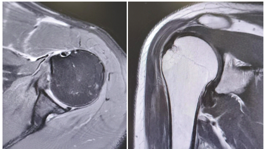
Figure 1: Magnetic resonance imaging demonstrating Bankart lesion and Hill-Sachs defect.
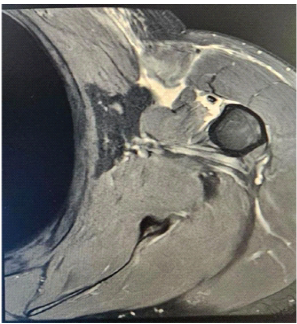
Figure 2: Magnetic resonance imaging demonstrating complete pectoralis major tendon rupture.
Surgical technique
Given the patient’s symptomatic anterior instability and complete pectoralis major tendon rupture, surgical treatment was recommended.
The patient was positioned in the beach-chair position under general anesthesia with an interscalene regional block. Examination under anesthesia demonstrated anterior instability with increased anterior translation and apprehension in abduction and external rotation.
The entire upper extremity and hemithorax were prepared and draped in standard sterile fashion.
Open pectoralis major repair
The open pectoralis major repair was performed first, prior to the arthroscopic portion of the procedure, to avoid operating through edematous tissue resulting from arthroscopic fluid extravasation. Fluid-related soft tissue swelling may obscure tissue planes, hinder identification of anatomic structures, and potentially compromise the ability to perform a stable anatomic repair while increasing the risk of injury to adjacent structures.
A standard deltopectoral approach was utilized. Dissection through the deltopectoral interval exposed the ruptured pectoralis major tendon. Significant hematoma and surrounding soft tissue edema were encountered and evacuated.
Complete avulsion of the clavicular head of the pectoralis major tendon from its humeral insertion lateral to the bicipital groove was confirmed intraoperatively, whereas the sternocostal head remained intact. The ruptured clavicular head was retracted medially with early tendon involution into the muscle belly. Careful mobilization of the tendon–muscle unit was performed to restore adequate excursion for anatomic repair. Adhesions were released to restore tendon excursion while protecting the pectoral neurovascular structures.
Because the sternocostal head remained intact, it served as an important anatomic landmark for identification of the pectoralis major insertional footprint.
The humeral insertion site was prepared to bleeding bone using a curette and rasp. Two 3,2 mm pectoralis major cortical buttons (Pec Button; Arthrex, Naples, FL, USA) were inserted at the anatomic footprint of the pectoralis major insertion lateral to the bicipital groove. The ruptured clavicular head was repaired using two strands of high-strength surgical tape (FiberTape; Arthrex, Naples, FL, USA) and two No. 5 high-strength nonabsorbable sutures (FiberWire; Arthrex, Naples, FL, USA) placed in a Krackow running locking configuration through the tendon–muscle unit to provide secure fixation and optimize load distribution (Fig. 3). The tendon was anatomically reduced to its insertion site with the arm positioned in slight adduction and internal rotation to minimize repair tension. Repair stability and restoration of the anterior axillary contour were confirmed intraoperatively.
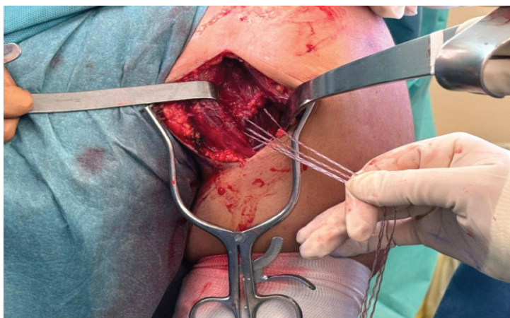
Figure 3: Intraoperative image of pectoralis major repair with suture anchors.
Diagnostic arthroscopy
Following completion of the open pectoralis major repair, attention was directed to the arthroscopic portion of the procedure.A standard posterior portal was established, followed by an anterior working portal through the rotator interval.
Diagnostic arthroscopy revealed:
- Detachment of the anterior-inferior labrum from the glenoid rim extending from approximately the 3 o’clock to 6 o’clock position (Fig. 4).
- Capsular stretching and disruption of the anterior band of the inferior glenohumeral ligament complex
- A small Hill-Sachs lesion involving the posterolateral humeral head that was non-engaging during dynamic assessment in abduction and external rotation.
- Intact rotator cuff tendons and biceps anchor
- No significant glenoid bone loss.
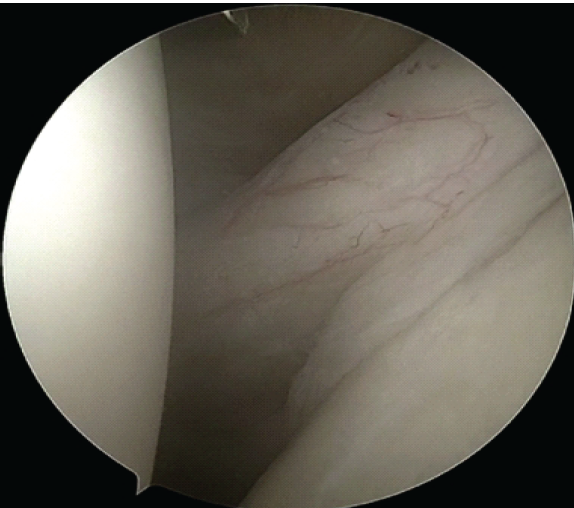
Figure 4: Arthroscopic view of the avulsion of the anteroinferior labrum (Bankart lesion).
Arthroscopic bankart repair
Two 2.9-mm PushLock suture anchors (Arthrex, Naples, FL, USA) were sequentially placed along the anterior-inferior glenoid rim beginning at the 5:30 o’clock position and progressing superiorly. Suture passage was performed using a suture-passing device through the capsulolabral tissue.
The capsulolabral complex was advanced superiorly and laterally to restore the anterior bumper effect and retension the inferior glenohumeral ligament complex.
Arthroscopic knot tying was then performed sequentially from inferior to superior, restoring stability of the anterior labrum and centering of the humeral head (Fig. 5).
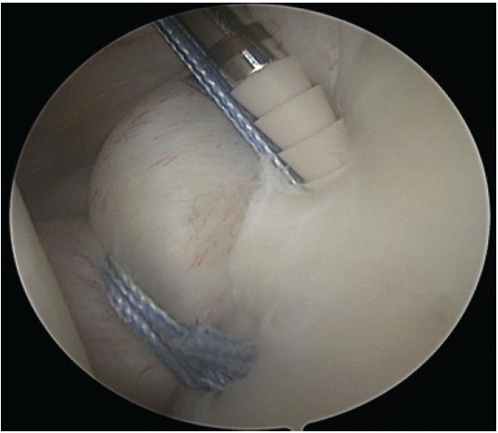
Figure 5: Arthroscopic view of Bankart repair.
Arthroscopic remplissage
Attention was subsequently directed to the Hill-Sachs lesion. The humeral head defect was visualized from both intra-articular and subacromial views. The lesion was noted to be small and non-engaging during dynamic examination.
Although the lesion was not engaging, remplissage was performed as an adjunct to the Bankart repair based on emerging evidence suggesting that the addition of remplissage may improve clinical and functional outcomes and may reduce rates of recurrent instability compared with isolated Bankart repair in selected patients. Given the patient’s high functional demands and participation in recreational athletic activity, augmentation of the stabilization procedure was considered appropriate.
Two 1.8-mm Knotless FiberTak soft suture anchors (Arthrex, Naples, FL, USA) were inserted into the Hill-Sachs defect through a posterior approach. Sutures were passed through the posterior capsule and infraspinatus tendon (Fig. 6). After completion of the Bankart repair, the remplissage sutures were tied in the subacromial space, advancing the posterior capsule and infraspinatus tendon into the humeral head defect.
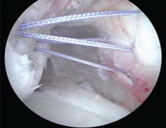
Figure 6: Arthroscopic remplissage procedure.
Postoperative rehabilitation
The arm was immobilized in a sling for 6 weeks postoperatively.
Passive range-of-motion exercises were initiated during the early postoperative period while avoiding excessive external rotation and abduction. Active-assisted motion was gradually introduced after 6 weeks.
Strengthening exercises focusing on rotator cuff and scapular stabilizers were initiated at approximately 10–12 weeks postoperatively. Progressive pectoralis major strengthening was introduced thereafter.
Return to non-contact athletic activity was permitted at approximately 6 months postoperatively, while unrestricted sports participation was allowed following restoration of strength and range of motion.
Outcome and follow-up
At final follow-up 1 year postoperatively, the patient reported excellent clinical recovery with no recurrent instability episodes, no pain during daily activities, and successful return to recreational sports participation including water polo.
Physical examination demonstrated:
- Symmetric restoration of the anterior axillary fold
- Excellent cosmetic appearance of the pectoralis major contour
- Full active forward elevation
- Stable glenohumeral joint without apprehension
- Restoration of strength during resisted adduction and internal rotation.
Objective clinical outcome measures at 1-year follow-up were as follows:
- Constant Score: 98/100
- Rowe Score: 100/100
- Oxford shoulder score: 48/48
- Abduction strength: 20 kg
- External rotation at 0° abduction: 92°
- External rotation at 90° abduction: 88°.
The patient demonstrated excellent shoulder stability and functional recovery with near-complete restoration of motion and strength.
No postoperative complications, rerupture, infection, or recurrent instability events were observed during the follow-up period.
Combined anterior shoulder dislocation and pectoralis major tendon rupture is an exceptionally uncommon injury pattern that may be overlooked during the initial evaluation of shoulder instability. Pectoralis major rupture itself is uncommon and has historically been underrecognized because attention is frequently directed toward the glenohumeral injury or anterior chest pain. Following successful reduction of a traumatic anterior shoulder dislocation, persistent anterior chest pain, ecchymosis, weakness of shoulder adduction and internal rotation, or loss of the anterior axillary fold should prompt careful examination for associated pectoralis major injury and advanced imaging.
A review of the English-language literature identified only three previously published reports describing this combined injury pattern, making the present case, to our knowledge, the fourth reported case. Arciero and Cruser first reported simultaneous anterior shoulder dislocation with pectoralis major rupture in 1997, Vaughan et al. subsequently described a comparable injury following weightlifting, and Wolfley et al. reported a pectoralis major tear associated with anterior labral injury and HAGL lesion. All of these authors emphasized the importance of maintaining a high index of suspicion for associated tendon injuries and obtaining advanced imaging when clinical findings are not fully explained by shoulder instability alone [1,2,3]. Our case differs from previously reported cases because the injury occurred during recreational water polo rather than resistance training and involved an isolated rupture of the clavicular head of the pectoralis major associated with a Bankart lesion and Hill-Sachs lesion.
Most pectoralis major ruptures occur in men between 20 and 40 years of age and are typically associated with resistance training, particularly the bench press, during eccentric loading of the muscle with the shoulder in extension and external rotation [4,5,6,7]. In contrast, our patient was a 47-year-old recreational water polo athlete who sustained the injury during forced abduction and external rotation while contesting the ball overhead. Furthermore, the rupture involved only the clavicular head of the pectoralis major tendon, whereas the sternocostal head remained intact, representing a relatively uncommon injury pattern.
MRI played a crucial role in establishing the diagnosis by simultaneously identifying the Bankart lesion, Hill-Sachs lesion, and complete pectoralis major rupture, thereby facilitating comprehensive preoperative planning. Previous studies have demonstrated that MRI accurately defines the location, extent, and degree of tendon retraction in pectoralis major injuries and remains the imaging modality of choice for surgical planning [8,9,10].
The sequence of surgical treatment also deserves consideration. Open repair of the pectoralis major tendon was performed before shoulder arthroscopy to avoid fluid extravasation into the anterior chest wall, which may obscure tissue planes, increase soft-tissue edema, and complicate tendon mobilization and identification. This sequence allowed optimal visualization of the ruptured tendon and facilitated an anatomic repair before proceeding with arthroscopic stabilization.
Although remplissage has traditionally been recommended for engaging or off-track Hill-Sachs lesions, the concept of bipolar bone loss and the glenoid track has substantially refined surgical indications for instability surgery [11,12]. In the present case, the Hill-Sachs lesion was small, on-track, and non-engaging during dynamic arthroscopic assessment. Nevertheless, remplissage was performed because of the patient’s high functional demands, traumatic instability mechanism, and desire to return to overhead sporting activity. Emerging evidence suggests that selected patients with on-track Hill-Sachs lesions may benefit from adjunctive remplissage, demonstrating lower rates of recurrent instability without clinically significant loss of external rotation [13,14,15,16,17]. In particular, the recent randomized controlled trial by Woodmass et al. demonstrated significantly fewer redislocations and recurrent instability events following combined Bankart repair and remplissage compared with isolated Bankart repair at medium-term follow-up [15].
At 1-year follow-up, our patient demonstrated restoration of glenohumeral stability, recovery of pectoralis major strength and anterior axillary contour, excellent patient-reported outcome scores, and successful return to recreational water polo without recurrent instability or tendon rerupture. This case illustrates that early recognition of associated injuries, comprehensive MRI evaluation, and single-stage surgical management can result in excellent functional outcomes in this rare combined injury pattern.
Concomitant pectoralis major rupture associated with traumatic anterior shoulder dislocation represents a rare but clinically important injury pattern. Early diagnosis and comprehensive surgical management can provide excellent functional outcomes and facilitate return to athletic activity.
Concomitant pectoralis major rupture should be suspected in patients with traumatic anterior shoulder dislocation and persistent anterior chest symptoms, as early diagnosis and comprehensive surgical management can restore excellent shoulder function.
References
- 1. Vaughan A, Bhalla S, Chen A, Traumatic anterior shoulder dislocation with concomitant pectoralis major rupture treated surgically: A case report. Cureus 2022;14:???. [Google Scholar] [PubMed]
- 2. Wolfley CN, DeFoor MT, Antosh IJ, Parada SA. Treatment of a combined pectoralis major tear, anterior labral tear, and humeral avulsion of the glenohumeral ligament (HAGL) in an active duty soldier. Mil Med 2022;187:e530-4. [Google Scholar] [PubMed]
- 3. Arciero RA, Cruser DL. Pectoralis major rupture with simultaneous anterior dislocation of the shoulder. J Shoulder Elbow Surg 1997;6:318-20.</p> [Google Scholar] [PubMed]
- 4. <p>4. Bak K, Cameron EA, Henderson IJ. Rupture of the pectoralis major: A meta-analysis of 112 cases. Knee Surg Sports Traumatol Arthrosc 2000;8:113-9. [Google Scholar] [PubMed]
- 5. Tietjen R. Closed injuries of the pectoralis major muscle. J Trauma 1980;20:262-4. [Google Scholar] [PubMed]
- 6. Schepsis AA, Grafe MW, Jones HP, Lemos MJ. Rupture of the pectoralis major muscle. Outcome after repair of acute and chronic injuries. Am J Sports Med 2000;28:9-15. [Google Scholar] [PubMed]
- 7. Aarimaa V, Rantanen J, Heikkilä J, Helttula I, Orava S. Rupture of the pectoralis major muscle. Am J Sports Med 2004;32:1256-62. [Google Scholar] [PubMed]
- 8. Connell DA, Potter HG, Sherman MF, Wickiewicz TL. Injuries of the pectoralis major muscle: Evaluation with MR imaging. Radiology 1999;210:785-91. [Google Scholar] [PubMed]
- 9. Qylafi K, Alkhalfan Y. Pectoralis major rupture: A case report. Cureus 2022;14:e29512. [Google Scholar] [PubMed]
- 10. Durant EJ, De Cicco FL. Pectoralis major tear. In: StatPearls. Treasure Island, FL: StatPearls Publishing; 2024. [Google Scholar] [PubMed]
- 11. Di Giacomo G, Itoi E, Burkhart SS. Evolving concept of bipolar bone loss and the Hill-Sachs lesion: From ”engaging/non-engaging” lesion to “on-track/off-track” lesion. Arthroscopy 2014;30:90-8. [Google Scholar] [PubMed]
- 12. Yamamoto N, Itoi E, Abe H, Kikuchi K, Seki N, Minagawa H, et al. Contact between the glenoid and the humeral head in abduction, external rotation, and horizontal extension: A new concept of glenoid track. J Shoulder Elbow Surg 2007;16:649-56. [Google Scholar] [PubMed]
- 13. Yu W, Kim H, Seo JH, Jeon IH, Koh KH. Remplissage in addition to arthroscopic Bankart repair for shoulder instability with on-track Hill-Sachs lesions reduces residual apprehension without external rotation limitation. Arthroscopy 2023;39:692-702. [Google Scholar] [PubMed]
- 14. Boileau P, O’Shea K, Vargas P, Pinedo M, Old J, Zumstein M. Anatomical and functional results after arthroscopic Hill-Sachs remplissage. J Bone Joint Surg Am 2012;94:618-26. [Google Scholar] [PubMed]
- 15. Woodmass JM, McRae S, Lapner P, Kamikovski I, Jong B, Old J, et al. Arthroscopic Bankart repair with remplissage in anterior shoulder instability results in fewer redislocations than Bankart repair alone at medium-term follow-up of a randomized controlled trial. Am J Sports Med 2024;52:2055-62. [Google Scholar] [PubMed]
- 16. MacDonald P, McRae S, Old J, Dubberley J, Stranges G, Marsh J, et al. Arthroscopic Bankart repair with and without arthroscopic infraspinatus remplissage in anterior shoulder instability with a Hill-Sachs defect: A randomized controlled trial. J Shoulder Elbow Surg 2021;30:1288-98. [Google Scholar] [PubMed]
- 17. Fares MY, Salhab HA, Khachfe HH, Fares J, Baydoun H. Arthroscopic remplissage: History, indications, and clinical perspectives. EFORT Open Rev 2023;8:424-34. [Google Scholar] [PubMed]
Related Articles in Journal of Orthopaedic Case Reports
 September 1, 2026 A Prospective Study Showing Functional Outcome of the Modified Boytchev Procedure for Recurrent Anterior Shoulder Dislocation
 August 1, 2026 Repair of a Chronic Pectoralis Major Rupture with Achilles Tendon–Calcaneal Allograft: A Case Report
 July 1, 2026 Humeral Avulsion of Glenohumeral Ligament Lesion – A Case Report and Surgical Considerations
 July 1, 2026 Single-Screw versus Two-Screw Fixation in the Latarjet Procedure: A Prospective Comparative Study