


Introduction
The methods for reconstructing the anterior cruciate ligament (ACL) have evolved from isometric reconstruction to grafts that more anatomically restore the natural morphology and biomechanics of the native ligament. Among the various technical aspects involved in ACL reconstruction, femoral tunnel creation continues to remain one of the most critical determinants of graft orientation and intra-articular biomechanics [1,2].
The femoral tunnel is a manmade intraosseous tract through the distal femur. The shape of a tunnel depends not just on the technique a surgeon uses, but also on the shape of the lateral femoral condyle, the shape of the anatomy of the intercondylar notch, and the angle of the distal femur [3]. A change in the angle of a tunnel can greatly affect the distance that it travels through the lateral femoral condyle, which affects the length of the tunnel, and the containment, fixation strength, and bending characteristics of the graft [4,5].
Anatomical ACL reconstruction is gaining increasing emphasis. To try to achieve the native anatomy of the footprint, ACL surgeons try to create more oblique femoral tunnels. However, as tunnel obliquity increases, that interferes with the intraosseous tunnel lengthening, perhaps augmenting the risk of posterior cortical compromise or inadequate graft-bone interface [6]. In contrast, overtly vertical tunnels might lengthen the tunnel but jeopardize rotational biomechanics [7].
The geometrical assessment of femoral tunnels has become quite critical. According to conventional post-operative radiographs, tunnel inclination and orientation can be assessed. However, computed tomography (CT) offers a superior visualization of the tunnel trajectory and the intraosseous dimensions of the distal femur. Despite several studies evaluating graft bending angle, tunnel widening, and tunnel positioning, limited literature exists assessing femoral tunnel angle in relation to intraosseous tunnel length anatomically.
The current study was undertaken to assess the anatomical relationship between femoral tunnel angle and intraosseous tunnel length within the distal femur following arthroscopic ACL reconstruction using radiographic and CT evaluation.
Materials and Methods
Study design
This was a hospital-based radiological observational study conducted in patients who underwent arthroscopic ACL reconstruction.
Study population
A total of 70 patients who underwent arthroscopic ACL reconstruction using quadrupled hamstring autograft were included in the study.
Inclusion criteria
-
Patients aged between 18 and 45 years
-
Primary ACL tear confirmed clinically and radiologically
-
Patients who underwent arthroscopic single-bundle ACL reconstruction
-
Availability of post-operative radiographs and CT scans.
Exclusion criteria
-
Revision ACL reconstruction
-
Multiligamentous knee injuries
-
Previous distal femoral fractures
-
Severe osteoarthritis
-
Inadequate radiological imaging.
Surgical technique
All surgeries were performed arthroscopically using standard anteromedial and anterolateral portals. Femoral tunnel drilling was performed through the anteromedial portal with the knee in hyperflexion. The native femoral ACL footprint was identified arthroscopically before tunnel creation. Quadrupled hamstring autograft was used in all patients.
Radiological assessment
Measurement of femoral tunnel angle
The femoral tunnel angle was determined from the true lateral radiograph as the angle formed between:
The long axis of the distal femoral shaft and the axis of the femoral tunnel, which is the centreline drawn through the shadow of the tunnel (Fig. 1) [1,5,8,9].
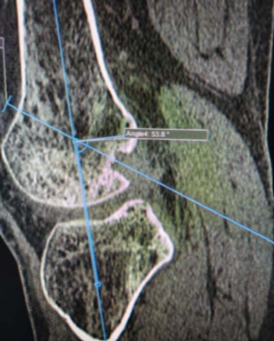
We recorded angles in degrees and grouped them along the reported corridor (50°-57°). This made it possible to look at clustering patterns and the consistency of the surgical tunnel’s orientation.
Measurement of femoral tunnel length
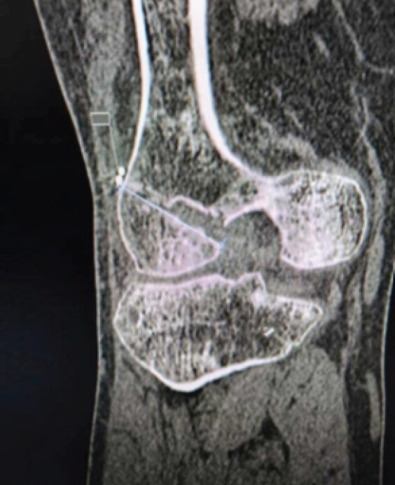
The length of the femoral tunnel was mainly measured by CT multiplanar reconstruction. This gave the most precise estimate of the length of the tunnel in millimeters [8,9].
Radiographically, the length of the femoral tunnel was secondarily corroborated by verifying the position of the Endobutton at the lateral femoral cortex and evaluating the anticipated tunnel trajectory on the lateral radiograph. For the sake of consistency in the analysis, femoral tunnel length values were grouped into millimeter ranges (35–40 mm) (Fig. 2).
Statistical analysis
Data were analyzed using the Statistical Package for the Social Sciences software. Both Pearson and Spearman correlation analyses were performed. Because the variables were not normally distributed, Spearman correlation was considered the primary analysis. Continuous variables were expressed as mean ± standard deviation. P < 0.05 was considered statistically significant.
Results
Femoral tunnel length and tunnel angle on sagittal
The length of the femoral tunnel ranged from 35 mm to 40 mm, with most (22.86%) measuring 40 mm. The more uniform distribution across the remaining length categories indicates consistent proximal femur intraosseous tunnel morphology. The angles of the femoral tunnels were mostly found between 51° and 55° (58.6%), indicating a possible reproducible orientation of the tunnel through the lateral femoral condyle. To sum up, findings reveal that femoral tunnel geometry, as indicated by tunnel length and tunnel angle, remains relatively unchanged following anatomical ACL reconstruction and may reflect distal femoral osseous morphology rather than a mere technical consequence of tunnel drilling [Tables 1, 2, 3, 4, 5, 6].
Femoral tunnel length
| Femoral tunnel length | Frequency | Percentage | Cumulative frequency | Cumulative percentage |
|---|---|---|---|---|
| 35 | 11 | 15.71 | 11 | 15.71 |
| 36 | 12 | 17.14 | 23 | 32.86 |
| 37 | 11 | 15.71 | 34 | 48.57 |
| 38 | 10 | 14.29 | 44 | 62.86 |
| 39 | 10 | 14.29 | 54 | 77.14 |
| 40 | 16 | 22.86 | 70 | 100 |
Tunnel angle on sagittal
| Tunnel angle on sagittal | Frequency | Percentage | Cumulative frequency | Cumulative percentage |
|---|---|---|---|---|
| 50 | 8 | 11.43 | 8 | 11.43 |
| 51 | 10 | 14.29 | 18 | 25.71 |
| 52 | 8 | 11.43 | 26 | 37.14 |
| 53 | 11 | 15.71 | 37 | 52.86 |
| 54 | 8 | 11.43 | 45 | 64.29 |
| 55 | 12 | 17.14 | 57 | 81.43 |
| 56 | 7 | 10 | 64 | 91.43 |
| 57 | 6 | 8.57 | 70 | 100 |
Descriptive statistics
| Statistic | Tunnel angle (°) | Tunnel length (mm) |
|---|---|---|
| n | 70 | 70 |
| Mean±standard deviation | 53.36±2.17 | 37.81±1.64 |
| Median | 53 | 38 |
| Minimum–Maximum | 50–57 | 35–40 |
| Range | 7 | 5 |
| Q1 (25%) | 51.25 | 37 |
| Q3 (75%) | 55 | 39 |
| Interquartile range | 3.75 | 2 |
| 95% confidence interval | 52.84–53.88 | 37.42–38.21 |
Shapiro–Wilk normality test
| Variable | W Statistic | P-value | Interpretation |
|---|---|---|---|
| Tunnel angle (°) | 0.937 | 0.00161 | Not normally distributed |
| Tunnel length (mm) | 0.911 | 0.000108 | Not normally distributed |
Correlation analysis
| Parameter | Value |
|---|---|
| Pearson correlation | |
| Pearson r | –0.3379 |
| R2 | 0.1142 |
| t -statistic | –2.961 |
| Degrees of freedom | 68 |
| P -value | 0.00422 |
| Spearman rank correlation | |
| Spearman p | –0.3285 |
| t -statistic | –2.868 |
| P -value | 0.00549 |
| Interpretation | Significant negative rank correlation |
Linear regression analysis
| Parameter | Value |
|---|---|
| Slope (b) | −0.2556 |
| Intercept (a) | 51.4545 |
| R2 | 0.1142 |
| Standard error of estimate | 1.559 |
| Standard error of slope | 0.0863 |
| t- statistic | −2.961 |
| P -value | 0.00422 |
Regression equation:
Length = 51.455 – 0.256 × Angle
Descriptive analysis of femoral tunnel morphometry
The study included 70 patients who underwent arthroscopic ACL reconstruction. The mean femoral tunnel angle as measured on sagittal radiographs was 53.36 ± 2.17° (50°–57°). On post-operative computed tomography, the average intraosseous femoral tunnel length was found to be 37.819 ± 1.64 mm (35–40 mm). The median angle of the tunnel was 53°. The median tunnel length was 38 mm. The tunnel angle’s interquartile range is 3.75°, indicating moderate scatter around the average. The distribution of the length of the tunnel is most closely related to the interquartile range of 2 mm. The angle of the tunnel, with a 95% confidence interval, was 52.84°–53.88°, and the length of the tunnel, with a 95% confidence interval, was 37.42 –38.21 mm, indicating satisfactory precision.
Assessment of data distribution
The normality of the data was assessed using the Shapiro–Wilk test. Tunnel angle demonstrated a W statistic of 0.937 (P = 0.0016), whereas tunnel length showed a W statistic of 0.911 (P = 0.0001). Both variables exhibited statistically significant deviation from a normal distribution. Consequently, non-parametric correlation analysis was considered appropriate for evaluating the relationship between tunnel angle and tunnel length.
Correlation between femoral tunnel angle and tunnel length
Spearman rank correlation analysis revealed a statistically significant negative correlation between femoral tunnel angle and intraosseous femoral tunnel length (Ρ = −0.329, P = 0.005). This finding indicates that increasing tunnel obliquity was associated with a reduction in the intraosseous tunnel length within the distal femur.
Pearson correlation analysis produced similar findings, demonstrating a significant inverse relationship between tunnel angle and tunnel length (r = −0.338, P = 0.004). Although the magnitude of the correlation was modest, the consistency of the findings across both parametric and non-parametric methods supports the existence of a genuine anatomical association between tunnel orientation and tunnel length.
Linear regression analysis
The relationship between tunnel angle and tunnel length was further investigated through linear regression analysis. The regression model revealed a significant negative correlation (P = 0.004), with a coefficient of determination of 0.114. Thus, tunnel angle explains only about 11.4% of the variation in tunnel length. Thus, variations in tunnel angle and tunnel length are poorly predictable.
The negative coefficient of the regression slope means that more oblique tunnels were associated with shorter intraosseous tunnels. While changes to the tunnel angle were sufficient to cause variation in tunnel length, the low coefficient of determination indicates that other anatomical factors related to the distal femur may influence the tunnel geometry.
Radiological anatomical interpretation
We found a weak correlation between the femoral tunnel angle and the available intraosseous corridor in the distal femur. Higher tunnel obliquity correlated with shorter tunnel length, but tunnel angle accounted for only 11.4% of the variation seen, suggesting other anatomical and technical factors also contribute substantially to tunnel length. Shorter intraosseous trajectories through the lateral femoral condyle were associated with more oblique tunnel orientations. This relationship may be explained by interaction between tunnel trajectory, distal femoral morphology, and technical factors during tunnel creation. Conversely, less oblique tunnels had a longer intraosseous course. The fact that femoral tunnel morphometry is directly related to distal femoral bone morphology and not merely due to surgical techniques could be seen from these observations.
Discussion
The present study evaluated the anatomical relationship between femoral tunnel angle and intraosseous femoral tunnel length within the distal femur following arthroscopic ACL reconstruction. The key finding was a statistically significant negative correlation between tunnel angle and tunnel length, indicating that increasing tunnel obliquity was associated with a reduction in the available intraosseous tunnel corridor. Although the strength of correlation was modest, the finding is anatomically meaningful, as it suggests that femoral tunnel length is influenced not only by surgical drilling technique but also by the osseous geometry of the distal femur. Table 7 compares the present study with previously published studies evaluating femoral tunnel geometry, tunnel length, and tunnel orientation in ACL reconstruction.
Comparison of the present study with previously published studies evaluating femoral tunnel geometry, tunnel length, and tunnel orientation in ACL reconstruction
| Study | Primary focus | Verified finding | Relationship to present study |
|---|---|---|---|
| Ahn et al. [1] | Post-operative graft angle and tunnel orientation | Quantified graft angle and tunnel placement after ACL reconstruction using imaging | Supports the importance of radiological evaluation of tunnel orientation, which was also analyzed in the present study. |
| Gali et al. [8] | Effect of knee flexion angle on femoral tunnel length | Demonstrated that femoral tunnel length varies according to drilling conditions and knee flexion angle during tunnel creation | Consistent with our finding that tunnel geometry influences intraosseous tunnel length. Our study further evaluated this relationship using post-operative tunnel angle measurements. |
| Bedi et al. [5] | Anteromedial portal versus transtibial drilling | Demonstrated that tunnel orientation changes with drilling technique and influences anatomical tunnel placement | Supports the concept that tunnel trajectory is a major determinant of femoral tunnel geometry. |
| Nishimoto et al. [2] | Graft bending angle at femoral tunnel aperture | Showed that tunnel orientation affects graft bending angle at the femoral aperture | Supports the anatomical relevance of tunnel geometry beyond tunnel creation alone. |
| Tashiro et al. [6, 7] | Dynamic graft bending angle and tunnel orientation | Demonstrated that graft bending behavior changes according to tunnel orientation and reconstruction technique | Indicates that femoral tunnel orientation influences graft mechanics and tunnel-related biomechanics. |
| Wein et al. [9] | Tunnel anteversion and tunnel length | Demonstrated that tunnel direction and instrumentation influence femoral tunnel length | Similar to our findings showing that tunnel orientation affects intraosseous tunnel length within the distal femur. |
| Tompkins et al. | Femoral tunnel length and lateral femoral condylar dimensions | Demonstrated that tunnel length correlates with lateral femoral condylar morphology and anthropometric parameters. | Strengthens our interpretation that tunnel length reflects distal femoral osseous anatomy rather than being solely a technical measurement. |
| Present study | Distal femoral tunnel morphometry | Significant negative correlation between tunnel angle and tunnel length (ρ = −0.329, P = 0.005) | One of the few studies specifically evaluating tunnel angle and tunnel length as a distal femoral morphometric relationship using combined radiographic and computed tomography assessment. |
Modern ACL reconstruction has moved toward anatomical femoral tunnel placement, with the intention of restoring the native femoral footprint and improving rotational control. Bedi et al. demonstrated that anteromedial portal drilling allows more anatomical and oblique femoral tunnel placement when compared with the transtibial technique [5]. Their work supports the current understanding that tunnel orientation is central to anatomical ACL reconstruction. However, our study adds a further anatomical perspective by showing that such tunnel orientation has a measurable relationship with intraosseous tunnel length. In other words, the tunnel is not only a surgical pathway but also a morphometric expression of the lateral femoral condylar corridor.
Gali et al. reported that femoral tunnel length changes with knee flexion angle during anatomical ACL reconstruction [8]. Their findings suggested that tunnel length is sensitive to the direction of drilling and the spatial orientation of the femur at the time of tunnel creation. The present study is in agreement with this concept, as more oblique tunnels were associated with shorter intraosseous tunnel lengths. However, while Gali et al. focused mainly on the influence of knee flexion angle, the present study specifically examines the relationship between post-operative tunnel angle and CT-measured tunnel length as a distal femoral morphometric phenomenon.
Wein et al. also highlighted the relationship between femoral tunnel anteversion, instrumentation, and tunnel length in ACL reconstruction [9]. Their study supports the idea that femoral tunnel geometry is affected by the three-dimensional direction of tunnel creation. The present study similarly indicates that the intraosseous path available within the distal femur is not fixed. It changes according to the angular orientation of the tunnel. This reinforces the view that femoral tunnel length should not be interpreted as a simple technical measurement. It reflects the interaction between tunnel trajectory, condylar morphology, and the cancellous osseous corridor of the lateral femoral condyle.
The distal femur has a complex anatomical architecture. The femoral tunnel begins at the anatomical femoral ACL footprint, passes through cancellous bone in the lateral femoral condyle, and exits toward the lateral cortex. As tunnel obliquity increases, the tunnel takes a more horizontal course through the condyle. This reduces the available intraosseous distance and may bring the tunnel closer to the posterior cortical boundary. This anatomical explanation is consistent with our observation that greater tunnel obliquity was associated with shorter tunnel length. It also explains why a balance is required between anatomical footprint restoration and maintenance of an adequate tunnel length for graft containment.
Tunnel geometry also has implications for graft bending. Nishimoto et al. analyzed the graft bending angle at the femoral tunnel aperture in anatomical double-bundle ACL reconstruction and demonstrated that tunnel orientation affects graft bending at the aperture [2]. Tashiro et al. further showed that graft bending angle varies during knee motion and differs according to reconstruction technique [6,7]. These studies indicate that femoral tunnel orientation does not end as an isolated bony measurement. It affects the course of the graft, the bending angle at the aperture, and potentially the stress distribution across the graft-tunnel interface. The present study complements these observations by showing that the same tunnel orientation also affects the intraosseous length available within the distal femur.[10].
The biomechanical implications of femoral tunnel orientation are not limited to tunnel length. Graft bending angle was shown by Chen et al. to affect early allograft tendon maturation after ACL reconstruction [11]. Zaffagnini et al. investigated the influence of the orientation of the femoral tunnel on knee stability and the relevance of the positioning of the graft for the restoration of the biomechanics of the joint [12]. Similarly, Kato et al. demonstrated that different femoral graft positions result in different biomechanical responses, indicating the importance of anatomical tunnel placement [13]. The findings suggest that the geometry of the femoral tunnel should not only be considered as an anatomical measurement, but also in relation to the behaviour of the graft and postoperative knee biomechanics.
Chung et al. further showed that the knee flexion angle during transportal femoral tunnel drilling affects the graft bending angle, tunnel length, and risk of posterior cortical compromise [14]. Guglielmetti et al. investigated the influence of femoral tunnel length on graft rupture after ACL reconstruction and emphasised the clinical significance of measuring tunnel length [15]. Collectively, the results of these studies emphasise the need to balance anatomical graft positioning with sufficient intraosseous tunnel length and good graft mechanics. However, the present study was confined to radiological morphometry and did not assess graft maturation, fixation strength, clinical stability, or graft failure. Therefore, the relationship between tunnel angle and tunnel length observed here should not be interpreted as direct evidence of altered graft biomechanics or clinical outcomes.
CT was useful in the present study because it allowed better assessment of the tunnel course within the distal femoral bone. Conventional radiographs provided a practical method for assessing tunnel angle, but CT defined the intraosseous tunnel length and its relationship to the distal femoral cortex more clearly. This combined radiographic and CT-based approach strengthens the anatomical relevance of the study. It allows the femoral tunnel to be studied as a three-dimensional osseous corridor rather than merely as a post-operative surgical mark.
An important strength of this study is its anatomical framing. Most previous studies have discussed femoral tunnel position, tunnel length, graft bending angle, or drilling technique mainly from a surgical or biomechanical perspective [2,5,6,7,8,9]. The present study specifically examines the correlation between femoral tunnel angle and tunnel length as a distal femoral morphometric relationship. This makes the work relevant to applied anatomy, particularly because the femoral tunnel is created within the lateral femoral condyle and is directly influenced by the osseous architecture of the distal femur (Table 7).
The modest correlation observed in this study also deserves attention. Tunnel angle explained only part of the variation in tunnel length. This is expected, as tunnel length may also be influenced by individual variation in condylar size, intercondylar notch morphology, lateral femoral wall thickness, knee flexion during drilling, and subtle differences in portal placement. Therefore, the findings should not be interpreted as a purely mechanical rule. Rather, they suggest that tunnel angle is one important anatomical determinant of intraosseous tunnel length within a broader distal femoral morphometric framework.
Overall, the present study supports the view that femoral tunnel length should not be considered only as an intraoperative technical parameter. It is a radiological reflection of distal femoral osseous anatomy and tunnel orientation. Recognition of this relationship may help surgeons achieve a more balanced reconstruction, where anatomical tunnel placement is obtained without compromising tunnel length, graft containment, or cortical safety.
Limitations of the study
The study carries multiple limitations. The single-center observational design and relatively small sample size may limit the generalisability of the findings. The cohort was sufficient to demonstrate the observed association between femoral tunnel angle and tunnel length, but was not powered for subgroup analyses based on age, sex, graft characteristics, or variations in femoral morphology.
The study was mostly designed to evaluate the post-operative tunnel geometry. There was no pre-operative three-dimensional assessment of the distal femur nor a non-operated control group. Therefore, the relative contribution of native femoral anatomy and surgically determined tunnel orientation cannot be clearly disentangled. The analysis also did not include detailed morphometric parameters such as the dimensions of the lateral femoral condyle, notch morphology, thickness of the posterior wall, cortical thickness, and overall femoral size.
The femoral tunnel angle was measured on true lateral radiographs and therefore is a two-dimensional projection of a three-dimensional trajectory. Although only technically adequate radiographs were included, small rotational variations during imaging as well as differences in identification of the tunnel axis may have affected the measured angle. Tunnel orientation in axial and coronal planes was not quantified independently. Reliability analyses for intraobserver and interobserver were also not done. Future studies with three-dimensional CT-based measurements and formal assessment of measurement reliability would yield more comprehensive assessments of tunnel geometry.
Furthermore, the interpretation of the results has to take into account the rather limited range of tunnel angles (50°–57°) and tunnel lengths (35–40 mm). The observed relationship may not necessarily hold for substantially more vertical or more oblique tunnel orientations. Furthermore, a number of anatomical and technical variables that might influence tunnel length were excluded from multivariate analysis. Factors include knee flexion during drilling, portal position, guidewire placement, tunnel starting position, reamer trajectory, and individual skeletal morphology. The general method of reconstruction was consistent, but these individual operative variables were not standardized in a quantitative manner.
Most importantly, the correlation observed was modest, and tunnel angle explained only a small amount of the variability in tunnel length. Thus, the results should be interpreted as an association and not as a causal or predictive relationship. The length of the tunnel is probably a function of an interaction between the tunnel orientation, the distal femoral morphology, and the surgical technique.
Finally, clinical- and graft-related outcomes were beyond the scope of the present radiological morphometric study. Graft stability, post-operative laxity, functional scores, return to sport, graft bending, fixation-related parameters, tunnel widening, graft failure, and revision surgery were not assessed. However, the clinical and biomechanical implications of this morphometric relationship are yet to be established. Larger prospective multicenter studies including pre-operative three-dimensional morphometry, standardized surgical parameters, reliability testing, multivariable analysis, and longitudinal clinical outcomes would help to clarify the clinical relevance of these findings.
Conclusion
The relationship between the intraosseous tunnel length after arthroscopic ACL reconstruction and the femoral tunnel angle was statistically significant but weakly inverse. Tunnel angle accounted for only a small proportion of tunnel-length variability, but more oblique tunnel orientations were associated with shorter intraosseous trajectories. Femoral tunnel angle demonstrated a statistically significant but modest inverse association with intraosseous tunnel length. This finding should be interpreted as a morphometric relationship rather than evidence that increasing tunnel obliquity directly causes tunnel shortening. Therefore, femoral tunnel length is a multifactorial parameter influenced both by tunnel orientation and distal femoral anatomy.
Clinical Message
In anatomical ACL reconstruction, femoral tunnel orientation should be evaluated in conjunction with available intraosseous tunnel length, not in isolation. An understanding of this relationship may assist the surgeon in achieving an anatomical tunnel placement while being aware of the anatomical limitations of the distal femoral osseous corridor.
Conflict of Interest:
Nil
Source of Support:
Nil
Consent
The authors confirm that informed consent was obtained from the patient for publication of this article
How to Cite this Article
Rajadurai ORJW, Bellamkonda S, Saminathan T, Kumar R, Sathesh Kumar M, Purushothaman K. Intraosseous Femoral Tunnel Morphometry of the Distal Femur: Correlation between Tunnel Angle and Tunnel Length following Arthroscopic Anterior Cruciate Ligament Reconstruction. J Orthop Case Rep. 2026 November;16(11): 532-540. doi: 10.13107/jocr.2026.v16.i11.8544.
References
- Ahn JH, Lee SH, Yoo JC, Ha HC. Measurement of the graft angles for the anterior cruciate ligament reconstruction with transtibial technique using postoperative magnetic resonance imaging in comparative study. Knee Surg Sports Traumatol Arthrosc 2007;15:1293-300. [Google Scholar] | [PubMed]
- Nishimoto K, Kuroda R, Mizuno K, Hoshino Y, Nagamune K, Kubo S. Analysis of the graft bending angle at the femoral tunnel aperture in anatomic double bundle anterior cruciate ligament reconstruction: A comparison of the transtibial and the far anteromedial portal technique. Knee Surg Sports Traumatol Arthrosc 2009;17:270-6. [Google Scholar] | [PubMed]
- Xu Y, Ao Y, Wang J, Yu J, Cui G. Relation of tunnel enlargement and tunnel placement after single-bundle anterior cruciate ligament reconstruction. Arthroscopy 2011;27:923-32. [Google Scholar] | [PubMed]
- Zantop T, Ferretti M, Bell KM, Brucker PU, Gilbertson L, Fu FH. Effect of tunnel-graft length on the biomechanics of anterior cruciate ligament-reconstructed knees: Intra-articular study in a goat model. Am J Sports Med 2008;36:2158-66. [Google Scholar] | [PubMed]
- Bedi A, Musahl V, Steuber V, Kendoff D, Choi D, Allen AA. Transtibial versus anteromedial portal reaming in anterior cruciate ligament reconstruction: An anatomic and biomechanical evaluation of surgical technique. Arthroscopy 2011;27:380-90. [Google Scholar] | [PubMed]
- Tashiro Y, Sundaram V, Thorhauer E, Gale T, Anderst W, Irrgang JJ. In vivo analysis of dynamic graft bending angle in anterior cruciate ligament-reconstructed knees during downward running and level walking: Comparison of flexible and rigid drills for transportal technique. Arthroscopy 2017;33:1393-402. [Google Scholar] | [PubMed]
- Tashiro Y, Okazaki K, Uemura M, Matsuda S. Comparison of graft bending angle during knee motion after outside-in, transportal, and transtibial anterior cruciate ligament reconstruction techniques. Am J Sports Med 2017;45:2584-91. [Google Scholar] | [PubMed]
- Gali JC, Resina AF, Pedro G, Moraes FB, Fernandes TD. Femoral tunnels' length changes with knee flexion angle in anatomical anterior cruciate ligament reconstruction. Rev Bras Ortop 2015;50:436-41. [Google Scholar] | [PubMed]
- Wein F, Osemont B, Goetzmann T, Jacquot A, Valluy J, Saffarini M. Anteversion and length of the femoral tunnel in ACL reconstruction: In-vivo comparison between rigid and flexible instrumentation. J Exp Orthop 2019;6:26. [Google Scholar] | [PubMed]
- Lee DK, Kim JH, Lee BH, Kim H, Jang MJ, Lee SS. Influence of graft bending angle on femoral tunnel widening after double-bundle ACL reconstruction: Comparison of transportal and outside-in techniques. Orthop J Sports Med 2021;9:23259671211035780. [Google Scholar] | [PubMed]
- Chen L, Wu Y, Lin G, Wei P, Ye Z, Wang Y. Graft bending angle affects allograft tendon maturity early after anterior cruciate ligament reconstruction. Knee Surg Sports Traumatol Arthrosc 2018;26:3048-54. [Google Scholar] | [PubMed]
- Zaffagnini S, Bignozzi S, Martelli S, Lopomo N, Marcacci M. Does ACL reconstruction restore knee stability in combined lesions? An in vitro study on the influence of femoral tunnel orientation. Knee Surg Sports Traumatol Arthrosc 2008;16:219-25. [Google Scholar] | [PubMed]
- Kato Y, Maeyama A, Lertwanich P, Wang JH, Ingham SJ, Kramer S. Biomechanical comparison of different graft positions for single-bundle anterior cruciate ligament reconstruction. Knee Surg Sports Traumatol Arthrosc 2013;21:816-23. [Google Scholar] | [PubMed]
- Chung K, Choi CH, Kim SH, Kim SJ, Choi HC, Jung M. Influence of knee flexion angle on graft bending angle during anterior cruciate ligament reconstruction using the transportal technique. Sci Rep 2023;13:13638. [Google Scholar] | [PubMed]
- Guglielmetti LG, Camanho GL, Severino NR. The influence of femoral tunnel length on graft rupture after anterior cruciate ligament reconstruction. Rev Bras Ortop 2017;52:35-40. [Google Scholar] | [PubMed]
© 2026 Journal of Orthopaedic Case Reports - Published by Indian Orthopaedic Research Group
About the Authors
-

Jeff Walter OR Rajadurai
-

Sravya Bellamkonda
-

T Saminathan
-

Ram Kumar
-

M Sathesh Kumar
-

K Purushothaman
Related Articles in Journal of Orthopaedic Case Reports
 November 1, 2026 A Study of Functional Outcome of Arthroscopic Anterior Cruciate Ligament Reconstruction Using Hamstring Graft
 November 1, 2026 Functional Outcome in Arthroscopic Anterior Cruciate Ligament Reconstruction Using an Autologous Quadrupled Hamstring Tendon Versus Peroneus Longus Tendon Graft: A Comparative Study
 November 1, 2026 Hoffa Fracture Non-union Treated with Medial Parapatellar Approach and Headless Compression Screw Fixation: A Case Report
 October 1, 2026 Retrograde Intramedullary Nailing Versus Locking Plate Fixation in Extra-Articular Distal Femur Fractures: A Prospective Study