


Introduction
Intertrochanteric (IT) fractures of the femur are among the most common hip fractures encountered in older adults and are frequently associated with low-energy falls in the setting of age-related skeletal fragility. In younger individuals, these fractures are more commonly caused by high-energy trauma. Unlike intracapsular fractures of the neck of the femur, IT fractures are not typically complicated by avascular necrosis of the femoral head. However, delayed or inadequate treatment may result in malunion, limb shortening, impaired mobility, and prolonged functional disability [1].
Early surgical stabilization and mobilization are therefore important in the management of these fractures. Various operative techniques have been developed, including extramedullary fixation with a dynamic hip screw (DHS) and intramedullary fixation with proximal femoral nails. Comparative studies have evaluated these two approaches, with proximal femoral nailing offering biomechanical and technical advantages in selected fracture patterns [2].
Hip fractures represent a significant and increasing health burden, particularly among older adults. Epidemiological studies have demonstrated the substantial occurrence of hip fractures in elderly populations, emphasizing the importance of effective surgical management and early restoration of mobility [3].
The proximal femoral nail (PFN) was developed by the Arbeitsgemeinschaft für Osteosynthesefragen (AO)/Association for the Study of Internal Fixation as an intramedullary device for the treatment of unstable proximal femoral fractures. Its design provides intramedullary load sharing, reduced soft-tissue disruption, and improved biomechanical stability. The incorporation of two proximal screws provides fixation of the proximal fragment and is intended to enhance rotational stability [4].
Although PFN offers several potential advantages over conventional extramedullary fixation, successful treatment depends on appropriate fracture reduction, correct implant positioning, maintenance of alignment, fracture union, and restoration of hip function. Standard orthopedic principles emphasize the importance of stable fixation and preservation of function in the management of these fractures [5]. Therefore, assessment of both functional and radiological outcomes is essential to determine the effectiveness of PFN fixation.
Hence, the present study was undertaken to evaluate the functional and radiological outcomes of IT fractures of the proximal femur in adults treated with proximal femoral nailing at M. G. M. Medical College and L. S. K. Hospital, Kishanganj, Bihar.
Aims
This study aimed to evaluate the functional and radiological outcomes of IT fractures of the proximal femur in adults treated with proximal femoral nailing.
Objectives
-
To evaluate the functional outcome of patients with IT fractures treated with proximal femoral nailing using the Harris Hip Score (HHS), and to document post-operative complications.
-
To assess the radiological outcome following proximal femoral nailing, including fracture union, alignment, and implant position.
Materials and Methods
Study design and setting
A prospective observational, single-center study was conducted in the Department of Orthopaedics, M. G. M. Medical College and L. S. K. Hospital, Kishanganj, Bihar, over a period of 18 months. The study evaluated functional and radiological outcomes among adult patients with IT fractures treated with PFN. There was no randomization and no DHS, alternative intramedullary nail, or other fixation control group; therefore, the study was designed to describe outcomes after PFN rather than compare the relative efficacy of fixation methods.
Sample size
The sample size was calculated using the single population proportion formula:
n=(Z2*p*(1-p))/E2
Using the parameters specified in the study protocol, Z = 1.96, expected proportion p = 0.40, and absolute precision d = 0.09, the calculated sample size was 48. Accordingly, 48 eligible patients were enrolled in the study.
Patient selection
Patients aged 18–80 years with radiologically confirmed IT fractures of the proximal femur who were treated with PFN were considered eligible. Patients with pathological fractures, multiple fractures, associated head injuries, or those admitted for reoperation were excluded. Patients younger than 18 years or older than 80 years and those with inadequate follow-up were also excluded. These criteria produced a selected cohort and should be considered when applying the findings to broader real-world trauma populations.
Fractures were classified according to the AO classification system. In the study cohort, 18 fractures were classified as A2.2, 14 as A2.3, 4 as A3.1, and 12 as A3.3. No A1, A2.1, or A3.2 fractures were included.
Pre-operative evaluation
A detailed clinical assessment was performed for each patient. Routine investigations included hemoglobin, total and differential leucocyte counts, erythrocyte sedimentation rate, blood grouping and Rh typing, renal function tests, blood glucose, glycated hemoglobin, human immunodeficiency virus, hepatitis B surface antigen and hepatitis C virus testing, electrocardiography, and chest radiography. Echocardiography and serum Vitamin D estimation were performed when clinically indicated. Radiographic evaluation included anteroposterior radiographs of the pelvis with both hips.
Patients were assessed and optimized preoperatively, with appropriate physician and anesthetic fitness obtained before surgery. Pre-operative antibiotic prophylaxis was administered approximately 1 h before the procedure.
Surgical technique
All procedures were performed under spinal or epidural anesthesia with the patient positioned supine on a radiolucent fracture table. The affected limb was placed under traction, and the contralateral limb was positioned to permit fluoroscopic access. Fracture reduction was primarily achieved using longitudinal traction and internal rotation, with abduction or adduction as required. Reduction was confirmed on anteroposterior and lateral fluoroscopic views.
A minimally invasive approach was used through an incision proximal to the tip of the greater trochanter. Following identification of the greater trochanter, the entry point was established at its tip, and the guidewire was advanced into the femoral shaft under fluoroscopic guidance. Proximal reaming was performed, followed by insertion of the PFN to the appropriate depth.
Proximal targeting was performed using the nail’s targeting jig under C-arm guidance. Guidewires for the lag screw and derotation screw were positioned appropriately within the femoral neck and head, followed by insertion of the proximal screws. The derotation screw was positioned shorter than the lag screw to reduce the risk of screw migration and the Z-effect. Distal locking was subsequently performed using static or dynamic interlocking bolts according to the fracture configuration and the implant used. Final reduction and implant position were confirmed fluoroscopically in both anteroposterior and lateral views.
Three implant configurations were used: 135° PFN in 12 patients (25.0%), short 135° PFN in 26 patients (54.17%), and short 130° PFN in 10 patients (20.83%). The study was not designed or powered to compare outcomes among these three implant configurations, and implant-specific comparative outcome analysis was therefore not performed.
Perioperative and post-operative management
The duration of surgery and estimated intraoperative blood loss were documented for each patient. Blood loss was estimated according to the number of blood-soaked surgical mops used during the procedure.
Postoperatively, patients received analgesia and antibiotic therapy according to the departmental protocol. Early mobilization and physiotherapy included quadriceps and hip exercises. Patients were initially mobilized with non-weight-bearing ambulation, as tolerated, followed by progressive partial weight bearing; full weight bearing was permitted after satisfactory clinical and radiological evidence of fracture union. A more granular, prespecified rehabilitation schedule with fixed progression criteria at each post-operative time point was not recorded, which limits assessment of rehabilitation-related variation in outcomes.
Follow-up and outcome assessment
Patients were evaluated clinically and radiologically during follow-up. Assessment included pain, gait, deformity, limb shortening, hip and knee range of motion, ability to squat and sit cross-legged, walking ability with or without support, and return to pre-injury occupation. Radiological assessment included progression of fracture union, fracture alignment, varus collapse, lateral migration of the proximal screws, screw cut-out, Z-effect, implant failure, and loss of fixation. Quantitative measurements of fracture collapse, neck-shaft angle, and screw tip-apex distance were not prospectively recorded and therefore could not be added retrospectively.
Functional outcome was assessed using the Modified HHS at 3 and 6 months. Scores were categorized as excellent (90–100), good (80–89), fair (70–79), and poor (<70) [5]. The available dataset contained the total HHS/category rather than complete domain-wise HHS data. Patient-reported quality-of-life instruments, separate pain-specific scales, and formal satisfaction measures were not collected.
Radiological healing was assessed using the Radiographic Union Score for Hip (RUSH) [6]. A RUSH score of ≥26 was considered indicative of good union, whereas a score of <26 was categorized as delayed union. Fracture alignment was classified as anatomical/acceptable, varus malalignment, or valgus malalignment, and implant position was categorized as optimal, acceptable, or poor.
Data collection and statistical analysis
Data were collected prospectively using a structured study proforma and subsequently compiled for analysis. Continuous variables were summarized using the mean and standard deviation, whereas categorical variables were expressed as frequencies and percentages. Functional and radiological outcomes were assessed at the specified follow-up points, and post-operative complications were summarized descriptively. Because the available dataset did not contain patient-level cross-tabulations linking fracture subtype, implant configuration, reduction/alignment quality, and individual mechanical outcomes, post hoc comparative tests for these relationships could not be performed reliably without reconstructing or inventing data; these analyses are identified as priorities for future studies.
Ethical considerations
Written informed consent for participation and surgery was obtained from the patients. The study’s consent documentation states that the nature and risks of the procedure were explained to the patient and that the procedure could be photographed or documented for academic and scientific purposes
Results
A total of 48 patients with IT fractures of the proximal femur treated with proximal femoral nailing were included in the study. The mean age of the patients was 56.37 years. The largest proportion of patients belonged to the 61–70-year age group (29.16%), followed by 41–50 years (20.83%). Males predominated, accounting for 62.50% of the study population. Accidental falls were the most common mechanism of injury, accounting for 64.58% of cases. The baseline demographic and clinical characteristics are presented in Table 1.
Baseline demographic and clinical characteristics of the study participants (n=48)
| Characteristic | Category | n (%) |
|---|---|---|
| Age group (years) | 26–30 | 4 (8.33) |
| 31–40 | 2 (4.16) | |
| 41–50 | 10 (20.83) | |
| 51–60 | 9 (18.76) | |
| 61–70 | 14 (29.16) | |
| 71–80 | 9 (18.76) | |
| Sex | Male | 30 (62.50) |
| Female | 18 (37.50) | |
| Mechanism of injury | Accidental fall | 31 (64.58) |
| Road traffic accident | 17 (35.42) | |
| Associated fracture | Present | 0 (0.00) |
| Diabetes mellitus | Present | 1 (2.08) |
| Hypertension | Present | 3 (6.25) |
| Seizure disorder | Present | 1 (2.08) |
| Cardiac disease | Present | 2 (4.17) |
| Chronic obstructive pulmonary disease | Present | 1 (2.08) |
The mean interval from injury to surgery was 15.89 days, with most patients undergoing surgery within 11–20 days (37.50%). According to the AO classification, A2.2 fractures were most common (37.50%), followed by A2.3 (29.16%), A3.3 (25.00%), and A3.1 (8.33%); no A1, A2.1, or A3.2 fractures were included. A short 135° PFN was the most frequently used configuration (54.17%). Operative duration was most commonly 45–60 and 61–75 min (29.16% each), with a mean operative time of 65.34 min. Intraoperative complications included fracture displacement during nail insertion, difficulty in derotation screw insertion, and varus angulation, each occurring in 12.50% of patients, whereas failure to achieve anatomical reduction and guidewire breakage occurred in 4.17% each; no drill-bit breakage was observed. Because patient-level outcome linkage was unavailable, outcomes could not be validly stratified by AO subtype or implant configuration (Table 2).
Fracture characteristics, implant selection, operative duration, and intraoperative complications (n=48)
| Variable | Category | n (%) |
|---|---|---|
| Time from injury to surgery | 0–5 days | 2 (4.17) |
| 6–10 days | 12 (25.00) | |
| 11–20 days | 18 (37.50) | |
| 21–30 days | 14 (29.16) | |
| 31–40 days | 2 (4.17) | |
| AO fracture classification | A1 | 0 (0.00) |
| A2.1 | 0 (0.00) | |
| A2.2 | 18 (37.50) | |
| A2.3 | 14 (29.16) | |
| A3.1 | 4 (8.33) | |
| A3.2 | 0 (0.00) | |
| A3.3 | 12 (25.00) | |
| PFN configuration | 135° PFN | 12 (25.00) |
| Short 135° PFN | 26 (54.17) | |
| Short 130° PFN | 10 (20.83) | |
| Operative duration | <45 min | 10 (20.84) |
| 45–60 min | 14 (29.16) | |
| 61–75 min | 14 (29.16) | |
| 76–90 min | 10 (20.84) | |
| Intraoperative complications | Fracture displacement during nail insertion | 6 (12.50) |
| Failure to achieve anatomical reduction | 2 (4.17) | |
| Difficulty in inserting derotation screw | 6 (12.50) | |
| Guidewire breakage | 2 (4.17) | |
| Drill-bit breakage | 0 (0.00) | |
| Varus angulation | 6 (12.50) |
AO: Arbeitsgemeinschaft für Osteosynthesefragen
Functional outcome
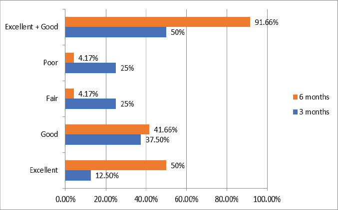
There was substantial improvement in functional status between 3 and 6 months. At 3 months, 24 patients (50.00%) had excellent or good functional outcomes. By 6 months, this increased to 44 patients (91.66%), with 24 patients (50.00%) achieving an excellent outcome and 20 (41.66%) achieving a good outcome. Only 2 patients each (4.17%) had fair or poor outcomes at 6 months. The mean HHS at 6 months was 88.75 (Fig. 1).
Radiological outcome and post-operative complications
The radiological outcome was assessed using the RUSH, which evaluates the radiographic progression of fracture healing. A RUSH score of ≥26, indicating good union, was observed in 34 patients (70.83%), whereas 14 (29.17%) had scores of <26, suggestive of delayed union. On detailed RUSH grading, 18 patients (37.50%) had excellent healing, 16 (33.33%) good, 8 (16.67%) fair, and 6 (12.50%) poor healing. Anatomical or acceptable fracture alignment was achieved in 83.34% of patients, whereas optimal or acceptable implant positioning was observed in 95.82% of patients. The most frequent post-operative complications were varus collapse and lateral migration of the proximal screws, each occurring in 25.00% of patients. Limb shortening, superficial or deep infection, Z-effect, and implant failure occurred in 4.17% each, whereas no cases of non-union or mortality were recorded (Table 3).
Radiological outcomes and post-operative complications following proximal femoral nailing (n=48)
| Parameter | Category | n (%) |
|---|---|---|
| RUSH score | ≥26 (good union) | 34 (70.83) |
| <26 (delayed union) | 14 (29.17) | |
| RUSH grading | Excellent (28–30) | 18 (37.50) |
| Good (24–27) | 16 (33.33) | |
| Fair (20–23) | 8 (16.67) | |
| Poor (<20) | 6 (12.50) | |
| Fracture alignment | Anatomical/acceptable | 40 (83.34) |
| Varus malalignment | 6 (12.50) | |
| Valgus malalignment | 2 (4.16) | |
| Implant position | Optimal | 38 (79.16) |
| Acceptable | 8 (16.66) | |
| Poor | 2 (4.16) | |
| Post-operative complications | Varus collapse | 12 (25.00) |
| Lateral migration of proximal screws | 12 (25.00) | |
| Limb shortening | 2 (4.17) | |
| Superficial infection | 2 (4.17) | |
| Deep infection | 2 (4.17) | |
| Z-effect | 2 (4.17) | |
| Implant failure | 2 (4.17) | |
| Non-union | 0 (0.00) | |
| Mortality | 0 (0.00) |
RUSH: Radiographic union score for hip
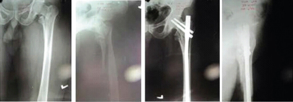
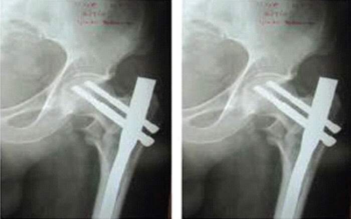
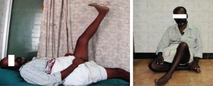
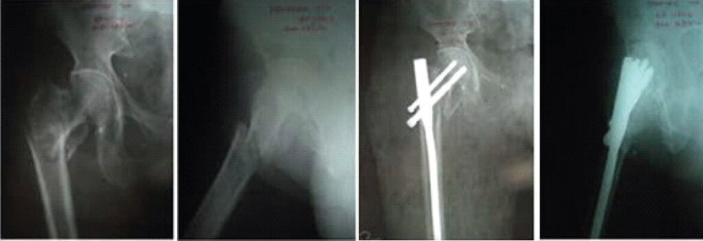
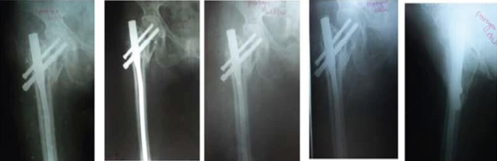
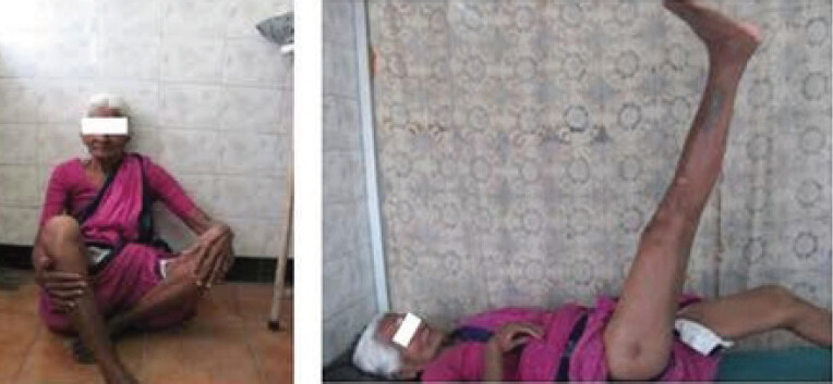
Additional functional complications during follow-up included abductor lurch in 18 patients (37.50%), which progressively decreased over time. Limb shortening was documented as 3 cm in one patient, 2 cm in two patients, 1 cm in five patients, and <1 cm in one patient; three patients were managed with a sole raise for residual limb-length discrepancy. All patients achieved partial weight bearing by approximately 2 weeks, and none required a walking aid beyond 3 months. For Case 1, pre-operative and immediate post-operative radiographs are shown in Fig. 2, follow-up radiographs in Fig. 3, and clinical photographs in Fig. 4. The corresponding images for Case 2 are shown in Fig. 5 (pre-operative and immediate post-operative radiographs), Fig. 6 (follow-up radiographs), and Fig. 7 (clinical photographs).
Discussion
IT fractures are influenced by patient-related factors, fracture stability, timing of treatment, and the quality of fixation. Both extramedullary and intramedullary implants are used in their management, with proximal femoral nailing offering biomechanical advantages related to its intramedullary position and shorter lever arm, particularly in unstable fracture patterns [7]. Radford et al. compared DHS fixation with gamma locking nail fixation in a prospective randomized study [8].
In the present study, the mean age was 56.37 years, with the highest proportion of patients belonging to the 61–70-year age group. The relatively younger age distribution observed in our series may reflect differences in population characteristics and mechanisms of injury compared with Western cohorts. Previous studies have reported a predominance of older patients with trochanteric fractures, particularly in series involving predominantly elderly populations [9, 10, 11, 12, 13]. Domingo et al. reported a mean age of 80 years, with three-fourths of their patients being female, whereas Banan et al. also reported a predominantly female study population [14, 15]. In contrast, the present study showed a male predominance (62.5%). This difference may be related to the relatively younger age distribution and the substantial contribution of road traffic accidents in the present series.
Accidental falls were the most common mechanism of injury (64.58%), followed by road traffic accidents (35.42%). This pattern is consistent with the general distinction between low-energy trauma in older individuals and higher-energy trauma in younger patients. The predominance of unstable fracture patterns in the present series is noteworthy, as fracture instability and loss of medial or lateral cortical support can make reduction more difficult and increase the risk of secondary displacement and mechanical failure [7, 16].
The mean interval between injury and surgery was 15.89 days, whereas the mean operative duration was 65.34 min. Previous studies evaluating PFN fixation have reported operative and perioperative outcomes within a broadly comparable range. Pajarinen et al. demonstrated that operation time was one of the principal perioperative variables differing between PFN and DHS fixation, whereas Pavelka et al. evaluated the complication profile of short PFN fixation in unstable proximal femoral fractures [1, 17]. The majority of fractures in the present study were unstable AO patterns, with A2.2, A2.3, and A3.3 accounting for most cases. Such unstable configurations are particularly relevant to PFN fixation because they place greater demands on reduction, implant positioning, and maintenance of mechanical alignment [7, 16].
Functional recovery was substantial during follow-up. The proportion of patients achieving excellent or good functional outcomes increased from 50.0% at 3 months to 91.66% at 6 months, with a mean HHS of 88.75 at 6 months. Boldin et al. reported favorable functional outcomes following PFN fixation in unstable proximal femoral fractures [18]. Pajarinen et al. demonstrated earlier restoration of pre-operative walking ability among patients treated with PFN compared with DHS fixation [1]. Prakash et al. similarly reported higher HHSs and better functional outcomes in patients treated with PFN compared with DHS [19]. The progressive improvement in HHS observed in the present study indicates satisfactory recovery of hip function following PFN fixation.
Radiological outcomes were also favorable. The RUSH was used to assess fracture healing. A RUSH score of ≥26 was observed in 70.83% of patients, whereas 29.17% had scores below 26. On detailed RUSH grading, 37.50% of patients had excellent healing, 33.33% had good healing, 16.67% had fair healing, and 12.50% had poor healing. The RUSH system was developed to improve the standardized radiographic assessment of hip fracture healing [6]. In the present study, anatomical or acceptable fracture alignment was achieved in 83.34% of patients, whereas optimal or acceptable implant positioning was observed in 95.82%. Appropriate reduction and implant placement are important for maintaining mechanical stability and minimizing complications such as varus collapse and proximal screw migration [7, 16].
Mechanical complications constituted the principal adverse outcomes in the present series. Varus collapse and lateral migration of the proximal screws were each observed in 12 of 48 patients (25.00%). This relatively high frequency is clinically important. Published literature identifies fracture instability, deficient cortical support, loss of reduction, varus alignment, and implant/screw positioning as potential contributors to mechanical failure after cephalomedullary fixation [7, 16]. However, the present dataset did not permit patient-level statistical correlation of reduction quality, AO subtype, implant configuration, or screw position with these complications. Consequently, specific causes of the observed varus collapse and screw migration cannot be established from this study. Their clinical consequences in this cohort included documented limb shortening in some patients and the need for continued radiological surveillance; no non-union or mortality was recorded.
Limb shortening was observed in a small proportion of patients, whereas abductor lurch was noted in 18 patients and gradually decreased during follow-up. Excessive fracture collapse and proximal screw migration can contribute to shortening and alteration of hip biomechanics [7, 20]. The findings emphasize the importance of maintaining appropriate fracture alignment and minimizing excessive post-operative collapse.
The Z-effect occurred in two patients in the present series. Differential migration of the proximal screws is a recognized mechanical complication of PFN fixation and may be influenced by fracture instability, varus alignment, medial comminution, and screw positioning [17,18]. Intraoperative technical difficulties, including fracture displacement during nail insertion, difficulty in inserting the derotation screw, and varus angulation, were also observed. These findings emphasize that PFN fixation requires careful reduction, accurate fluoroscopic positioning, and appropriate instrumentation. Khairnar and Patil similarly emphasized the importance of proper surgical technique and implant positioning in achieving satisfactory functional outcomes following PFN fixation [21]. Egol et al. examined mismatch between intramedullary nails and the anterior bow of the femur, highlighting an additional consideration in implant selection [22].
The complication profile of the present study was broadly comparable with previous reports. Simmermacher et al. described the PFN as an implant developed for unstable proximal femoral fractures, with technical complications remaining an important consideration [4]. Boldin et al. reported favorable outcomes with PFN in unstable proximal femoral fractures, whereas Domingo et al. reported technical intraoperative complications in 12%, immediate post-operative complications in 27%, and late complications in 4% of their 295 patients [14, 18]. Banan et al. also reported their experience with 60 patients treated with PFN for unstable extracapsular hip fractures [15]. These findings reinforce the importance of meticulous fracture reduction, appropriate implant positioning, and careful post-operative management.
No cases of non-union or mortality were observed in the present study. The absence of non-union and the favorable functional recovery are consistent with previous reports describing satisfactory union and functional outcomes following PFN fixation [16, 18, 23]. Furthermore, all patients achieved partial weight bearing by approximately 2 weeks, and none required a walking aid beyond 3 months. This progressive restoration of mobility is consistent with the earlier recovery of walking ability reported with PFN compared with DHS fixation [1].
Overall, the present cohort showed improvement in functional scores and satisfactory radiological healing after PFN during the available follow-up. More than 90% of patients were categorized as having excellent or good functional outcomes by 6 months, while varus collapse and proximal screw migration remained notable mechanical complications. These observations support careful reduction, implant and screw positioning, serial radiographic assessment, and structured rehabilitation. They do not establish superiority of PFN over DHS, other nails, or alternative fixation methods because no comparator group was included.
Limitations
This study has several limitations. The sample size was small (n = 48), the study was conducted at a single center, and the observational design did not include randomization or a control/comparator group. Follow-up focused mainly on outcomes up to 6 months, limiting assessment of long-term function and late mechanical complications. Several AO fracture subtypes and three PFN configurations were represented, but the available dataset did not support patient-level subgroup comparisons by fracture pattern or implant configuration. Similarly, the relationship between reduction/alignment quality and mechanical complications could not be statistically tested from the available data. Functional assessment relied on the Modified HHS without complete domain-wise reporting, and quality-of-life, separate pain-specific, and patient-satisfaction instruments were not collected. Radiological assessment did not include quantitative collapse, neck-shaft angle, or tip-apex distance measurements. The rehabilitation pathway was described generally rather than as a fully prespecified standardized protocol. Finally, exclusion of patients with multiple fractures, head injuries, pathological fractures, reoperations, and inadequate follow-up may have introduced selection bias and limits generalizability to broader trauma populations. These limitations should be considered when interpreting the findings, and larger multicenter comparative studies with longer follow-up and standardized functional, patient-reported, radiological, and rehabilitation measures are warranted.
Conclusion
In this cohort of 48 adults with IT fractures, PFN was associated with improvement in functional status and satisfactory radiological outcomes during 6 months of follow-up; 91.66% of patients had excellent or good Modified HHS outcomes at 6 months. Varus collapse and lateral migration of proximal screws, each occurring in 25.00%, remained important mechanical complications. Because this was a small, single-center observational study without a comparator group, no conclusion can be drawn regarding superiority of PFN over DHS, other nails, or alternative fixation methods. Larger comparative studies with longer follow-up and standardized radiological and patient-reported outcome measures are needed.
Clinical Message
In this observational cohort, PFN was associated with satisfactory short-term functional recovery and radiological healing. The observed rates of varus collapse and proximal screw migration emphasize the need for meticulous reduction, appropriate implant and screw positioning, serial assessment of alignment and healing, and clearly structured rehabilitation. These findings should be interpreted as descriptive outcomes after PFN rather than comparative evidence of superiority.
Conflict of Interest:
Nil
Source of Support:
Nil
Consent:
The authors confirm that informed consent was obtained from the patient for publication of this article
How to Cite this Article
Akhil M, Singh A, Gera Y. A Prospective Study of Functional Outcome of Intertrochanteric Fracture of Proximal Femur in Adults Treated with Proximal Femoral Nail. Journal of Orthopaedic Case Reports 2026 October;16(10): 625-634.
References
- Pajarinen J, Lindahl J, Michelsson O, Savolainen V, Hirvensalo E. Pertrochanteric femoral fractures treated with a dynamic hip screw or a proximal femoral nail. A randomised study comparing post-operative rehabilitation. J Bone Joint Surg Br 2005;87:76-81. [Google Scholar] | [PubMed]
- Karn NK, Jain A, Nepal P, Singh MP, Das N. A prospective randomized control trial comparing proximal femoral nail and sliding hip screw in the management of trochanteric fracture of the femur. Health Renaiss 2011;9:7-11. [Google Scholar] | [PubMed]
- Falch JA, Ilebekk A, Slungaard U. Epidemiology of hip fractures in Norway. Acta Orthop Scand 1985;56:12-6. [Google Scholar] | [PubMed]
- Simmermacher RK, Bosch AM, Van Der Werken C. The AO/ASIF-proximal femoral nail (PFN): A new device for the treatment of unstable proximal femoral fractures. Injury 1999;30:327-32. [Google Scholar] | [PubMed]
- Bucholz RW, Heckman JD, Koval KJ, Zuckerman JD. Rockwood and Green's Fractures in Adults. 6th ed. Philadelphia, PA: Lippincott Williams and Wilkins; 2006. [Google Scholar] | [PubMed]
- Bhandari M, Chiavaras MM, Parasu N, Choudur H, Ayeni O, Chakravertty R. Radiographic union score for hip substantially improves agreement between surgeons and radiologists. BMC Musculoskelet Disord 2013;14:70. [Google Scholar] | [PubMed]
- Schipper IB, Bresina S, Wahl D, Linke B, Van Vugt AB, Schneider E. Biomechanical evaluation of the proximal femoral nail. Clin Orthop Relat Res 2002;405:277-86. [Google Scholar] | [PubMed]
- Radford PJ, Needoff M, Webb JK. A prospective randomised comparison of the dynamic hip screw and the gamma locking nail. J Bone Joint Surg Br 1993;75:789-93. [Google Scholar] | [PubMed]
- Mohanty SP, Chacko V. A comparative analysis of operative and non-operative management of trochanteric fractures: A study of 135 consecutive cases. Indian J Orthop 1984;18:19-24. [Google Scholar] | [PubMed]
- Kyle RF. Fractures and dislocationsGustilo RB, Kyle RF, Templeman D. Mosby's Fracture and Dislocation Reference.. 2nd ed. St Louis: Mosby; 2023. 783-854. [Google Scholar] | [PubMed]
- Kulkarni GS. Treatment of trochanteric fractures of the hip by modified Richard's compressing and collapsing screw. Indian J Orthop 1984;18:30-4. [Google Scholar] | [PubMed]
- Boyd HB, Griffin LL. Classification and treatment of trochanteric fractures. Arch Surg (1920) 1949;58:853-66. [Google Scholar] | [PubMed]
- Fogagnolo F, Kfuri M Jr, Paccola CA. Intramedullary fixation of pertrochanteric hip fractures with the short AO-ASIF proximal femoral nail. Arch Orthop Trauma Surg 2004;124:31-7. [Google Scholar] | [PubMed]
- Domingo LJ, Cecilia D, Herrera A, Resines C. Trochanteric fractures treated with a proximal femoral nail. Int Orthop 2001;25:298-301. [Google Scholar] | [PubMed]
- Banan H, Al-Sabti A, Jimulia T, Hart AJ. The treatment of unstable, extracapsular hip fractures with the AO/ASIF proximal femoral nail (PFN)--our first 60 cases. Injury 2002;33:401-5. [Google Scholar] | [PubMed]
- Schipper IB, Steyerberg EW, Castelein RM, van der Heijden FH, den Hoed PT, Kerver AJ. Treatment of unstable trochanteric fractures. Randomised comparison of the gamma nail and the proximal femoral nail. J Bone Joint Surg Br 2004;86:86-94. [Google Scholar] | [PubMed]
- Pavelka T, Matejka J, Cervenková H. Complications of internal fixation by a short proximal femoral nail. Acta Chir Orthop Traumatol Cech 2005;72:344-54. [Google Scholar] | [PubMed]
- Boldin C, Seibert FJ, Fankhauser F, Peicha G, Grechenig W, Szyszkowitz R. The proximal femoral nail (PFN)--a minimal invasive treatment of unstable proximal femoral fractures: A prospective study of 55 patients with a follow-up of 15 months. Acta Orthop Scand 2003;74:53-8. [Google Scholar] | [PubMed]
- Prakash AK, Nagakumar JS, Shanthappa AH, Venkataraman S, Kamath A. A comparative study of functional outcome following dynamic hip screw and proximal femoral nailing for intertrochanteric fractures of the femur. Cureus 2022;14:e23803. [Google Scholar] | [PubMed]
- Pajarinen J, Lindahl J, Savolainen V, Michelsson O, Hirvensalo E. Femoral shaft medialisation and neck-shaft angle in unstable pertrochanteric femoral fractures. Int Orthop 2004;28:347-53. [Google Scholar] | [PubMed]
- Khairnar A, Patil L. A study of functional outcome in intertrochanteric femur fractures treated by proximal femoral nailing. Int J Med Res Rev 2018;6:335-40. [Google Scholar] | [PubMed]
- Egol KA, Chang EY, Cvitkovic J, Kummer FJ, Koval KJ. Mismatch of current intramedullary nails with the anterior bow of the femur. J Orthop Trauma 2004;18:410-5. [Google Scholar] | [PubMed]
- Thakur P, Khanal KR, Amatya I. Functional outcome of proximal femoral nailing in intertrochanteric fracture. J Nepal Health Res Counc 2022;19:805-8. [Google Scholar] | [PubMed]
© 2026 Journal of Orthopaedic Case Reports - Published by Indian Orthopaedic Research Group
About the Authors
-

M Akhil
-

Amrit Singh
-

Yakshit Gera
Related Articles in Journal of Orthopaedic Case Reports
 May 1, 2025 Fixation in Intertrochanteric Fractures Using Short Proximal Femoral Nail Anti-Rotation-2: A Functional and Radiological Prospective Study
 February 1, 2026 Functional and Radiological Outcomes of Proximal Femoral Nailing in Unstable Peritrochanteric Fractures: A Prospective Observational Study
 March 1, 2025 Evaluation of Functional and Radiological Outcome of Different Cephalomedullary Nail Designs in Intertrochanteric Fractures of Femur
 October 1, 2026 Posterior Hip Dislocation with Ipsilateral Subtrochanteric Femur Fracture: A Rare Case Report