


Introduction
Distal triceps tendon rupture is rare, accounting for <1% of all tendon ruptures [1,2]. It may result from direct trauma to the posterior aspect of the elbow or, more commonly, from sudden eccentric triceps contraction after a fall onto an outstretched hand [1]. Because this mechanism is also associated with other traumatic lesions, distal triceps rupture may coexist with fractures or ligamentous injuries around the elbow [2,3].
Combined distal triceps tendon rupture and radial head fracture is particularly uncommon [3,4,5]. The diagnosis may be missed initially because pain, swelling, and limitation of motion can mask the underlying tendon injury [3,5,6]. Clinical suspicion should be raised in the presence of inability to actively extend the elbow, a palpable gap at the triceps insertion, and the Fleck sign on plain radiographs [3, 7].
We present a rare case of distal triceps tendon rupture associated with a comminuted Mason type III radial head fracture, treated with anatomic triceps footprint repair and radial head arthroplasty in a single surgical session.
Case Report
A 60-year-old woman presented to the emergency department with diffuse pain and swelling of the left elbow after a fall onto an outstretched upper limb. She reported feeling a distinct “click” at the time of injury.
On examination, the elbow was swollen, with medial-sided effusion. The patient was unable to actively extend the elbow. Passive pronation and supination were painful. Tenderness over the radial head and a palpable gap at the triceps insertion were noted.
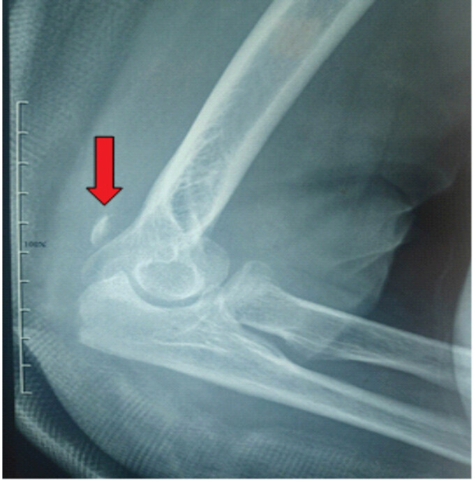
Plain radiographs demonstrated an intra-articular radial head fracture together with the characteristic Fleck sign, indicating distal triceps avulsion (Fig. 1) [3,7]. The elbow was initially immobilized in a cast at 90° of flexion with the forearm in a neutral position, and the patient was admitted for operative treatment.
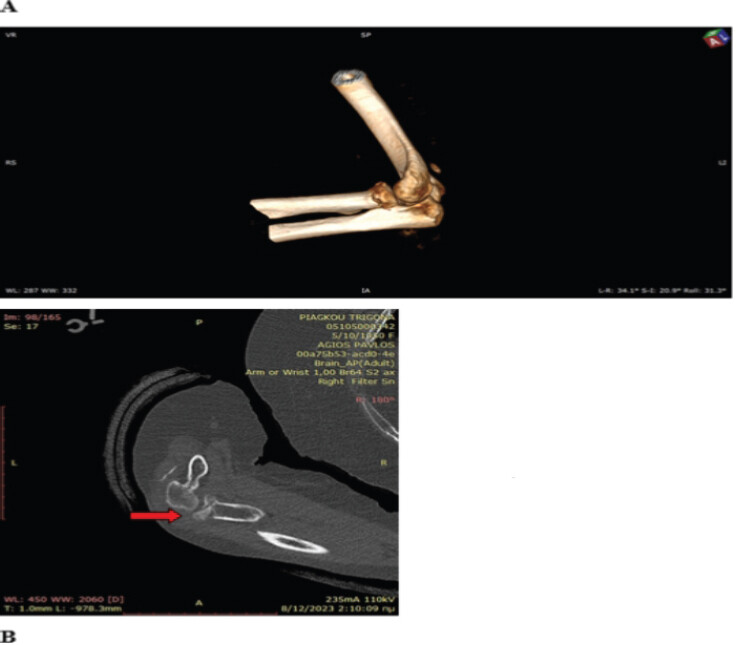
Computed tomography confirmed a comminuted intra-articular radial head fracture with displacement >2 mm, consistent with a Mason type III injury (Fig. 2) [8,9]. Taking into account the fracture pattern, the associated distal triceps rupture, and the patient’s age and functional demands, surgical treatment was planned. The operative strategy consisted of triceps tendon repair using the knotless anatomic footprint technique described by Paci et al. [10], combined with press-fit radial head arthroplasty.
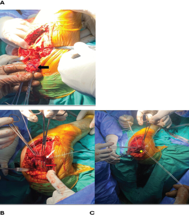
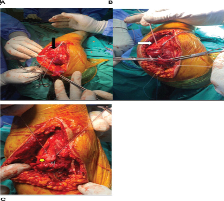
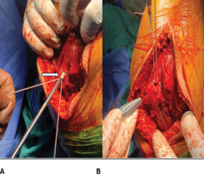
For the procedure, the patient was placed supine under general anesthesia with a tourniquet applied to the left arm. Through a standard posterior approach, the retracted distal triceps tendon was identified, mobilized, and debrided. The olecranon footprint was marked, and two Krackow sutures were placed, one medially and one laterally, with their limbs exiting anteriorly. Two FiberLoop sutures were also placed with posterior loop exit points (Fig. 3). Two parallel 2-mm transosseous tunnels were then drilled from the proximal footprint to the dorsal surface of the ulna. A pilot hole for anchor placement was created between the tunnels. The suture limbs were passed through the tunnels, and a “box-and-X” footprint configuration was created (Fig. 4). The free suture limbs were then secured with an anchor placed flush with the cortex under final tension (Fig. 5).
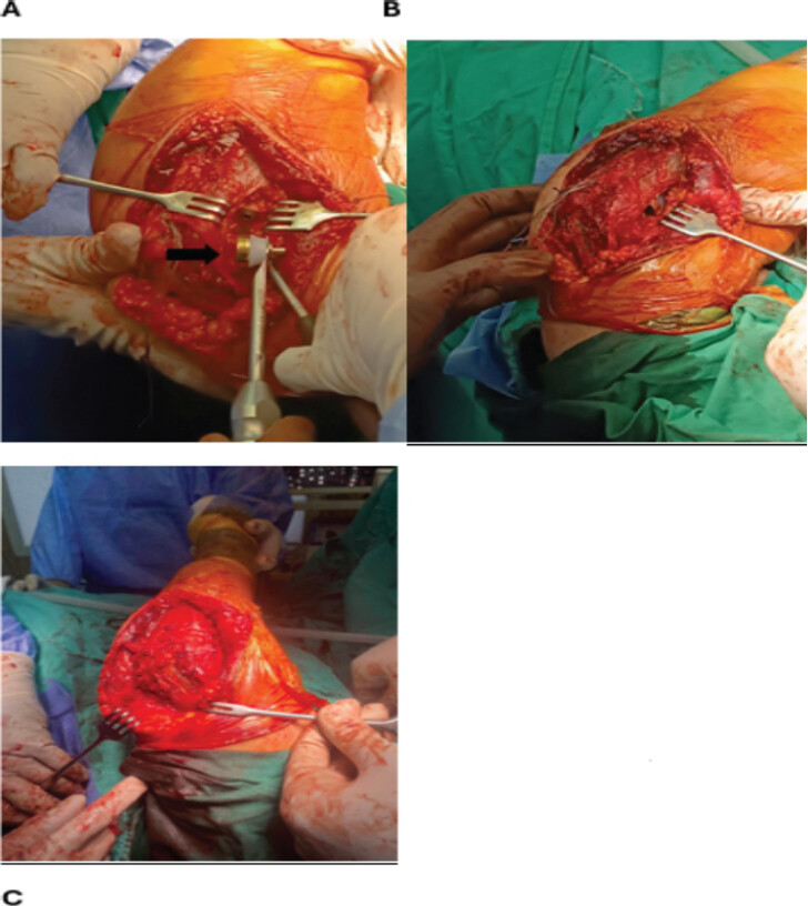
Attention was then directed to the radial head. Through an anterolateral elbow approach, the radial head and neck were exposed, osteotomized, and removed. The radial canal was prepared and a press-fit prosthesis was implanted under fluoroscopic guidance. Implant size and height were assessed dynamically to avoid overstuffing of the radiocapitellar joint (Fig. 6) [8, 9].
Post-operatively, the elbow was immobilized in a plaster cast at 90° of flexion with the forearm in neutral rotation for 2 weeks. At 2 weeks, sutures were removed and a functional splint was applied, initially allowing motion from 90° of flexion to 30° of extension. Range of motion was progressively increased by 20° every 2 weeks, aiming for full motion by 6–8 weeks. Full active mobilization was allowed after 8 weeks. At follow-up, the patient had a stable elbow with satisfactory functional recovery.
Discussion
Combined distal triceps tendon rupture and radial head fracture is an unusual injury pattern that requires a high index of suspicion [3,4,5,6]. Failure to diagnose the tendon injury may compromise elbow stability, extension strength, and overall functional outcome.
The Fleck sign is a useful radiographic clue for triceps avulsion and should prompt careful clinical assessment of the extensor mechanism [3,7]. In our patient, the inability to actively extend the elbow, the palpable defect at the triceps insertion, and the displaced comminuted radial head fracture led to recognition of the combined injury.
Anatomic footprint repair of the distal triceps has demonstrated encouraging biomechanical properties and satisfactory clinical results [7,10,11]. Compared with conventional transosseous repair, the knotless anatomic footprint technique may improve restoration of the native insertional anatomy and distribute load more effectively across the repair site [7,11]. However, it remains technically demanding and requires appropriate post-operative protection.
Radial head arthroplasty is a well-established treatment option for comminuted Mason type III fractures not amenable to stable fixation, especially in older patients or in the presence of associated instability [8, 9]. Proper implant sizing is crucial, as overstuffing of the radiocapitellar joint may lead to pain, stiffness, and altered biomechanics [8, 9]. The most commonly reported post-operative complications include joint instability, aseptic loosening, and osteolysis [8,9].
This case emphasizes the importance of treating all associated lesions during the same surgical setting. Combined management of the distal triceps rupture and radial head fracture allowed restoration of stability and facilitated rehabilitation.
Conclusion
Distal triceps tendon rupture associated with comminuted radial head fracture is a rare and potentially underdiagnosed complex elbow injury. Thorough physical examination and careful radiographic assessment are essential for timely diagnosis. Single-stage treatment with anatomic triceps footprint repair and radial head arthroplasty may provide stable reconstruction and satisfactory functional recovery.
Clinical Message
In patients with complex elbow trauma, the triceps tendon must be carefully assessed both clinically and radiographically. Recognition and treatment of all associated injuries in a single procedure are key to restoring a stable and functional elbow.
Conflict of Interest:
Nil
Source of Support:
Nil
Consent
The authors confirm that informed consent was obtained from the patient for publication of this article
How to Cite this Article
Georgiadis N, Manidakis N, Topaloglou A, Asteriadis A, Kalyvas E. Management of a Rare Complex Elbow Injury: Anatomic Reconstruction of the Triceps Footprint Combined with Radial Head Arthroplasty – A Case Report. J Orthop Case Rep. 2026 Nvovember;16(11)L93-103.doi: 10.13107/jocr.2026.v16.i11-8402.
References
- Dunn JC, Kusnezov N, Fares A, Rubin S, Orr J, Friedman D. Triceps tendon ruptures: A systematic review. Hand (N Y) 2017;12:431-8. [Google Scholar] | [PubMed]
- Alkhalfan YH, Jha G, Verma B, Coatwala A, Mishra A, Khan TM. The elbow's achilles heel: A systematic review and meta-analysis of triceps tendon rupture and repair techniques. Cureus 2023;15:e41584. [Google Scholar] | [PubMed]
- Canbora K, Ozyurek S, Gumussuyu G, Kose O. Triceps tendon avulsion and associated injuries of the elbow. BMJ Case Rep 2013;2013:bcr2013009460. [Google Scholar] | [PubMed]
- Levy M, Fishel RE, Stern GM. Triceps tendon avulsion with or without fracture of the radial head--a rare injury?. J Trauma 1978;18:677-9. [Google Scholar] | [PubMed]
- Lee JH, Ahn KB, Kwon KR, Kim KC, Rhyou IH. Differences in rupture patterns and associated lesions related to traumatic distal triceps tendon rupture between outstretched hand and direct injuries. Clin Orthop Relat Res 2021;479:781-9. [Google Scholar] | [PubMed]
- Kose O, Kilicaslan OF, Guler F, Acar B, Yuksel HY. Functional outcomes and complications after surgical repair of triceps tendon rupture. Eur J Orthop Surg Traumatol 2015;25:1131-9. [Google Scholar] | [PubMed]
- Yeh PC, Stephens KT, Solovyova O, Obopilwe E, Smart LR, Mazzocca AD. The distal triceps tendon footprint and a biomechanical analysis of 3 repair techniques. Am J Sports Med 2010;38:1025-33. [Google Scholar] | [PubMed]
- Jordan RW, Jones AD. Radial head fractures. Open Orthop J 2017;11:1405-16. [Google Scholar] | [PubMed]
- Al-Tawil K, Arya A. Radial head fractures. J Clin Orthop Trauma 2021;20:101497. [Google Scholar] | [PubMed]
- Paci JM, Clark J, Rizzi A. Distal triceps knotless anatomic footprint repair: A new technique. Arthrosc Tech 2014;3:e621-6. [Google Scholar] | [PubMed]
- Clark J, Obopilwe E, Rizzi A, Komatsu DE, Singh H, Mazzocca AD. Distal triceps knotless anatomic footprint repair is superior to transosseous cruciate repair: A biomechanical comparison. Arthroscopy 2014;30:1254-60. [Google Scholar] | [PubMed]
© 2026 Journal of Orthopaedic Case Reports - Published by Indian Orthopaedic Research Group
About the Authors
-

Nikolaos Georgiadis
-

Nikolaos Manidakis
-

Alexandros Topaloglou
-

Konstantinos Asteriadis
-

Efstathios Kalyvas
Related Articles in Journal of Orthopaedic Case Reports
 August 1, 2026 The “Terrible Tetrad” of the Elbow: A Case Series of Two Patients with a Rare Quadruple-Component Injury and a Proposed New Classification Term
 September 1, 2025 Capitellum Fracture with Distal Triceps Tendon Avulsion Injury: A Rare Presentation with Literature Review
 December 1, 2024 Displaced Jeffery Type 2 Radial Neck Fracture Managed by a Modified Métaizeau Technique: A Case Report
 December 10, 2021 Clinical and Surgical Insights on Bilateral Total Knee Arthroplasty in Ochronotic Arthropathy: A Case-based Review