


Introduction
Triplane fractures are transitional injuries of the distal tibia that occur during asymmetric physeal closure in skeletally immature adolescents [1,2]. The mechanism of injury involves supination with external rotation and axial compression, producing fracture lines that extend through the epiphysis, physis, and metaphysis in three planes [1,3]. Triplane fractures account for approximately 5–10% of pediatric intra-articular ankle fractures [2].
Because plain radiographs do not consistently show all fracture fragments and given the intra-articular nature of the fracture, computed tomography (CT) is routinely recommended for accurate fragment assessment and articular displacement [1,4,5]. The accepted threshold for surgical intervention is generally >2 mm of articular displacement, as residual incongruity has been associated with worse long-term functional outcomes and increased risk of post-traumatic arthritis [6,7].
Although most patients achieve favorable outcomes following appropriate treatment, persistent pain after fracture healing should prompt evaluation for occult intra-articular pathology, instability, impingement, or less commonly, peripheral nerve injury [8,9,10].
Superficial peroneal nerve (SPN) injury is an uncommon but recognized complication of ankle trauma [10,11]. Due to its variable anatomy and superficial course across the anterolateral ankle, the SPN and its branches are susceptible to traction, scar entrapment, and iatrogenic injury [10,12,13]. To our knowledge, symptomatic hardware-associated SPN entrapment following syndesmotic stabilization after a triplane fracture has not previously been reported. We present the case of an adolescent athlete who developed persistent neuropathic pain secondary to SPN entrapment following surgical treatment of post-traumatic ankle instability after a triplane fracture.
Case Report
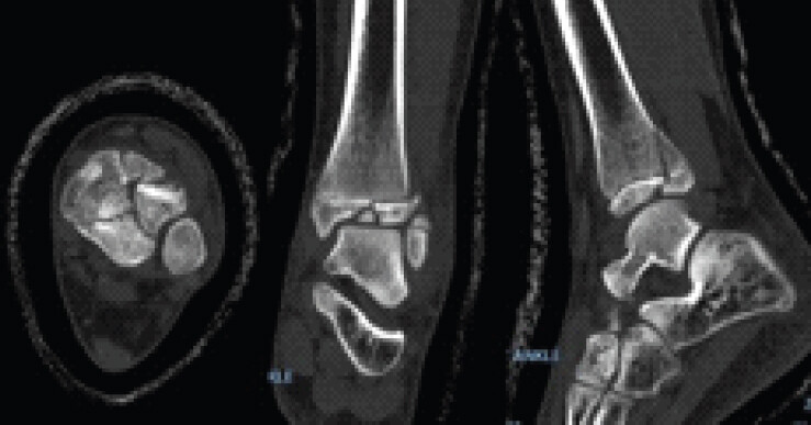
A 13-year-old female soccer player sustained a left triplane ankle fracture after falling from a golf cart. Initial radiographs and CT (Fig. 1) were obtained in the Emergency Department and were interpreted at an outside institution as demonstrating a non-displaced fracture. She was treated non-operatively with cast immobilization followed by progressive rehabilitation and return-to-sport protocols.
Approximately 8 months after injury, she developed persistent anterior and anterolateral ankle pain upon returning to full athletic activity. Symptoms failed to improve despite physical therapy, activity modification, and sport-specific rehabilitation. Radiographs demonstrated a healed fracture with maintained alignment; however, continued pain prompted referral to our institution.
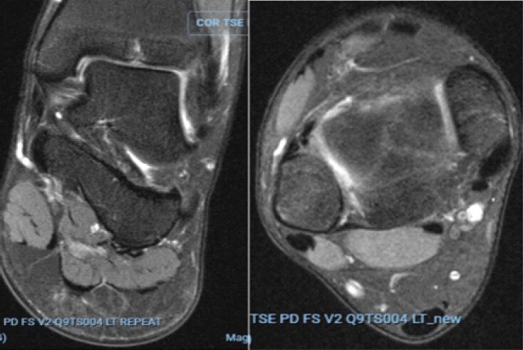
Physical examination demonstrated focal tenderness over the anterolateral ankle without instability. Review of the original CT scan revealed a comminuted intra-articular triplane fracture with approximately 5 mm of articular displacement. Magnetic resonance imaging (MRI) demonstrated residual post-traumatic changes within the distal tibial epiphysis, focal chondral irregularity, and signal abnormalities involving the anterior inferior tibiofibular ligament and anterior talofibular ligament (Fig. 2).
To further evaluate the source of her symptoms, the patient underwent a diagnostic and therapeutic intra-articular injection consisting of 40 mg triamcinolone acetonide (Kenalog) and lidocaine. The injection provided temporary relief, suggesting a component of post-traumatic synovitis; however, the patient continued to report ankle instability and apprehension during soccer participation. After failure of prolonged non-operative management, she elected to undergo left ankle arthroscopy, syndesmotic debridement and stabilization, and lateral ligament reconstruction.
Diagnostic arthroscopy demonstrated a prominent anterolateral distal tibial exostosis without significant chondral injury. Open inspection revealed impingement of the syndesmotic ligaments against the distal tibial exostosis, as well as a torn Bassett’s ligament, which was debrided. The distal tibia exostosis was excised, after which persistent syndesmotic instability was confirmed under direct visualization. Syndesmotic stabilization was performed using a lateral fibular plate with two suture-button fixation devices (TightRope®, Arthrex) placed under fluoroscopic guidance. The constructs were sequentially tensioned with the ankle held in dorsiflexion, resulting in anatomic reduction and restoration of syndesmotic stability on direct visualization and external rotation stress testing. Concomitant lateral ankle instability was addressed with a Broström-Gould lateral ligament reconstruction using knotless all-suture anchors and suture tape augmentation.
Following surgery, the patient’s pre-operative anterior ankle pain completely resolved. However, she subsequently developed new dorsal foot pain associated with paresthesias, allodynia, and intermittent electric shock-like pain that worsened with athletic activity. Examination demonstrated altered sensation in the distribution of the medial dorsal cutaneous branch of the SPN without recurrent ankle instability.
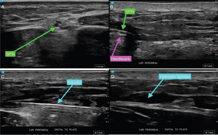
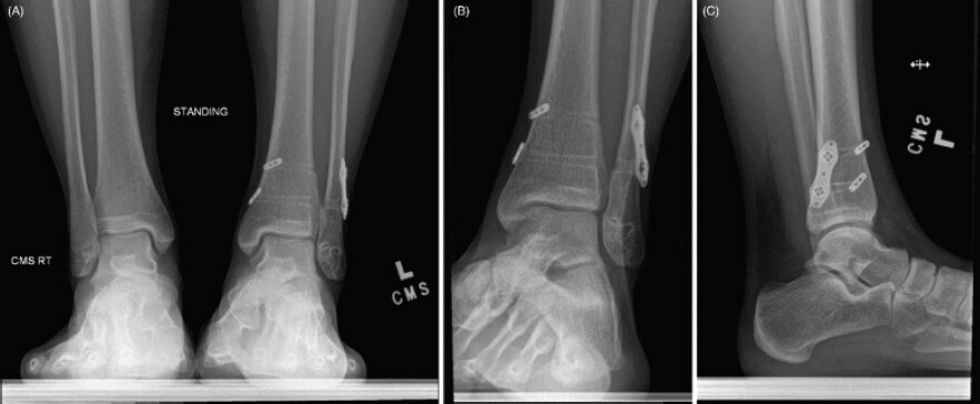
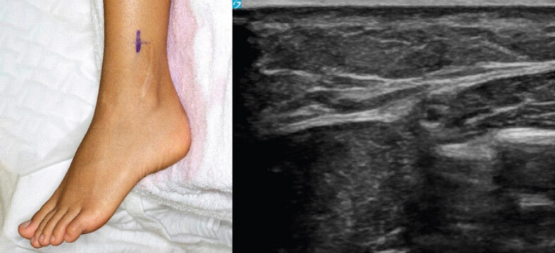
Given concern for peripheral nerve entrapment, ultrasound evaluation was performed. Visualization of the SPN was significantly limited by acoustic shadowing from the syndesmotic hardware, preventing reliable assessment of the nerve’s relationship to the implant (Fig. 3). The nerve could not be continuously traced. Despite these limitations, ultrasound-guided diagnostic injection and hydrodissection were performed. Real-time visualization demonstrated distal spread of the injectate along the course of the SPN, supporting nerve continuity. The injection resulted in complete but transient resolution of symptoms. Although hydrodissection temporarily alleviated the patient’s neuropathic pain, symptoms recurred, suggesting persistent mechanical compression or irritation of the SPN. Pre-operative radiographs obtained before surgery demonstrated maintained syndesmotic reduction with the retained lateral fibular plate and dual suture-button fixation construct (Fig. 4). These findings supported the diagnosis of hardware-associated SPN entrapment and prompted surgical exploration.
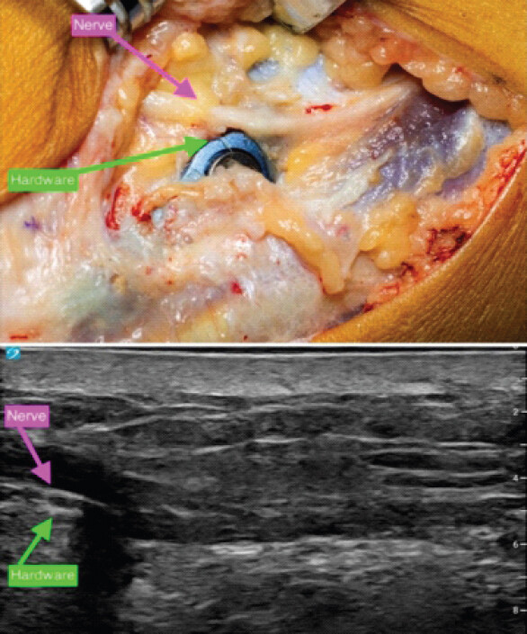
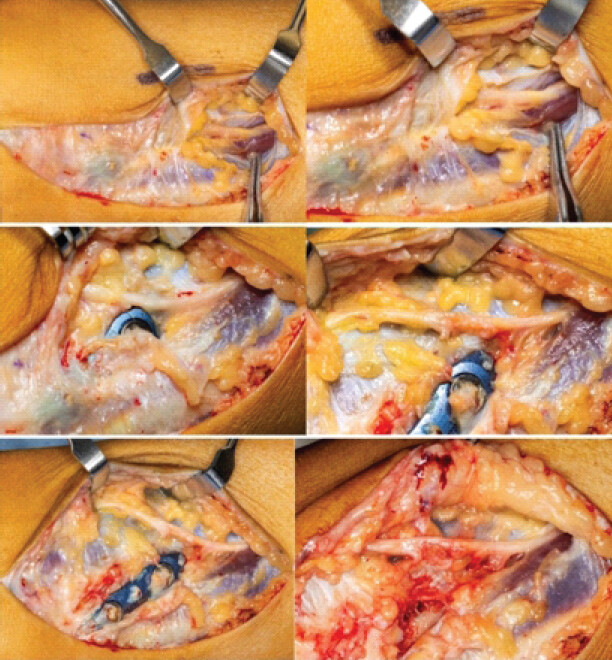
Three years after the index procedure, the patient’s neuropathic symptoms persisted despite non-operative management. Pre-operatively, ultrasound was again utilized to localize the course of the SPN relative to the syndesmotic construct (Fig. 5). Across the patient’s previous lateral incision, the SPN was identified proximally and carefully dissected distally to the level of the syndesmotic construct. The nerve was found immediately adjacent to the implant, consistent with ultrasound (Fig. 6), and was decompressed throughout its course. The nerve appeared structurally intact, and the suture-button fixation construct was removed (Fig. 7).
At the most recent follow-up, approximately 3 weeks after SPN exploration and hardware removal, the patient’s neuropathic pain had completely resolved. She had resumed competitive soccer with only 1 day of post-operative downtime. Although these early clinical outcomes are encouraging, longer-term follow-up is necessary to confirm maintenance of syndesmotic stability and evaluate for the development of post-traumatic degenerative changes.
Discussion
Triplane fractures generally demonstrate favorable outcomes when anatomic reduction is achieved and residual articular displacement is minimized [1,6,14]. Nevertheless, persistent pain following apparent fracture healing should prompt careful evaluation for occult pathology. In the present case, residual articular incongruity, anterolateral impingement, syndesmotic pathology, and lateral ankle instability contributed to ongoing symptoms and ultimately required operative intervention. Although these mechanical symptoms were successfully addressed, the patient subsequently developed dorsal foot paresthesias, allodynia, and electric shock-like pain that worsened with athletic activity.
Unlike her initial mechanical symptoms, the patient’s post-operative complaints were highly suggestive of neuropathic pain. The SPN, particularly its medial dorsal cutaneous branch, is vulnerable to injury because of its superficial course and substantial anatomic variability. Previous studies have demonstrated considerable variation in the location and branching pattern of the SPN, increasing its susceptibility to traction injury, scar entrapment, and iatrogenic injury during ankle surgery [11,13,15,16]. Although SPN injury has been described following ankle trauma, arthroscopy, Broström procedures, and syndesmotic fixation, symptomatic hardware-associated entrapment remains uncommon and may be difficult to recognize [11,17,18,19]. To the author’s knowledge, symptomatic SPN entrapment caused by syndesmotic stabilization following treatment of a triplane fracture has not previously been reported. Although the SPN was identified and protected during the index procedure, its variable course and the close proximity of the suture-button construct to the nerve likely contributed to delayed post-operative entrapment. This case illustrates that meticulous intraoperative nerve protection does not completely eliminate the risk of subsequent delayed post-operative hardware-associated nerve irritation.
In the present case, SPN exploration and removal of the suture-button syndesmotic fixation were performed 3 years after the index procedure. At the time of hardware removal, the syndesmosis had healed, and clinical and radiographic assessment demonstrated maintained reduction without evidence of recurrent instability. Therefore, removal of the suture-button construct to facilitate nerve decompression was not expected to compromise syndesmotic stability [20].
A notable feature of this case was the diagnostic complexity. The patient had multiple potential symptom generators throughout her course, including residual articular incongruity, post-traumatic synovitis, syndesmotic instability, and anterolateral impingement. Consequently, her post-operative neuropathic symptoms could have been attributed to persistent instability or incomplete rehabilitation. Instead, the development of dorsal foot paresthesias, allodynia, and exertional neuropathic pain raised suspicion for peripheral nerve pathology. Recognition of the change in symptom quality from mechanical ankle pain to neuropathic dorsal foot pain proved critical in establishing the diagnosis.
A particularly unique aspect of this case was the role of ultrasound in the diagnostic workup. Visualization of the SPN was significantly limited by acoustic shadowing from the metallic syndesmotic hardware, preventing continuous visualization of the nerve and obscuring its relationship to the implant. Consequently, the site of compression could not be directly identified on imaging. Despite these limitations, ultrasound remained clinically valuable by facilitating targeted diagnostic injection and hydrodissection. Complete temporary symptom resolution following injection strongly supported the diagnosis of SPN entrapment despite the inability to directly visualize the site of compression. This finding is particularly relevant in post-operative patients, where metallic hardware may limit the diagnostic utility of conventional imaging modalities. Although pre-operative ultrasound successfully localized the SPN before surgery in this case, this single case does not support routine ultrasound mapping before primary syndesmotic stabilization. Rather, ultrasound may be a useful adjunct in patients with suspected post-operative neuropathic symptoms to aid diagnosis and facilitate pre-operative planning for SPN exploration and hardware removal.
Ultrasound-guided nerve injections have demonstrated excellent outcomes in the evaluation and treatment of foot and ankle neuropathies [21]. Furthermore, ultrasound-guided hydrodissection has emerged as a safe and effective technique for separating entrapped nerves from surrounding scar tissue, with particular utility in surgical entrapment neuropathies [22]. In the present case, hydrodissection resulted in complete but temporary symptom relief, supporting a dynamic compressive neuropathy rather than intrinsic nerve injury. However, because the SPN remained immediately adjacent to the syndesmotic fixation construct, hydrodissection alone did not eliminate the underlying mechanical source of irritation. Symptom recurrence therefore prompted definitive surgical decompression and hardware removal.
This case underscores the diagnostic challenges of evaluating post-operative neuropathic pain after syndesmotic stabilization. Although metallic hardware limited direct visualization of the SPN on ultrasound, ultrasound-guided diagnostic injection provided critical functional information to support the diagnosis, which was confirmed intraoperatively and successfully treated with SPN decompression and hardware removal.
Conclusion
Hardware-associated SPN entrapment is an uncommon but important cause of post-operative neuropathic pain following syndesmotic stabilization. In patients who develop dorsal foot paresthesias, allodynia, or exertional neuropathic pain following syndesmotic fixation, SPN entrapment should be included in the differential diagnosis. Even when metallic artifact limits direct visualization, ultrasound-guided diagnostic injection can provide valuable functional information to support the diagnosis and guide definitive surgical management.
Clinical Message
Persistent neuropathic pain following syndesmotic stabilization should prompt consideration of SPN entrapment, even when routine imaging is inconclusive. Ultrasound-guided diagnostic injection can establish the diagnosis despite metallic artifact limiting nerve visualization and guide definitive surgical management.
Conflict of Interest:
Nil
Source of Support:
Nil
Consent:
The authors confirm that informed consent was obtained from the patient for publication of this article
How to Cite this Article
Meli GM, Henderson AP, Rakauskas T, Strohecker JB, Clendenen SR, Haupt ET. Hardware-Associated Superficial Peroneal Nerve Entrapment Following Syndesmotic Stabilization: A Case Report. J Orthop Case Rep. 2026 November;16(11):152-158. doi: 10.13107/jocr.2026.v16.i11.8422.
References
- Crawford AH.. Triplane and Tillaux fractures: Is a 2 mm residual gap acceptable?. J Pediatr Orthop 2012;32 Suppl 1:S69-73. [Google Scholar] | [PubMed]
- Schnetzler KA, Hoernschemeyer D.. The pediatric triplane ankle fracture. J Am Acad Orthop Surg 2007;15:738-47. [Google Scholar] | [PubMed]
- Hadad MJ, Sullivan BT, Sponseller PD.. Surgically relevant patterns in triplane fractures: A mapping study. J Bone Joint Surg Am 2018;100:1039-46. [Google Scholar] | [PubMed]
- Greenhill DA, Mundluru SN, Gomez RW, Romero J, Riccio AI.. Metaphyseal fracture displacement is predictive of intra-articular diastasis in adolescent triplane ankle fractures. J Pediatr Orthop 2024;44:94-8. [Google Scholar] | [PubMed]
- Eismann EA, Stephan ZA, Mehlman CT, Denning J, Mehlman T, Parikh SN. Pediatric triplane ankle fractures: Impact of radiographs and computed tomography on fracture classification and treatment planning. J Bone Joint Surg Am 2015;97:995-1002. [Google Scholar] | [PubMed]
- Lurie B, Van Rysselberghe N, Pennock AT, Upasani VV.. Functional outcomes of tillaux and triplane fractures with 2 to 5 millimeters of intra-articular gap. J Bone Joint Surg Am 2020;102:679-86. [Google Scholar] | [PubMed]
- Gaudiani MA, Knapik DM, Liu RW.. Clinical outcomes of triplane fractures based on imaging modality utilization and management: A systematic review and meta-analysis. J Pediatr Orthop 2020;40:e936-41. [Google Scholar] | [PubMed]
- Kay RM, Matthys GA.. Pediatric ankle fractures: Evaluation and treatment. J Am Acad Orthop Surg 2001;9:268-78. [Google Scholar] | [PubMed]
- Kim HN, Park YJ, Kim GL, Park YW.. Arthroscopy combined with hardware removal for chronic pain after ankle fracture. Knee Surg Sports Traumatol Arthrosc 2013;21:1427-33. [Google Scholar] | [PubMed]
- Falciglia F, Basiglini L, Aulisa AG, Toniolo RM.. Superficial peroneal nerve entrapment in ankle sprain in childhood and adolescence. Sci Rep 2021;11:15123. [Google Scholar] | [PubMed]
- Redfern DJ, Sauvé PS, Sakellariou A.. Investigation of incidence of superficial peroneal nerve injury following ankle fracture. Foot Ankle Int 2003;24:771-4. [Google Scholar] | [PubMed]
- Tomaszewski KA, Graves MJ, Vikse J, Pękala PA, Sanna B, Henry BM. Superficial fibular nerve variations of fascial piercing: A meta-analysis and clinical consideration. Clin Anat 2017;30:120-5. [Google Scholar] | [PubMed]
- O'Neill PJ, Parks BG, Walsh R, Simmons LM, Miller SD.. Excursion and strain of the superficial peroneal nerve during inversion ankle sprain. J Bone Joint Surg Am 2007;89:979-86. [Google Scholar] | [PubMed]
- Ertl JP, Barrack RL, Alexander AH, VanBuecken K.. Triplane fracture of the distal tibial epiphysis. Long-term follow-up. J Bone Joint Surg Am 1988;70:967-76. [Google Scholar] | [PubMed]
- Lee KL, Kim SB, Yoon K, Kim HJ.. Topographical anatomy of the superficial peroneal nerve: A cadaveric study on anatomical patterns and clinical implications. Clin Anat 2026;39:77-82. [Google Scholar] | [PubMed]
- Correia A, Vieira JG, Rodrigues BC, Fortes LH, Garção DC.. Branching patterns of the superficial fibular nerve: An anatomical study with meta-analysis. Surg Radiol Anat 2022;44:1419-25. [Google Scholar] | [PubMed]
- Malagelada F, Vega J, Guelfi M, Kerkhoffs G, Karlsson J, Dalmau-Pastor M.. Anatomic lectures on structures at risk prior to cadaveric courses reduce injury to the superficial peroneal nerve, the commonest complication in ankle arthroscopy. Knee Surg Sports Traumatol Arthrosc 2020;28:79-85. [Google Scholar] | [PubMed]
- Corte-Real NM, Moreira RM.. Arthroscopic repair of chronic lateral ankle instability. Foot Ankle Int 2009;30:213-7. [Google Scholar] | [PubMed]
- Goss DAJr, Reb CW, Philbin TM.. Anatomic structures at risk when utilizing an intramedullary nail for distal fibular fractures: A cadaveric study. Foot Ankle Int 2017;38:916-20. [Google Scholar] | [PubMed]
- Kim J, Kwon M, Day J, Aspang JS, Shim J, Cho J.. The impact of suture button removal in syndesmosis fixation. J Clin Med 2021;10:3726. [Google Scholar] | [PubMed]
- Walter WR, Burke CJ, Adler RS.. Ultrasound-guided therapeutic injections for neural pathology about the foot and ankle: A 4 year retrospective review. Skeletal Radiol 2017;46:795-803. [Google Scholar] | [PubMed]
- Colorado B, McNeill D, Norbury J.. Ultrasound-guided nerve hydrodissection for peripheral entrapment neuropathies. Muscle Nerve 2025;72:1052-9. [Google Scholar] | [PubMed]
© 2026 Journal of Orthopaedic Case Reports - Published by Indian Orthopaedic Research Group
About the Authors
-

Gabrielle M Meli
-

Adam P Henderson
-

Taylor Rakauskas
-

Jeffrey B Strohecker
-

Steven R Clendenen
-

Edward T Haupt
Related Articles in Journal of Orthopaedic Case Reports
 September 1, 2025 Chronic Saphenous Nerve Entrapment Neuropathy after Closed Blunt Trauma: A Case Report
 September 1, 2026 Extensive Post-Traumatic Heterotopic Ossification with Multi-joint Involvement: A Case Report
 December 1, 2025 Ulnar Nerve Cross-sectional Area among Ulnar Neuropathy Patients and Healthy Subjects – A Study in Hospital-based Population of Eastern India
 July 8, 2017 Retained Drain after Anterior Cruciate Ligament Surgery : A Silent Threat to an Athlete’s Career: A Case Report