


Introduction
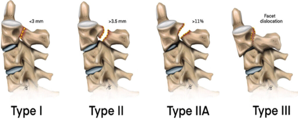
Traumatic spondylolisthesis of the axis (Hangman’s fracture) accounts for 4–7% of cervical spine fractures [1, 2]. The Levine–Edwards classification [3, 4] divides these fractures into four types based on displacement, angulation, and facet integrity (Fig. 1). Type IIa fractures, characterized by significant angulation (>11°) with minimal translation (<3 mm) from a flexion-distraction mechanism, are considered unstable and typically require surgical stabilization [3, 5].
Standard surgical options include anterior C2–C3 fusion (anterior cervical discectomy and fusion [ACDF]) and posterior C1–C3 fusion, both of which permanently sacrifice motion segments. The C1–C2 joint provides approximately 50% of cervical rotation, and C2–C3 contributes 5–10° of rotation and 8–10° of flexion-extension per side [6,7]. Fusion may also accelerate adjacent segment degeneration [8].
Direct isthmus screw fixation, first described by Judet et al. [9] in 1970, offers anatomic fracture reduction while preserving all upper cervical motion segments. Clinical studies have demonstrated favorable fracture union rates following direct pars/isthmus screw fixation, whereas preservation of the C2/3 motion segment represents a potential advantage over fusion-based techniques [10, 11]. Li et al. [12] systematically reviewed the management of Hangman’s fractures, providing broader context for treatment selection. Despite these advantages, the technique remains underutilized due to concerns regarding technical complexity and vertebral artery injury risk [13,14]. Costa et al. [15] described intraoperative three-dimensional navigation for upper cervical fracture fixation, emphasizing the importance of precise anatomical assessment and instrumentation.
This case report describes the successful application of bilateral dorsal isthmus screw fixation in a young patient with an unstable Type IIa Hangman’s fracture, with emphasis on surgical technique, clinical outcomes, and functional motion assessment at 13-month follow-up.
Case Report
A 26-year-old male presented to the emergency department following a 3-m fall from a construction platform. The Glasgow Coma Scale was 15/15 with stable hemodynamics. Neurological examination was intact (Medical Research Council grade 5/5 bilaterally) with focal cervical paraspinal tenderness.
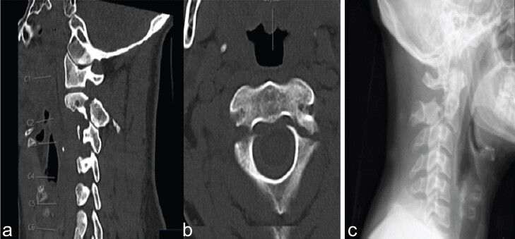
Contrast-enhanced computed tomography (CT) with angiography revealed a Type IIa Hangman’s fracture (Levine–Edwards classification) [3] with bilateral C2 isthmus fractures, 12.4° kyphotic angulation, and approximately 3 mm ventral translation (Fig. 2a, Fig. 2b, c). CT angiography confirmed normal bilateral vertebral artery anatomy. Axial imaging demonstrated bilateral pars fractures with adequate isthmus dimensions for screw placement (Fig. 2b).
Given the patient’s young age, high functional demands, and unstable fracture pattern, a motion-preserving approach was favored over conventional fusion [3,5]. Pre-operative planning confirmed anatomical suitability: Adequate isthmus dimensions for 3.5 mm screws, normal vertebral artery course, sufficient spinal canal diameter, and good bone quality on CT.
Surgical technique
Under general anesthesia with fiberoptic intubation and manual inline stabilization, Mayfield pins were applied, and the patient was positioned prone. Closed reduction was performed under lateral fluoroscopy with incremental axial traction (2.3–5.4 kg), achieving partial reduction of kyphotic angulation from 12.4° to approximately 5–7° (Fig. 3a) [16].
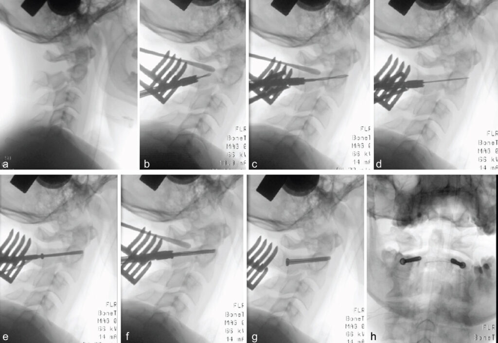
Through a midline posterior cervical incision (~6 cm), subperiosteal dissection exposed the C2 posterior elements bilaterally while preserving C2–C3 facet capsules. Under biplanar fluoroscopic guidance, bilateral entry points were identified at the inferior C2 lateral mass, approximately 3–4 mm lateral to the medial border [17]. Intermittent oscillating drilling (2.5 mm drill bit) was performed through the C2 isthmus bilaterally.
During drilling, reduction was augmented using a Penfield dura dissector to apply controlled anterior-inferior pressure to the C2 spinous process with countertraction on C3 posteriorly, progressively closing the residual fracture gap (Fig. 3b and c). This combined traction and Penfield-assisted technique achieved near-anatomic alignment with fracture gaps reduced to <1 mm and angulation corrected to approximately 3–5° (Fig. 3b and c).
Guidewires (2.0 mm) were placed bilaterally (Fig. 3d and e), followed by cannulated 3.5 mm cortical screws (Fig. 3f): right side 34 mm (11 mm thread), left side 36 mm (12 mm thread). Progressive tightening achieved interfragmentary compression with bicortical purchase. Following traction release, alignment remained stable, confirming adequate fixation (Fig. 3g and h). Operative time was 70 min; estimated blood loss was <50 mL with no intraoperative complications.
A rigid Aspen cervical collar was applied postoperatively for 6 weeks.
Post-operative course and follow-up
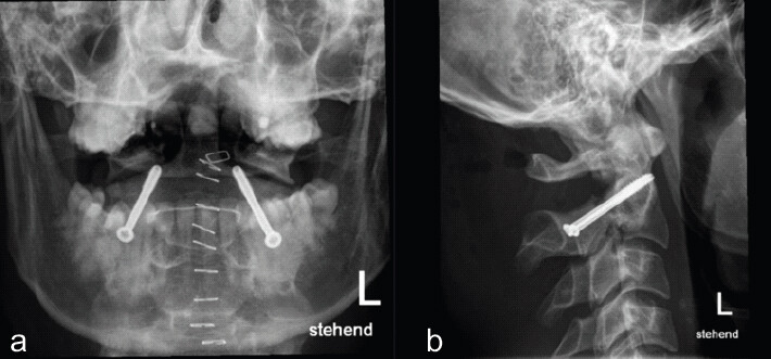
The patient was extubated without complications and discharged on post-operative day 6. Post-operative radiographs confirmed bilateral screw placement with kyphotic angulation corrected to <5° and fracture gap reduced to <1 mm (Fig. 4a and b).
Recovery was uneventful. The cervical orthosis was discontinued at 6 weeks, 50% work capacity was achieved at 3 months, and full work capacity (100%) with near-normal cervical rotation (70–75° bilaterally, >85% of normative values) was restored by 6 months. Clearance for contact sports, including ice hockey, was granted at 6 months (Fig. 5a and b).
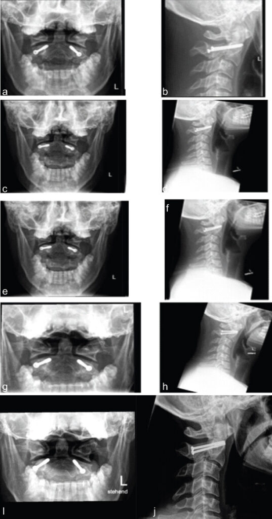
Sequential radiographs demonstrated progressive healing, with early callus formation at 6 weeks (Fig. 5c and d), bridging consolidation at 3 months (Fig. 5e and f), and solid bony union at 6 months (Fig. 5g and h). Mild physiologic settling (2–3° increased kyphosis) was observed at 3 months but stabilized within acceptable parameters. No hardware failure, screw loosening, or implant migration occurred.
At 1-year follow-up, the patient remained asymptomatic with full work capacity and unrestricted athletic activity. Physical examination demonstrated painless cervical range of motion (chin-sternum distance 1–13 cm, bilateral head rotation 90°). Radiographs confirmed stable hardware and maintained consolidation but showed progressive C2/3 degenerative changes including anterior osteophyte formation and disc space narrowing (Fig. 5i and j).
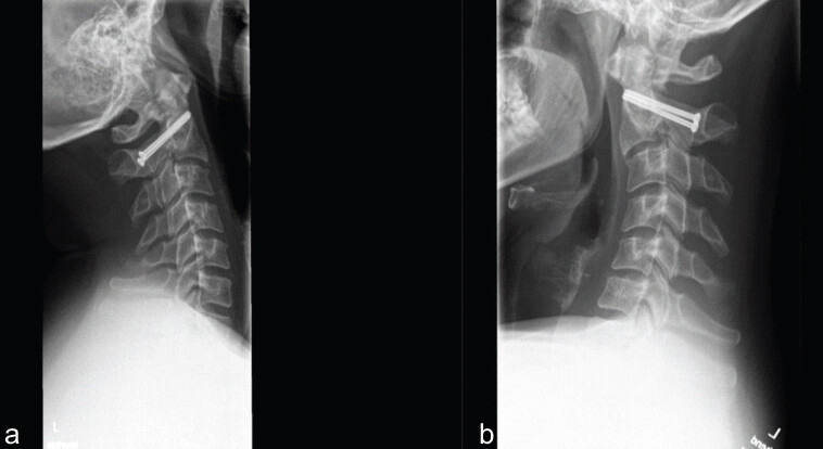
Functional flexion-extension radiographs at 13 months (Fig. 6) demonstrated preserved segmental mobility at C2/3 without pathological instability. A 2 mm anterolisthesis at C2/3 was noted but remained stable during dynamic imaging, confirming that the motion-preserving objective was achieved despite degenerative changes.
Discussion
We present a rare case of bilateral isthmus screw fixation for an unstable Type IIa Hangman’s fracture, with objective documentation of preserved C2/3 motion at 13-month follow-up. Our patient achieved solid osseous union at 6 months and excellent functional outcomes, consistent with the high fracture union rates reported in previous clinical series of direct pars/isthmus screw fixation [10,11]. No complications occurred, including no vertebral artery injury, hardware failure, or neurological deterioration.
Compared with the published literature, the present case is most consistent with posterior direct pars/isthmus fixation as originally described by Judet et al. [9] and subsequently reported by Verheggen and Jansen [18] and Bristol et al. [19]. Bristol et al. [19] described direct C2 pars screw fixation without intervertebral fusion, supporting the feasibility of motion-preserving stabilization in selected patients. ElMiligui et al. [10] reported transpedicular fixation, while Li et al. [12] systematically reviewed the management of Hangman’s fractures and Schleicher et al. [5] reviewed traumatic spondylolisthesis of the axis.
What distinguishes the present case is the addition of objective dynamic flexion–extension assessment at 13 months, documenting preserved C2/3 motion despite progressive C2/3 degenerative changes.
Recent reviews and clinical studies discuss direct isthmus fixation as a safe option for appropriately selected patients [11,20,21,22]. However, the evidence base consists primarily of small clinical series and observational studies, and objective range-of-motion data are rarely reported.
Successful application requires careful patient selection. Pre-operative CT should assess individual C2 isthmus dimensions and the feasibility of the planned screw trajectory [23], whereas spinal canal dimensions and vertebral artery anatomy require careful evaluation [13,24,25]. Ideal candidates are selected according to fracture stability, anatomical suitability, bone quality, and functional requirements [3,5].
Our hybrid reduction technique – combining Mayfield traction with intraoperative Penfield dissector–assisted open reduction – was essential for achieving near-anatomic alignment in this Type IIa fracture with significant angulation, where traction alone proved insufficient. In contrast to prolonged skeletal traction, reduction was performed intraoperatively under fluoroscopic guidance, with controlled traction supplemented by direct Penfield-assisted manipulation to correct residual kyphotic angulation. Compared to ACDF, direct isthmus fixation avoids intentional C2–C3 fusion but demands technical expertise and meticulous pre-operative planning [8, 21].
Importantly, although functional flexion-extension imaging at 13 months confirmed preserved segmental mobility at C2/3, progressive degenerative changes with osteophyte formation were observed radiographically. This post-traumatic disc degeneration – despite successful non-fusion fixation – highlights a potential limitation of this technique in Type IIa fractures where the initial flexion-distraction mechanism may cause irreversible disc and ligamentous injury. Whether the degree of initial displacement correlates with long-term degeneration warrants further study.
Key limitations include the single-case design. Multicenter registries with standardized outcome measures and long-term dynamic imaging are needed to validate motion preservation claims and establish whether this technique offers durable advantages over fusion.
Conclusion
Bilateral dorsal isthmus screw fixation achieved solid bony union, excellent functional outcomes, and confirmed motion preservation at 13 months in a 26-year-old patient with an unstable Type IIa Hangman’s fracture. Progressive C2/3 degenerative changes despite preserved segmental mobility warrant further investigation, particularly regarding long-term implications in fractures with significant initial angulation.
Clinical Message
In young, active patients with unstable Type IIa Hangman’s fractures and favorable anatomy, bilateral dorsal isthmus screw fixation should be considered a motion-preserving alternative to fusion, though patients should be counseled about possible progressive post-traumatic degenerative changes.
Conflict of Interest:
Nil
Source of Support:
Nil
Consent
The authors confirm that informed consent was obtained from the patient for publication of this article
How to Cite this Article
Lupi VL, Tinner C, Deml MC, Häckel S. Motion-Preserving Bilateral Isthmus Screw Fixation for Unstable Hangman’s Fracture: A Case Report. J Orthop Case Rep. 2026 November;16(11):201-207. doi: 10.13107/jocr.2026.v16.i11.8440.
References
- Greene KA, Dickman CA, Marciano FF, Drabier JB, Hadley MN, Sonntag VK. Acute axis fractures. Analysis of management and outcome in 340 consecutive cases. Spine (Phila Pa 1976) 1997;22:1843-52. [Google Scholar] | [PubMed]
- National Library of Medicine. Hangman's Fractures. StatPearls. Treasure Island, FL: StatPearls Publishing; 2024; Available from: https://www.ncbi.nlm.nih.gov/books/nbk448084. [Last accessed on 2025 Oct 25]. [Google Scholar] | [PubMed]
- Levine AM, Edwards CC. The management of traumatic spondylolisthesis of the axis. J Bone Joint Surg Am 1985;67:217-26. [Google Scholar] | [PubMed]
- McDermott M, Quinteros G, Landriel F, Stastny C, Raskin D, Ricciardi G. Management of C2 fractures following multiple classifications, a narrative review. Brain Spine 2024;4:102928. [Google Scholar] | [PubMed]
- Schleicher P, Scholz M, Pingel A, Kandziora F. Traumatic spondylolisthesis of the axis vertebra in adults. Global Spine J 2015;5:346-58. [Google Scholar] | [PubMed]
- White AA, Panjabi MM. Clinical Biomechanics of the Spine 2nd ed Philadelphia, PA: Lippincott Williams and Wilkins;1990. [Google Scholar] | [PubMed]
- Werne S. Studies in spontaneous atlas dislocation. Acta Orthop Scand Suppl 1957;23:1-150. [Google Scholar] | [PubMed]
- Hilibrand AS, Robbins M. Adjacent segment degeneration and adjacent segment disease: The consequences of spinal fusion?. Spine J 2004;4 Suppl 6:190S-4. [Google Scholar] | [PubMed]
- Judet R, Roy-Camille R, Saillant G. Fractures du rachis cervical: Ostéosynthèse par vissage direct des pédicules de C2. Rev Chir Orthop Reparatrice Appar Mot 1970;56:133-5. [Google Scholar] | [PubMed]
- ElMiligui Y, Koptan W, Emran I. Transpedicular screw fixation for type II Hangman's fracture: A motion preserving procedure. Eur Spine J 2010;19:1299-305. [Google Scholar] | [PubMed]
- Liu Y, Zhu Y, Li X, Chen J, Yang S, Yang H. A new transpedicular lag screw fixation for treatment of unstable Hangman's fracture: A minimum 2-year follow-up study. J Orthop Surg Res 2020;15:372. [Google Scholar] | [PubMed]
- Li XF, Dai LY, Lu H, Chen XD. A systematic review of the management of Hangman's fractures. European Spine Journal. 2006;15:257-269. [Google Scholar] | [PubMed]
- Duan S, Lv S, Ye F, Lin Q. Imaging anatomy and variation of vertebral artery and bone structure at craniocervical junction. Eur Spine J 2009;18:1102-8. [Google Scholar] | [PubMed]
- Neo M, Fujibayashi S, Miyata M, Takemoto M, Nakamura T. Vertebral artery injury during cervical spine surgery: A survey of more than 5600 operations. Spine (Phila Pa 1976) 2008;33:779-85. [Google Scholar] | [PubMed]
- Costa F, Ortolina A, Attuati L, Cardia A, Tomei M, Riva M. Management of C1–2 traumatic fractures using an intraoperative 3D imaging-based navigation system. J Neurosurg Spine 2015;22:128-33. [Google Scholar] | [PubMed]
- Vieweg U, Grochulla F. Reduction techniques in cervical spine trauma. Unfallchirurg 2000;103:852-60. [Google Scholar] | [PubMed]
- Ludwig SC, Kramer DL, Vaccaro AR, Albert TJ. Transpedicle screw fixation of the cervical spine. Clin Orthop Relat Res 1999;359:77-88. [Google Scholar] | [PubMed]
- Verheggen R, Jansen J. Hangman's fracture: Arguments in favor of surgical therapy for type II and III according to Edwards and Levine. Surg Neurol 1998;49:253-61. [Google Scholar] | [PubMed]
- Bristol R, Henn JS, Dickman CA. Pars screw fixation of a hangman's fracture: technical case report. Neurosurgery 2005;56Suppl 1:E204. [Google Scholar] | [PubMed]
- Mahmoud A, Shanmuganathan K, Montgomery A. Surgical management of hangman's fracture: A systematic review. Int J Spine Surg 2023;17:454-67. [Google Scholar] | [PubMed]
- Lee S, Hur JW, Oh Y, An S, Yun GY, Ahn JM. Current concepts in the treatment of traumatic C2 vertebral fracture: A literature review. J Korean Neurosurg Soc 2024;67:6-13. [Google Scholar] | [PubMed]
- Certo F, Altieri R, Garozzo M, Visocchi M, Barbagallo GM. Direct transpedicular C2 fixation for the surgical management of Hangman's fractures: A “second youth” for the judet approach. Acta Neurochir Suppl 2023;135:291-9. [Google Scholar] | [PubMed]
- Mandel IM, Kambach BJ, Petersilge CA, Johnstone B, Yoo JU. Morphologic considerations of C2 isthmus dimensions for the placement of transarticular screws. Spine (Phila Pa 1976) 2000;25:1542-7. [Google Scholar] | [PubMed]
- Pavlov H, Torg JS, Robie B, Jahre C. Cervical spinal stenosis: Determination with vertebral body ratio method. Radiology 1987;164:771-5. [Google Scholar] | [PubMed]
- Torg JS, Pavlov H, Genuario SE, Sennett B, Wisneski RJ, Robie BH. Neurapraxia of the cervical spinal cord with transient quadriplegia. J Bone Joint Surg Am 1986;68:1354-70. [Google Scholar] | [PubMed]
© 2026 Journal of Orthopaedic Case Reports - Published by Indian Orthopaedic Research Group
About the Authors
-

Valérie Laura Lupi
-

Christian Tinner
-

Moritz C Deml
-

Sonja Häckel
Related Articles in Journal of Orthopaedic Case Reports
 September 1, 2026 Pediatric Cervical Spine Aneurysmal Bone Cyst with Recurrent Atlantoaxial Rotatory Subluxation Requiring C1–C2 Fusion: A 7-Year Follow-Up Case Report
 September 1, 2026 Three-Grade Neurological Recovery (ASIA Impairment Scale A to D) Following Surgical Stabilization of Traumatic D11–D12 Meyerding Grade IV Spondylolisthesis: A Case Report
 September 1, 2026 Combined Ultrasound- and Fluoroscopy-guided Percutaneous Cementoplasty and Ablation for a C2 Pathologic Fracture: A Case Report
 August 1, 2026 O-arm Navigation-assisted Excision of Cervical Osteoid Osteoma Adjacent to the Vertebral Artery: Technical Note and Case Report