


Introduction
The acromioclavicular (AC) joint, a diarthrodial joint, accounts for 40–50% of shoulder injuries [1,2]. Dislocations of the AC joint occur more frequently in males than females (5:1 ratio) and are more often incomplete than complete (2:1 ratio) [3]. Patients with AC joint injuries often experience changes in scapular orientation, leading to rotator cuff injuries and chronic pain [4]. Biomechanical studies underscore the importance of anatomically reconstructing the coracoclavicular (CC) ligaments in cases of unstable AC joint injuries [5].
The treatment strategy for chronic AC joint instability (ACJI) differs from that of acute cases. In the acute phase, the AC and CC ligaments have the potential to heal, thus the treatment focuses on aligning the torn ligament ends during tissue healing. However, this healing potential is lost after 3 weeks [6]. Various techniques have been documented for treating AC joint dislocation, including primary joint fixation, CC ligament reconstruction, clavicle excision, dynamic muscle transfers, coracoacromial ligament transfer, and various fixation methods. Combinations of these techniques are now prevalent [7].
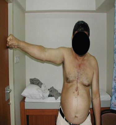
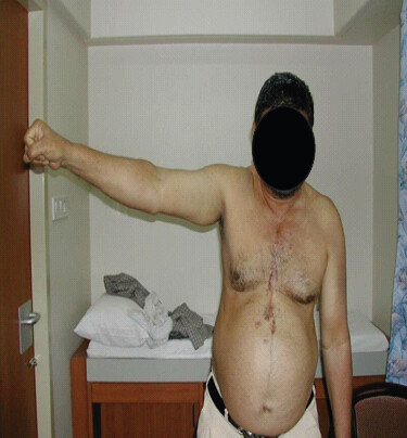
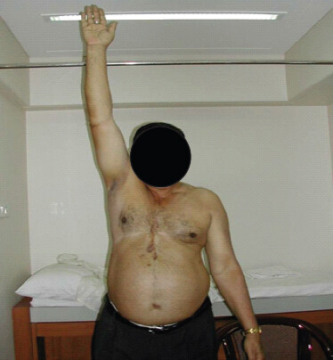
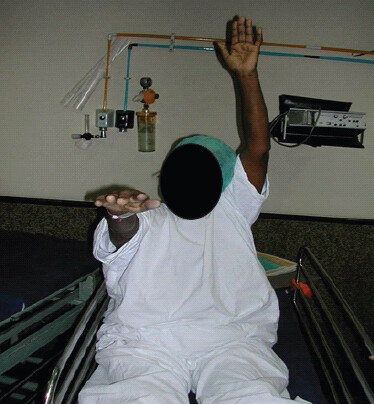
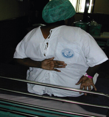
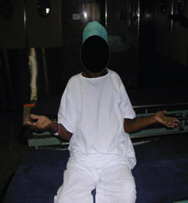
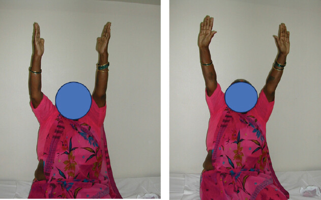
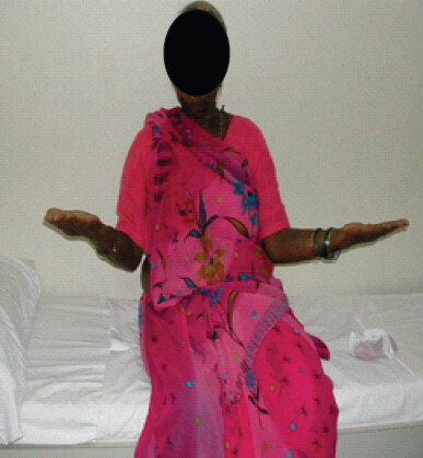
This case series presents a technique for AC joint reconstruction in chronic cases of Rockwood types 3, 4, and 5, utilizing a synthetic tape alongside lateral clavicle excision. This method is demonstrated to be rapid, economical, effective, and associated with low morbidity and favorable clinical outcomes (Fig.1–9).
Materials and Methods
In this retrospective study, we evaluated 11 patients with delayed presentation (6 weeks or more post-injury) who underwent surgery. Patients were excluded if they had associated fractures, brain injuries, cognitive impairments, or abnormal shoulder function before injury.
Technique
Our technique utilizes a simple 30 cm × 5 mm Mersilene tape (Polyethylene Terephthalate) (Ethicon- Johnson and Johnson) to replicate the function of the CC ligaments, without the need for a fixation device, hardware, or graft.
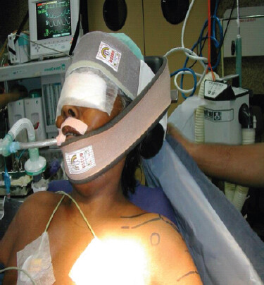
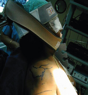
The patient, under general anesthesia, is placed in the beach-chair position, with a sandbag placed under the ipsilateral scapula. Two incisions are used in this technique. The first is a 3–4 cm horizontal incision over the lateral-most end of the clavicle. The second is a 3 cm vertical incision placed over the coracoid process. Across the first incision, the lateral clavicle end is exposed, and the lateral 7–10 mm of the clavicle is excised using an oscillating saw. Any deltoid-trapezial fascia/muscle fibers found interposed within the ACJ must be cleared, so that the clavicle can be reduced back to the level of the acromion. A 3.2 mm drill bit is used to drill a hole in the clavicle about 10 mm from the excised edge.
Across the second incision, the dissection is taken down to the base of the coracoid process. Two retractors are placed on either side of the coracoid, with care, avoiding overzealous retraction. Bluntly dissect down to the coracoid base.
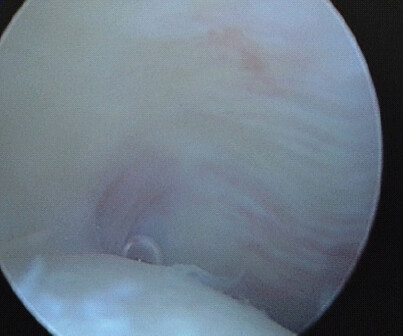
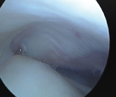
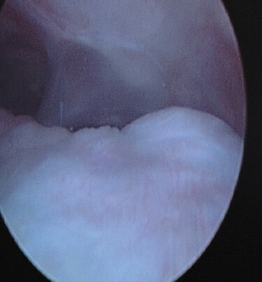
Next, using Mixter forceps, the Mersilene tape is passed under the coracoid near its base. The intervening tissue between the two incisions is bluntly dissected from the 1st incision above toward the 2nd incision below. This facilitates shuttling the two limbs of the Mersilene tape from the coracoid base to the clavicle. The medial limb of the tape is brought out posterior to the clavicle. This medial limb is then passed through the 3.2 mm clavicle hole, from superior to inferior, to exit below the clavicle. The assistant then reduces the clavicle down to the acromion, and while holding this reduction, the surgeon knots the two ends of the Mersilene tape under the coracoid base through the 2nd incision. At least 4 knots on the Mersilene tape are preferred. The assistant gently releases the force applied to the reduced clavicle, and the reduction is then maintained by the tape (Fig . 10-15).
Post-operative care
Patients’ upper limbs are supported in a sling for 3–4 weeks, with no weight lifting allowed. All patients were discharged on the 2nd post-operative day after dressing changes. Sutures were removed at 2 weeks, and gentle pendular exercises commenced based on pain tolerance. Shoulder-strengthening exercises were progressively intensified after a month.
Results
The procedure was performed on 11 patients with neglected AC joint dislocations of Rockwood grades 3 to 5, with a mean age of 46.6 years. The right shoulder was involved in 8 of 12 cases, with a male-to-female ratio of 3:1. Injuries were caused by falls in 10 cases and road traffic accidents in 2 cases. Pre- and post-operative assessments of pain, daily activities, cosmesis, and range of motion showed satisfactory functional outcomes without paracervical or trapezius pain.
One patient experienced a 20% vertical displacement of the clavicle’s lateral end, but had no clinical complaints. The mean post-operative ACJI score improved from 63.7 pre-operatively to 81.4 post-operatively at 2 years. The visual analog scale score improved from 6.41 pre-operatively to 2.68 at 6 months and 1.25 at 12 months. The Constant Murley score improved from 51 pre-operatively to 92.08 at 12 months. At final follow-up, all patients had satisfactory outcomes in terms of pain, cosmesis, and shoulder function. The AC joint was stable both clinically and radiologically in all cases (Table 1 & 2).
Paired Samples Statistics
| Mean | N | Std. Deviation | Std. Error Mean | ||
|---|---|---|---|---|---|
| Pair 1 | FLEXION RANGE OF MOTION-PRE OP | 80.75 | 20 | 13.599 | 3.041 |
| FLEXION RANGE OF MOTION POST | 158.75 | 20 | 9.301 | 2.08 | |
| Pair 2 | ABDUCTION_PRE | 58.5 | 20 | 9.881 | 2.209 |
| ABDUCTION POST | 141 | 20 | 12.732 | 2.847 | |
| Pair 3 | EXTRNAL_ROTATION_PRE | 17 | 20 | 5.938 | 1.328 |
| EXTERNAL_ROTATION POST | 58.5 | 20 | 9.881 | 2.209 | |
| Pair 4 | INTERNAL_ROTATION_PRE | 30.5 | 20 | 7.763 | 1.736 |
| POST OP INTERNAL | 64.25 | 20 | 6.742 | 1.508 | |
| Pair 5 | OSS PRE | 9.35 | 20 | 1.496 | 0.335 |
| OSS POST | 34.85 | 20 | 5.214 | 1.166 |
Paired Samples Test
| Paired Differences | t | Df | Significance | |||||||
|---|---|---|---|---|---|---|---|---|---|---|
| Mean | SD | SE | 95% Confidence Interval of the Difference | OneSided p | Two-Sided p | |||||
| Lower | Upper | |||||||||
| Pair 1 | FLEXION RANGE OF MOTION-PRE OP – FLEXION RANGE OF MOTION | -78 | 12.711 | 2.842 | -83.949 | -72.051 | -27.442 | 19 | < .001 | < .001 |
| Pair 2 | ABDUCTION_1 – ABDUCTION | -82.5 | 13.328 | 2.98 | -88.738 | -76.262 | -27.683 | 19 | < .001 | < .001 |
| Pair 3 | EXTRNAL_ROTATION_1 – EXTERNAL ROTATION | -41.5 | 7.797 | 1.743 | -45.149 | -37.851 | -23.804 | 19 | < .001 | < .001 |
| Pair 4 | INTERNAL_ROTATION_1 – POST OP | -33.75 | 7.759 | 1.735 | -37.381 | -30.119 | -19.454 | 19 | < .001 | < .001 |
| Pair 5 | RANGE OF MOTION PRE OP – POST OP | -25.5 | 5.643 | 1.262 | -28.141 | -22.859 | -20.209 | 19 | < .001 | < .001 |
Discussion
Despite various surgical techniques for treating these injuries, achieving long-term functional outcomes has been challenging [8]. Techniques include fixing the AC joint with K-wires or tension bands or reconstructing the CC ligaments with various materials. The clavicle hook plate is a simple and beneficial method, but complications, such as hardware prominence and fixation loss are significant [9]. Arirachakaran et al. found that loop suspensory fixation of the CC joint yields better functional scores than hook plates for AC joint dislocations [10].
Most techniques assume the CC ligaments will heal to their original length and strength, but if they do not, these methods may fail. Reconstruction can avoid this issue by using local tissue sources or tendon grafts, either autografts or allografts, to align the clavicle and coracoid. Initial reconstruction techniques by Weaver and Dunn involved ligament transfer [11], but this method had higher risks of dislocation compared to anatomical ligaments. Dewar and Barrington [12] described coracoid bone transposition, which carries high risks of bone fracture and nerve injury.
Autologous graft use for CC ligament reconstruction loses up to 30–40% of its original strength [13], and early graft weakness during necrosis and revascularization can lead to failure [14]. In addition, the graft remains weak post-fixation, increasing the risk of stress fractures [15].
Studies have shown that CC ligaments provide superior stability, while AC ligaments provide anterior-posterior stability [8,16,17]. Beitzel et al. concluded that both ligaments should be anatomically repaired for optimal function, but our case did not include AC ligament repair and resulted in no functional outcome changes.
Anatomical AC joint reduction and CC ligament reconstruction are essential for achieving good clinical and imaging results, regardless of the material used. Mersilene tape avoids implant-related complications and does not require removal, offering favorable functional outcomes and pain-free shoulder movement. This method is cost-effective compared to imported alternatives, making it more accessible and providing good objective and subjective outcomes with minimal complications.
Conclusion
This technique offers a cost-effective, rapid, robust, and reproducible solution with durable and predictable outcomes for treating chronic AC joint dislocations.
Clinical Message
Chronic and neglected AC joint dislocations (Rockwood grades 3–5) can be reliably managed using a simple, low-cost Mersilene tape reconstruction of the CC ligaments combined with lateral clavicle excision, avoiding the need for grafts or fixation hardware. This technique offers durable joint stability, significant pain relief, and improved shoulder function, making it a reproducible and accessible option, particularly in resource-limited settings.
Conflict of Interest:
Nil
Source of Support:
Nil
Consent
The authors confirm that informed consent was obtained from the patient for publication of this article
How to Cite this Article
Rathod V, Munshi P. An Innovative Approach to Chronic and Neglected Acromioclavicular Joint Dislocation: A Cost-Effective and Reliable Solution. J Orthop Case Rep. 2026 November;16(11):568-573. doi: 10.13107/jocr.2026.v16.i11.8554.
References
- Kaplan LD, Flanigan DC, Norwig J, Jost P, Bradley J. Prevalence and variance of shoulder injuries in elite collegiate football players. Am J Sports Med 2005;33:1142-6. [Google Scholar] | [PubMed]
- Thorndike AJ. Injuries to the acromioclavicular joint: A plea for conservative treatment. Am J Surg 1942;55:250-61. [Google Scholar] | [PubMed]
- Rockwood CJ, Williams G, Young D. Disorders of the acromioclavicular joint Rockwood CJ, Matsen FA3rd. The Shoulder.. 2nd ed. Philadelphia, PA. WB Saunders 1998:483-553. [Google Scholar] | [PubMed]
- Lee SJ, Nicholas SJ, Akizuki KH, McHugh MP, Kremenic IJ, Ben-Avi S. Reconstruction of the coracoclavicular ligaments with tendon grafts: A comparative biomechanical study. Am J Sports Med 2003;31:648-54. [Google Scholar] | [PubMed]
- Rios CG, Arciero RA, Mazzocca AD. Anatomy of the clavicle and coracoid process for reconstruction of the coracoclavicular ligaments. Am J Sports Med 2007;35:811-7. [Google Scholar] | [PubMed]
- Weinstein DM, McCann PD, McIlveen SJ, Flatow EL, Bigliani LU. Surgical treatment of complete acromioclavicular dislocations. Am J Sports Med 1995;23:324-31. [Google Scholar] | [PubMed]
- Dumontier C, Sautet A, Man M, Apoil A. Acromioclavicular dislocations: Treatment by coracoacromial ligamentoplasty. Shoulder Elbow Surg 1995;4:130-4. [Google Scholar] | [PubMed]
- Lee KW, Debski RE, Chen CH, Woo SL, Fu FH. Functional evaluation of the ligaments at the acromioclavicular joint during anteroposterior and superoinferior translation. Am J Sports Surg 1997;25:858-62. [Google Scholar] | [PubMed]
- Cadenat FM. The treatment of dislocations and fractures of the outer end of the clavicle. Int Clin 1917;1:145-69. [Google Scholar] | [PubMed]
- Arirachakaran A, Boonard M, Piyapittayanun P, Phiphobmongkol V, Chaijenkij K, Kongtharvonskul J. Comparison of surgical outcomes betweenfixation with hook plate and loop suspensory fixation for acute unstable acromioclavicular joint dislocation: A systematic review and meta-analysis. Eur J Orthop Surg Traumatol 2016;26:565-74. [Google Scholar] | [PubMed]
- Weaver JK, Dunn HK. Treatment of acromioclavicular injuries, especially complete acromioclavicular separation. J Bone Joint Surg Am 1972;54:1187-94. [Google Scholar] | [PubMed]
- Dewar FP, Barrington TW. The treatment of chronic acromio-clavicular dislocation. J Bone Joint Surg Br 1965;47:32-5. [Google Scholar] | [PubMed]
- Jackson DW, Grood ES, Goldstein JD, Rosen MA, Kurzweil PR, Cummings JF. A comparison of patellar tendon autograft and allograft used for anterior cruciate ligament reconstruction in the goat model. Am J Sports Med 1993;21:176-85. [Google Scholar] | [PubMed]
- Choi NH, Lim SM, Lee SY, Lim TK. Loss of reduction and complications of coracoclavicular ligament reconstruction with autogenous tendon graft in acute acromioclavicular dislocations. J Shoulder Elb Surg 2017;26:692-8. [Google Scholar] | [PubMed]
- Turman KA, Miller CD, Miller MD. Clavicular fractures following coracoclavicular ligament reconstruction with tendon graft: A report of three cases. J Bone Joint Surg Am 2010;92:1526-32. [Google Scholar] | [PubMed]
- Fukuda K, Craig EV, An KN, Cofield RH, Chao EY. Biomechanical study of the ligamentous system of the acromioclavicular joint. J Bone Joint Surg Am 1986;68:434-40. [Google Scholar] | [PubMed]
- Urist MR. Complete dislocations of the acromioclavicular joint: The nature of the traumatic lesion and effective methods of treatment with an analysis of forty-one cases. J Bone Joint Surg 1946;28:813-37. [Google Scholar] | [PubMed]
© 2026 Journal of Orthopaedic Case Reports - Published by Indian Orthopaedic Research Group
About the Authors
-

Vishal Rathod
-

Parag Munshi
Related Articles in Journal of Orthopaedic Case Reports
 November 1, 2026 Functional Outcome of Double Endobutton Technique in Acute Acromioclavicular Joint Dislocation: A Prospective Observational Study
 August 1, 2026 Outcome Analysis of Loop Endobutton Repair for Acromioclavicular Joint Dislocation
 July 1, 2025 Late Presentation of Acromioclavicular Joint Dislocation with Double Fracture of Coracoid Process Managed by Arthroscopic Stabilization Technique: A Case Report
 November 1, 2024 Pediatric AC Joint Fracture Dislocation: Arthroscopy-assisted Reduction and Internal Bracing