
Pain, deformity, or stiffness following mega-prosthesis reconstruction in the presence of normal inflammatory markers may indicate metallosis arising from various mechanisms; early recognition of both metallosis and its underlying cause can help avoid extensive revision surgery.
Dr Nikita Jajodia, Department of Orthopaedics, Marengo Asia Hospitals, Golf Course Ext Rd, Sushant Lok II, Sector 56, Gurugram - 122 011, Haryana, India. E-mail: jalannikita@gmail.com
Abstract
Introduction: Tumour mega-prostheses are widely used for limb salvage following resection of bone tumours. Although infection and aseptic loosening are the most common causes of failure, metallosis secondary to structural failure is uncommon and may mimic other complications. We describe a case series of two cases of severe metallosis following tumour mega-prosthesis reconstruction.
Case Report: The first case involved a 34-year-old man who presented with valgus deformity, pain, and restricted knee motion 3 years after proximal tibial reconstruction for a giant cell tumour. Inflammatory markers were normal, and radiographs showed no loosening or implant breakage. Surgical exploration revealed extensive metallosis caused by polyethylene bushing failure of the hinge mechanism. Radical debridement and isolated bushing exchange restored alignment and function while preserving well-fixed components. A second case involved a 24-year-old man with pain in the leg 2 years after distal tibial replacement for osteosarcoma. Radiographs demonstrated osteolysis and stem loosening. However, markers of inflammation were within normal limits. This scenario raised concerns of possible infection or a structural failure. Intraoperatively, severe metallosis with implant loosening was identified. Extensive debridement and revision reconstruction with a longer cemented stem were performed.
Conclusion: Metallosis is a rare but potentially devastating complication of tumour mega-prosthesis reconstruction. Polyethylene bushing failure and implant loosening can result in metal-on-metal articulation and extensive tissue damage. Early recognition, exclusion of infection, and timely surgical intervention may permit limited revision and prevent catastrophic implant failure.
Keywords: tumour prosthesis, metallosis, hinge knee prosthesis, polyethylene bushing failure, limb salvage, mega-prosthesis revision.
Lower limb reconstruction with a tumour mega-prosthesis is an accepted treatment worldwide for tumours involving the distal femur, proximal tibia, and distal tibia. However, there is a high risk of complications. The literature says loosening and infection are the two most common causes of failure. Furthermore, there is a higher failure rate in the proximal tibial reconstruction prosthesis compared to that of the femur [1]. The failure modes of a tumour prosthesis can be categorised into mechanical and non-mechanical types. Mechanical failures include periprosthetic fractures, soft-tissue failures such as wound dehiscence and extensor mechanism failure, and structural failures like aseptic loosening and component failure. Non-mechanical failures are typically related to haematoma formation and infection, with infection being the most common cause [2]. The overall incidence of revision procedures for any cause has been reported in the literature to be as high as 28% at 5 years and 32% at 8 years [3]. Whereas causes of mechanical failure, such as aseptic loosening, warrant complete revision of the prosthesis, structural failure of implants may or may not require a total revision of the prosthesis for instance breakage of the prosthesis requires a total revision of the prosthesis. However, there are less common causes of structural failures, such as polyetheretherketone (PEEK) failure and polyethylene bushing wear, which seldom demand a total revision of the prosthesis. The PEEK and bushing are important components of the hinge, as the locking mechanism relies completely on them [3]. Although structural failures account for around 16% [4], hinge failure rates are reported to be around 1–7% in various studies [3]. Aseptic loosening of the hinged knee tumour prosthesis usually presents with pain and swelling of the limb, with difficulty in bearing weight. Structural failure may present with a wide variety of symptoms such as pain, reduced range of motion, dislocated knee, deformity at the knee, and so on. Metallosis after joint arthroplasty is a rare occurrence that may lead to chronic inflammatory synovitis, progressive osteolysis, aseptic loosening, and eventual implant failure [5]. We herein present two cases of severe metallosis after tumour mega-prosthesis surgery.
Case 1:
A 34-year-old male presented to us with the inability to bear weight and valgus deformity at the knee. He had a history of a giant cell tumour in the right proximal tibia, for which he was operated on elsewhere with a hinged knee tumour prosthesis 3 years back. The patient was advised to undergo a total revision of the prosthesis in another hospital due to prosthesis failure. We took a detailed history from the patient. There was no history of trauma, fever, or any other clinical symptoms suggestive of infection. Blood investigations, including the total leukocyte count (TLC), erythrocyte sedimentation rate (ESR), and C-reactive protein (CRP), were within normal limits. His X-ray revealed valgus deformity (Fig. 1). We examined the patient’s knee, and there was an obvious valgus deformity in his right knee. He walked with a valgus thrust. There was no redness, local rise of temperature, or swelling over the knee. The range of motion was restricted terminally as well. From the history and examination findings, it appeared to us as a case of structural failure of the prosthesis. We decided to surgically explore the knee. Our pre-operative planning expected us to face two scenarios. First, a change of the hinge mechanism only. This would save the patient from an extensive surgery. The other is a total revision of the prosthesis.
Surgery:
After taking valid consent, under combined spinal and epidural anaesthesia and all septic conditions, a midline incision and medial parapatellar arthrotomy were done. The prosthesis was exposed, and severe metallosis was found in the adjacent tissues. On further examination, there were no signs of infection or loosening in the femoral or tibial component. The hinge of the prosthesis was then examined in detail, and the bushing was found to be broken (Fig. 2). Hence, we decided to change the polyethylene bushing only. Radical debridement of tissue having metallosis was done, and the bushing was changed. The deformity was corrected, and the range of motion improved significantly compared to the pre-operative condition. Fig. 3 shows the post-operative radiograph. The patient ambulated with a walker on the 1st day after surgery. During the course of their stay in the hospital, the patient was given intravenous antibiotics, analgesics, anti-inflammatory drugs, physiotherapy, and other supportive treatment.
Case 2:
A 24-year-old male presented to us with complaints of persistent weight-bearing pain in the left leg. The patient had a history of osteosarcoma of the left distal tibia for which he underwent limb-salvage surgery with cemented tumour prosthesis reconstruction 2 years before presentation. The post-operative course was initially uneventful. However, over the preceding 2 months, the patient developed progressive pain while walking, which gradually worsened and limited daily activities. Radiographic evaluation revealed osteolysis and loosening of the cemented stem of the left distal tibia tumour prosthesis, raising concern for mechanical failure and possible infection. Blood investigations, including TLC, ESR, and CRP, were all within normal limits, which contradicted our differential diagnosis of infection as a cause of loosening. We decided to explore and revise. Here, we had anticipated two scenarios. First, infection, where we would remove the implant, debride, and put an antibiotic spacer in the first stage, and replace it with a prosthesis at a later stage. Second, aseptic loosening, where we would remove the existing prosthesis and revise with a new prosthesis in a single stage.
Surgery:
After taking valid consent, under combined spinal and epidural anaesthesia and all aseptic conditions, we operated on the patient. Intraoperatively, features consistent with severe metallosis were noted, including dark-stained periprosthetic tissues and evidence of implant loosening (Fig. 4). The procedure involved removal of the failed distal tibia implant, radical debridement of metallotic tissues (Fig. 5), and reconstruction with a cemented distal tibia with a longer stem (Fig. 6). Prophylactic encirclage was done to reduce the risk of iatrogenic fracture. Thorough debridement of metallotic tissue was performed to reduce inflammatory burden and prevent further soft tissue compromise. The post-operative course was uneventful. Intravenous antibiotics, analgesics, anti-inflammatory drugs, physiotherapy, and other supportive treatment were continued, and the patient was allowed ambulation on the next day.
Metallosis is an uncommon but well-recognised complication of total joint arthroplasty and surgeries involving mega prostheses, resulting from deposition of metallic debris within periprosthetic tissues and leading to chronic inflammatory synovitis, progressive osteolysis, aseptic loosening, and eventual implant failure [5]. Its occurrence in hinge knee tumour prostheses is rare and potentially catastrophic due to the high mechanical demands and constrained design of these implants. Clinical features include persistent pain, swelling, instability, grinding sensation, deformity, and reduced range of motion [5,6,7,8,9]. As the symptoms are non-specific and mimic infection, it needs to be ruled out. Usually, routine tests such as complete blood count, ESR, and CRP are normal, and culture sensitivity of the joint aspirate is negative, pointing toward the absence of an infection [6,7]. Radiographically, classic hallmarks of metallosis are the “cloud sign” – amorphous fluffy radiodensities in periarticular soft tissues – and the “bubble sign” – curvilinear radiodense outlines along the synovium – strongly suggesting extensive metallic debris infiltration. The metal-line sign, representing linear radiodensity, further supports the diagnosis [5,6]. Recognition of these signs is crucial, particularly in complex reconstructions where early symptoms may be attributed to mechanical failure alone. Histopathology shows the picture of foreign-body reactions with the presence of fibrosis, histiocytes, and multinucleated giant cells [5,6]. The underlying pathophysiology centres on metal-on-metal articulation. In conventional arthroplasty, polyethylene liner failure is a common initiating event, permitting direct contact between cobalt-chromium femoral and titanium tibial components. Tibial insert dissociation or progressive polyethylene wear can generate significant debris, amplifying the inflammatory cascade. In tumour prostheses with rotating-hinge mechanisms, failure of the polyethylene bushing represents a similar biomechanical breach. Loss of the interposing polyethylene interface converts intended constrained articulation into uncontrolled metal-on-metal contact at the hinge axis, accelerating debris generation under high torsional and axial loads. Historically, other aetiologies have been proposed in the literature, including metallic contamination from surgical instruments such as saw blades, where synovial tissue analysis demonstrated predominant iron deposition [7]. Although iron-predominant debris may suggest iatrogenic introduction, most contemporary cases are attributable to implant-derived cobalt-chromium and titanium particles secondary to polyethylene breakdown [5,8]. The dominant metal composition can therefore provide insight into the source of wear, though clinical correlation remains essential. Material science advancements have sought to mitigate wear-related complications. Conventional polyethylene (CPE), highly cross-linked polyethylene (HXLPE), and vitamin E-infused polyethylene (VEPE) have all been introduced to improve wear characteristics. However, current evidence does not demonstrate clear superiority of HXLPE or VEPE over CPE in preventing osteolysis or revision, although HXLPE may confer a modest reduction in infection risk [9]. Similarly, ceramic coatings such as titanium nitride aim to reduce metal ion release, yet meta-analyses have shown no consistent clinical advantage or improved long-term survival compared with uncoated implants [9]. These findings underscore that material modification alone cannot eliminate failure if mechanical integrity – such as that of a hinge bushing – is compromised. Metallosis susceptibility is therefore multifactorial and is influenced by implant design, bearing surface characteristics, component positioning, polyethylene wear, corrosion at modular junctions, and patient-specific biomechanical and biological factors that together determine the generation of metallic debris and the subsequent inflammatory response [10,11,12]. Management of severe metallosis requires aggressive surgical intervention. Treatment usually consists of complete synovectomy, extensive debridement of stained and necrotic tissues, removal of all prosthetic components, and revision with long stems, constrained reconstruction, and augments to address bone loss [6,8]. Use of an oxidised zirconium (Oxinium) femoral component was intended to reduce future wear potential, given its improved surface hardness and lower metal ion release compared with traditional cobalt-chromium alloys [5]. In this article, in the first case, we changed the broken bushing only, as there was no osteolysis or loosening of the implant, whereas complete revision of the distal tibia replacement was mandatory in the second case, as osteolysis and loosening had already occurred. The two cases described here not only represent two different mechanisms leading to metallosis but also highlight the importance of early recognition of bushing wear in hinged knee tumour prostheses. Once polyethylene failure occurs, rapid progression to metallosis can lead to extensive bone loss and soft-tissue destruction, complicating revision surgery.
Metallosis is an uncommon complication after joint arthroplasties and surgeries involving tumour mega-prostheses. It may present many years after implantation with catastrophic bone loss if not identified early. Early recognition is therefore critical. Distinct radiographic signs – such as the cloud and bubble signs – facilitate diagnosis. Prompt revision surgery remains the definitive treatment once extensive debris-related damage has occurred. These cases underscore the importance of vigilant follow-up, particularly in constrained or tumour prostheses subjected to high mechanical stresses. Periodic surveillance enables early detection of polyethylene wear or hinge mechanism compromise, preventing progression to severe osteolysis and technically demanding revision procedures. Severe metallosis secondary to bushing failure in a hinged knee tumour prosthesis can rapidly progress to extensive synovitis, osteolysis, and structural compromise. Importantly, metallosis is most often secondary to polyethylene failure rather than inherent flaws in metal design. Hence, prevention relies more on monitoring wear progression than on newer coatings or alternative polyethylene formulations alone. Vigilant follow-up, careful assessment of new mechanical symptoms, and prompt imaging evaluation are critical in preventing advanced structural compromise. In addition, in early post-arthroplasty synovitis with well-fixed implants and infection excluded, rare causes such as instrument-related metallic debris should also be considered.
New-onset pain, deformity, or loss of function in patients with tumour mega-prostheses warrants thorough evaluation even when infection markers are normal. Metallosis should be included in the differential diagnosis of painful prostheses with a negative infection workup, as it may result from potentially reversible mechanical failures such as polyethylene bushing or hinge component wear. Careful identification of the underlying cause of prosthetic failure is essential, as not all failed tumour prostheses require complete revision; early diagnosis may permit implant-preserving procedures, reduce surgical morbidity, and improve patient outcomes.
References
- 1. Mazaleyrat M, Le Nail LR, Auberger G, Biau D, Rosset P, Waast D, et al. Survival and complications in hinged knee reconstruction prostheses after distal femoral or proximal tibial tumour resection: A retrospective study of 161 cases. Orthop Traumatol Surg Res 2020;106:403-7. [Google Scholar] [PubMed]
- 2. Smith TH, Gad BV, Klika AK, Styron JF, Joyce TA, Barsoum WK. Comparison of mechanical and nonmechanical failure rates associated with rotating hinged total knee arthroplasty in nontumor patients. J Arthroplasty 2013;28:62-7.e1. [Google Scholar] [PubMed]
- 3. El Ghoneimy AM, Shehab AM, Farid N. What is the cumulative incidence of revision surgery, and what are the complications associated with stemmed cementless nonextendable endoprostheses in patients 18 years or younger with primary bone sarcomas about the knee? Clin Orthop Relat Res 2022;480:1329-38. [Google Scholar] [PubMed]
- 4. Bus MP, Van De Sande MA, Fiocco M, Schaap GR, Bramer JA, and Dijkstra PD. What are the long-term results of MUTARS® modular endoprostheses for the reconstruction of tumour resection of the distal femur and proximal tibia? Clin Orthop Relat Res 2017;475:708-18. Erratum in: Clin Orthop Relat Res 2017;475:922. [Google Scholar] [PubMed]
- 5. Vivegananthan B, Shah R, Karuppiah AS, Karuppiah SV. Metallosis in a total knee arthroplasty. BMJ Case Rep 2014;2014:bcr2013202801. [Google Scholar] [PubMed]
- 6. Sharareh B, Phan DL, Goreal W, Schwarzkopf R. Metallosis presenting as knee pain 26 years after primary total knee arthroplasty. J Orthop Case Rep 2015;5: 62-5. [Google Scholar] [PubMed]
- 7. Klontz KC, Smith WI, Jonathan CK. Acute metallosis following total knee replacement – a case report. J Orthop Case Rep 2014;4:21-3. [Google Scholar] [PubMed]
- 8. Bara A, Singh A, Patel K, Herlekar D. Extensive metallosis in a primary knee arthroplasty as a result of polyethylene wear: Is it avoidable?. Cureus 202;16:e57888. [Google Scholar] [PubMed]
- 9. Bogdonoff YM, Amirouche F. Addressing metallosis in knee arthroplasty: From diagnostic challenges to innovative treatments. World J Orthop 2024;15:386-9. [Google Scholar] [PubMed]
- 10. Bormann T, Jäger S, Kretzer JP, Nebel L, Clarius L, Omlor G, et al. Retrieval analysis of a modern knee tumour megaendoprosthesis shows considerable volumetric metal wear generated at the rotating hinge. Materials (Basel) 2020;13:1519. [Google Scholar] [PubMed]
- 11. Ude CC, Esdaille CJ, Ogueri KS, Ho-Man K, Laurencin SJ, Nair LS, et al. The mechanism of metallosis after total hip arthroplasty. Regen Eng Transl Med 2021;7:247-61. [Google Scholar] [PubMed]
- 12. Zhu YH, Chiu KY, Tang WM. Review article: Polyethylene wear and osteolysis in total hip arthroplasty. J Orthop Surg (Hong Kong) 2001;9:91-9. [Google Scholar] [PubMed]


Related Articles in Journal of Orthopaedic Case Reports
 July 1, 2026 Radiation-Associated Femoral Nonunion and Fixation Failure Following Limb-Salvage Surgery for Thigh Soft-tissue Sarcoma: Two Case Reports and a Review of the Literature
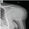 July 1, 2026 Short-term Functional Outcomes of Shoulder Girdle Limb Salvage in Primary Bone Tumors: A 2-year Follow-up Study
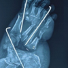 June 1, 2026 Limb Salvage in a Mangled Foot with Severe Midfoot Bone Loss: A Case Report and Review of Principles of Reconstruction
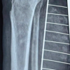 May 1, 2026 Repurposing Avulsed Skin as a Primary Full Thickness Autograft in Extensive Lower Limb Degloving: A Case Report