
Posterior proximal femoral osteochondroma should be considered in patients presenting with chronic buttock pain and unexplained sciatic symptoms, even in adulthood, as delayed neurological manifestations may occur despite the benign and longstanding nature of the lesion.
Dr M. Mohammed Tavfiq, Department of Orthopaedic Surgery, Sri Ramachandra Institute of Higher Education and Research, Chennai, Tamil Nadu, India. E-mail: tavfiqmm@gmail.com
Abstract
Introduction: Osteochondroma is the most common benign bone tumour and is usually asymptomatic. Neurovascular complications are uncommon, particularly in lesions involving the proximal femur. Sciatic nerve involvement secondary to a posterior proximal femoral osteochondroma is rare and may present with atypical symptoms, leading to delayed diagnosis.
Case Report: A 35-year-old male presented with a 3-year history of progressive right gluteal pain and discomfort associated with intermittent numbness of the right lower limb, which was aggravated by prolonged sitting. Clinical examination revealed a deep-seated bony swelling in the gluteal region with sensory deficits in the superficial and deep peroneal nerve distributions. Radiographs and magnetic resonance imaging demonstrated a large posteriorly projecting proximal femoral osteochondroma measuring 4.8 × 5.5 × 7.4 cm with corticomedullary continuity and posteromedial displacement of the sciatic nerve. The lesion was excised completely through a posterior approach with careful identification and protection of the sciatic nerve. Histopathological examination confirmed a benign osteochondroma. Postoperatively, the patient experienced complete resolution of neurological symptoms and significant improvement in gluteal pain.
Conclusion: Posterior proximal femoral osteochondromas can remain clinically silent for decades before presenting with symptoms related to sciatic nerve irritation. The presence of proximal femoral remodelling in this case suggested a longstanding lesion likely present since skeletal growth. Advanced imaging is essential for diagnosis and surgical planning. Complete excision provides excellent symptomatic relief and prevents further neurological compromise.
Keywords: Osteochondroma, proximal femur, sciatic nerve compression, buttock pain, benign bone tumour, case report.
Osteochondroma is the most common benign bone tumour, accounting for approximately 20–50% of all benign bone neoplasms [1]. It is characterised by a cartilage-capped bony projection arising from the external surface of a bone and demonstrating continuity of both the cortical and medullary components with the parent bone. These lesions typically develop during childhood and adolescence and are most frequently encountered around the metaphyses of long bones, particularly the distal femur, proximal tibia, and proximal humerus. Although osteochondromas are generally asymptomatic and discovered incidentally, symptoms may arise secondary to mechanical irritation, fracture, bursa formation, cosmetic concerns, or compression of adjacent neurovascular structures. Osteochondromas involving the proximal femur are relatively uncommon, and lesions arising from the posterior aspect of the proximal femur are particularly rare. Due to their anatomical proximity to the sciatic nerve, such lesions may present with atypical symptoms, including buttock pain, paraesthesia, or features mimicking lumbar radiculopathy, potentially leading to delays in diagnosis. Neurovascular complications associated with solitary osteochondromas are infrequently reported in the literature, with sciatic nerve compression representing an especially uncommon manifestation. Recognition of this presentation is important, as timely diagnosis and surgical excision can result in complete symptom resolution while preventing progressive neurological dysfunction. We report the case of a 35-year-old male who presented with chronic buttock pain and intermittent neurological symptoms caused by a posterior proximal femoral osteochondroma adjacent to the sciatic nerve. The lesion was successfully managed with complete surgical excision, resulting in significant symptomatic improvement.
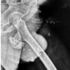
A 35-year-old male presented with complaints of pain and discomfort in the right gluteal region for the past 3 years. The symptoms were insidious in onset and gradually progressive, becoming particularly pronounced while sitting on hard surfaces and during prolonged periods of sitting. The patient also reported intermittent numbness involving the right lower limb, especially after maintaining a seated position for extended durations. There was no history of antecedent trauma, constitutional symptoms, weight loss, or similar complaints elsewhere in the body. On physical examination, a firm, non-tender, immobile bony swelling was palpable deep within the right gluteal region. The overlying skin was normal, and no local warmth or erythema was noted. Hip range of motion was preserved, although terminal movements elicited mild discomfort. Neurological examination revealed diminished sensation in the autonomous zones of the superficial and deep peroneal nerves. Motor power was preserved in all muscle groups, and distal pulses were palpable with no evidence of vascular compromise. Plain radiographs (Fig. 1) of the pelvis and right hip demonstrated a large exophytic osseous lesion arising from the proximal femur. Magnetic resonance imaging (MRI) was subsequently performed for further characterisation. MRI (Figs. 2 and 3) revealed a large bony exostosis arising from the greater trochanter of the right femur and projecting posteriorly. The lesion demonstrated continuity of both the cortex and medullary cavity with the proximal femur, consistent with an osteochondroma. It measured approximately 4.8 × 5.5 × 7.4 cm and had irregular margins without evidence of associated fracture or soft-tissue extension. The lesion was seen abutting the gluteus maximus muscle and closely related to the sciatic nerve, resulting in posteromedial displacement of the nerve. A thin cartilage cap measuring approximately 2 mm in thickness was identified on MRI, appearing hypointense on T1-weighted images and hyperintense on T2-weighted images. No radiological features suggestive of malignant transformation were observed. The hip joint was otherwise unremarkable. In view of the patient’s persistent symptoms and the close relationship of the lesion to the sciatic nerve, surgical excision and biopsy were planned. Under general anaesthesia, the patient was positioned in the lateral decubitus position, and a posterior approach to the proximal femur was utilised. A posteriorly curved lateral longitudinal incision was made centred over the tip of the greater trochanter. Skin, subcutaneous tissue, fascia, and gluteus maximus were incised to expose the mass. Exostosis was visualised as a large bony mass, which was irregular in shape with a stalk attached to the proximal femur posterior to the greater trochanter, covered by a pseudo-capsule. Due to its intimate relationship with the sciatic nerve (Fig. 4), meticulous dissection was performed to identify and protect the nerve throughout the procedure. The sciatic nerve was visualised and identified proximal and posterior to the mass but not isolated and tagged. Exostosis was removed in toto, osteotomising the base adjacent to normal bone. The specimen was sent for histopathological examination. Gross examination (Fig. 5) revealed a polypoidal osseocartilaginous lesion measuring 9.5 × 7 × 4.5 cm with a stalk measuring approximately 1 cm in length. The cut surface demonstrated a cartilage cap ranging from 0.2 to 0.8 cm in thickness. Microscopic examination showed lobules of mature hyaline cartilage containing chondrocytes within lacunae and areas of endochondral ossification transitioning into mature trabecular bone. No evidence of cytological atypia, increased mitotic activity, or necrosis was identified. Examination of the transition zone specimen revealed no residual lesion. These findings were consistent with a benign osteochondroma without evidence of malignant transformation. The post-operative period was uneventful. At the 1-month follow-up, the patient reported significant improvement in gluteal pain and complete resolution of the intermittent numbness experienced preoperatively. Clinical examination revealed no neurological deficits, and there was no evidence of local recurrence.
The proximal femur represents an uncommon site for osteochondroma compared with the more typical locations around the knee [2]. Furthermore, lesions arising from the posterior aspect of the proximal femur are particularly unusual because of their deep anatomical location and close relationship to important neurovascular structures [3,4,5]. As a result, patients may present with vague symptoms such as gluteal pain, sitting intolerance, restricted hip motion, or neurological complaints that can mimic lumbar spine pathology and delay diagnosis. The present case is notable because of the lesion’s location, size, and relationship to the sciatic nerve. Neurological manifestations secondary to osteochondromas are uncommon. When they occur, symptoms are generally attributable to chronic compression, displacement, or irritation of adjacent neural structures [6]. Nerve involvement has been most frequently described around the knee, particularly affecting the common peroneal nerve in association with proximal fibular osteochondromas [7]. In contrast, sciatic nerve involvement is rarely reported because posterior proximal femoral osteochondromas themselves are uncommon. Previous case reports have demonstrated that such lesions may produce symptoms ranging from buttock pain and paraesthesia to established sciatic neuropathy, depending on the severity and duration of neural involvement [3,4,8]. In the present case, the predominantly sensory and positional nature of the symptoms suggests intermittent mechanical irritation of the sciatic nerve rather than advanced neural compromise, which may explain the complete post-operative resolution of symptoms. Patient presented with symptoms only for the past 3 years, which was surprising considering that the maximal increase in the size of the exostoses occurs during the adolescent growth spurt. However, the patient was clear that the swelling and pain had been present only for the past 3 years, which was difficult to explain. The most noteworthy feature of this case was the remodelling and apparent enlargement of the proximal femoral neck region observed on radiographic evaluation (Fig. 6). Such bony remodelling is unlikely to occur over a short duration and strongly suggests that the lesion had been present for many years, likely originating during skeletal growth in keeping with the natural history of osteochondroma. Despite this probable long-standing presence, the patient remained asymptomatic until approximately 3 years before presentation. This observation illustrates that osteochondromas may remain clinically silent for decades before becoming symptomatic. In the present case, symptom onset was more likely related to progressive mechanical irritation of the surrounding soft tissues and sciatic nerve rather than recent acceleration of tumour growth. MRI demonstrated a thin cartilage cap measuring only 2 mm, while gross pathological examination revealed a cartilage cap ranging from 2 to 8 mm in thickness. The absence of aggressive radiological features further supports the diagnosis of a benign, long-standing lesion rather than malignant transformation [9,10]. This case, therefore, demonstrates that new-onset symptoms in adulthood do not necessarily indicate malignant change and may instead result from the gradual development of mechanical or neurological effects caused by a previously asymptomatic lesion. Histopathological findings further supported the benign and long-standing nature of the lesion. The presence of mature hyaline cartilage undergoing orderly endochondral ossification into trabecular bone, together with the absence of cytological atypia, increased mitotic activity, or necrosis, was consistent with a slowly evolving osteochondroma. These findings correlated well with the radiographic evidence of proximal femoral remodelling and support the hypothesis that the lesion had likely been present for many years before becoming symptomatic. Despite measuring more than 9.5 cm in maximal dimension, the lesion did not involve the hip joint, and the patient retained a near-normal range of hip motion. This finding highlights the ability of extra-articular osteochondromas arising from the proximal femur to attain considerable size before producing clinically significant symptoms, particularly when located posteriorly, where expansion can occur without immediate restriction of joint function. Surgical excision [11,12] remains the treatment of choice for symptomatic osteochondromas, lesions associated with neurovascular compromise, and cases in which malignant transformation cannot be confidently excluded [6,13]. Pre-operative MRI findings were especially valuable in the present case because they identified displacement of the sciatic nerve and facilitated appropriate surgical planning. Complete removal of the exostosis at its base is important to minimise recurrence while preserving surrounding neurovascular structures [13]. We used a posterior approach for excision of the mass, keeping in mind the risk of avascular necrosis of the femoral head, which the patient was warned about. Dissection was kept as close as possible to the bony base of the tumour to reduce the risk of damage to the ascending branch of the medial circumflex artery. Another option would have been a safe surgical dislocation and excision, but accessing this mass through a safe surgical dislocation would still require dissection distal and posterior to the lesser trochanter, negating the benefits of this approach.
This case highlights an uncommon presentation of a large posterior proximal femoral osteochondroma causing chronic buttock pain and sciatic nerve displacement in an adult patient. The presence of proximal femoral remodelling suggested a lesion that had likely existed since childhood but remained asymptomatic for decades before producing neurological symptoms. The favourable clinical outcome observed in this case supports previous reports demonstrating excellent symptomatic relief following excision of osteochondromas responsible for neural irritation or compression. Recognition of this entity is important, as careful clinical evaluation and advanced imaging can facilitate timely diagnosis, distinguish benign mechanical compression from malignant transformation, and enable successful surgical management with excellent functional outcomes.
Posterior proximal femoral osteochondromas may remain asymptomatic for decades and present in adulthood with atypical symptoms such as chronic buttock pain or intermittent sciatic nerve irritation. Careful radiological evaluation, particularly MRI, is essential to identify neural involvement and plan safe surgical excision. Complete excision of symptomatic lesions provides excellent clinical outcomes and prevents progressive neurological compromise.
References
- 1. Unni KK, Inwards CY. Dahlin’s Bone Tumours: General Aspects and Data on 11,087 Cases. 6th ed. Philadelphia, PA: Lippincott Williams & Wilkins; 2010. [Google Scholar] [PubMed]
- 2. Makhdom AM, Jiang F, Hamdy RC, Benaroch TE, Lavigne M, Saran N. Hip joint osteochondroma: Systematic review of the literature and report of three further cases. Adv Orthop 2014;2014:180254. [Google Scholar] [PubMed]
- 3. Yu K, Meehan JP, Fritz A, Jamali AA. Osteochondroma of the femoral neck: A rare cause of sciatic nerve compression. Orthopaedics 2010;33:597. [Google Scholar] [PubMed]
- 4. Aldashash F, Elraie M. Solitary osteochondroma of the proximal femur causing sciatic nerve compression. Ann Saudi Med 2017;37:166-9. [Google Scholar] [PubMed]
- 5. Naghizadeh H, Saberi S, Shooroki KK, Khabiri SS. Current concepts in the management of proximal femoral osteochondromas: A narrative review. J Orthop Surg Res 2025;20:734. [Google Scholar] [PubMed]
- 6. Kitsoulis P, Galani V, Stefanaki K, Paraskevas G, Karatzias G, Agnantis NJ, et al. Osteochondromas: Review of the clinical, radiological and pathological features. In Vivo 2008;22:633-46. [Google Scholar] [PubMed]
- 7. Göçmen S, Topuz AK, Atabey C, Şimşek H, Keklikçi K, Rodop O. Peripheral nerve injuries due to osteochondromas: Analysis of 20 cases and review of the literature. J Neurosurg. 2014;120(5):1105-1112. [Google Scholar] [PubMed]
- 8. Honwadkar NK, Pawar ED, Abhiram TV, Chanal A, Alaspure A. A rare and interesting case of sciatic nerve compression due to proximal femoral osteochondroma in a young adult: A case report. J Orthop Case Rep 2026;16:312-6. [Google Scholar] [PubMed]
- 9. Murphey MD, Choi JJ, Kransdorf MJ, Flemming DJ, Gannon FH. Imaging of osteochondroma: Variants and complications with radiologic-pathologic correlation. Radiographics 2000;20:1407-34. [Google Scholar] [PubMed]
- 10. Garrison RC, Unni KK, McLeod RA, Pritchard DJ, Dahlin DC. Chondrosarcoma arising in osteochondroma. Cancer 1982;49:1890-7. [Google Scholar] [PubMed]
- 11. Nag A. Surgical management of symptomatic solitary osteochondroma of the femur neck in an adult: a case report. J Bone Joint Dis 2022;37:183-7. [Google Scholar] [PubMed]
- 12. Mahajan NP, Gund A, Kondewar P, Chaudhari K, Bagimani P, Patel R. A case report on surgical excision of intracapsular osteochondroma of femur neck using a medial approach without hip dislocation in a young male. Int J Res Orthop. 2023;9(2):435-438. [Google Scholar] [PubMed]
- 13. Woertler K, Lindner N, Gosheger G, Brinkschmidt C, Heindel W. Osteochondroma: MR imaging of tumor-related complications. Eur Radiol 2000;10:832-40. [Google Scholar] [PubMed]


Related Articles in Journal of Orthopaedic Case Reports
 April 1, 2026 A Rare and Interesting Case of Sciatic Nerve Compression Due to Proximal Femoral Osteochondroma in a Young Adult – A Case Report
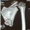 February 1, 2026 Symptomatic Ventromedial Scapular Osteochondroma Presenting with Restriction of Shoulder Movements: A Case Report
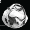 July 1, 2026 Extradigital Glomus Tumor of the Knee Mimicking Osteoarthritis: A Case Report
 July 1, 2026 Posterior Scapular Osteochondroma in a Pediatric Patient: An Uncommon Presentation of a Common Benign Tumor