

Prompt closed reduction, identification of persistent instability, and early mobilization with a structured rehabilitation program contribute significantly to favorable outcomes in isolated medial subtalar dislocations.
Dr. Adarsh Varghese Mathew, Department of Orthopaedics, JSS Hospital, Mysuru, Karnataka, India. E-mail: adarshvmathew@hotmail.com
Abstract
Introduction: Subtalar joint dislocations are rare clinical entities characterized by concurrent dislocations of the talonavicular and talocalcaneal joints. Most commonly, they occur as medial or lateral dislocations, with relatively good outcomes expected for medial dislocations and a worse prognosis for lateral dislocations.
Case Report: An 18-year-old athlete presented with pain and deformity in his left foot following a history of inversion and internal rotation twist injury while playing basketball. After initial evaluation in the casualty, he was taken up for closed reduction with Kirschner wire stabilization under anesthesia within 2 h of the injury and further immobilized with a posterior splint.
Conclusion: This case highlights the importance of identifying the mode of injury and of neutralizing any residual instability following the reduction procedure with supportive immobilization with or without internal fixation and splintage as early as possible.
Keywords: Subtalar dislocation, talus, calcaneus, isolated, medial.

Subtalar joint dislocations, also known as peritalar dislocations, are rare injuries (1% of all dislocations) associated with simultaneous dislocations of the talonavicular and talocalcaneal joints. The talus maintains its normal relation with respect to the ankle mortise while the subtalar joints are displaced beneath it [1]. Based on the direction of displacement with respect to the talus, such dislocations are classified into medial, lateral, anterior, and posterior. Most such injuries are associated with high-energy accidents, including but not limited to road traffic accidents, falls from height, and so on. Sports injuries have also been described in the etiology, with a lower incidence and usually resulting from a low-energy accident [2,3]. Moreover, isolated subtalar dislocations are unusual due to the inherent stability of the talus within the ankle mortise. They are frequently associated with fractures of either the malleoli, the talar neck, the calcaneus, or the fifth metatarsal, depending on the direction of the instability [4]. Hence, we describe a case of an 18-year-old male with an isolated medial subtalar dislocation.
An 18-year-old male presented to the casualty after suffering a twist injury to the outside edge of his right ankle and foot while playing basketball. He complained of pain, swelling, and deformity and was unable to bear weight over his foot. He had no prior history of ankle sprains. The ankle was found to be supinated and adducted, with prominent swelling over the dorsum of the midfoot. The neurovascular supply was intact with good capillary refill and no deficits in two-point discrimination and light touch sensation (Fig. 1).
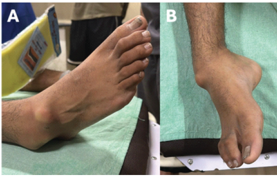
Figure 1: Clinical photograph showing the tibiotalar joint in normal angulation with displacement of the rest of the foot medially.
When compared to the opposite foot, there was a loss of the medial arch on the affected foot. Radiographic assessment identified medial-sided subtalar dislocation along with talo-navicular joint dislocation. The calcaneocuboid joint was found to be intact. No associated fractures were found on plain radiographs (Fig. 2).
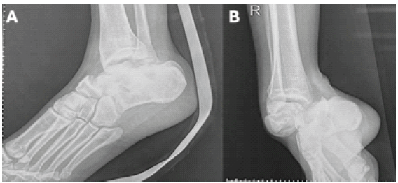
Figure 2: Pre-operative radiographs showing medial subtalar joint dislocation with talonavicular joint dislocation.
Considering the need for emergency relocation of the subtalar joint, the patient was taken up for surgery, and under general anesthesia, closed reduction was performed with the knee in flexion, manual traction, and an application of eversion force on the foot. The reduction was confirmed fluoroscopically. After reduction, the neurovascular structures were reassessed and found intact with palpable dorsalis pedis and posterior tibial arteries. Assessment of the stability of the subtalar joint found laxity in evidence. Intraoperative closed fixation with two Kirschner wires (K-wires) was performed across the talonavicular joint and tibio-talo-calcaneal joints. The foot and ankle were then immobilized with a posterior splint after a sterile dressing (Fig. 3).
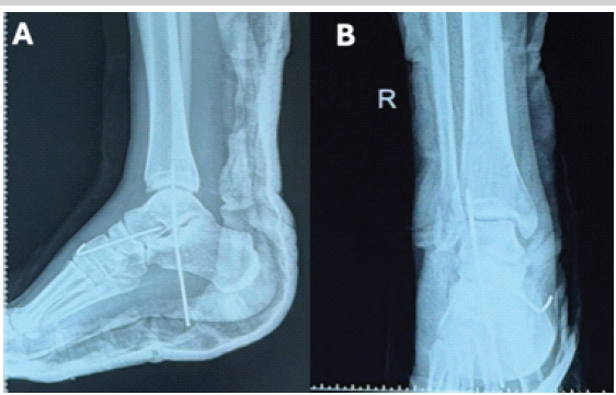
Figure 3: Post-operative radiographs showing reduction and position of the K-wires across the affected joints with immobilization in a posterior splint.
Computed tomography (CT) of the affected foot was done postoperatively. It revealed no fractures, osteochondral injuries, or loose bodies (Fig. 4).
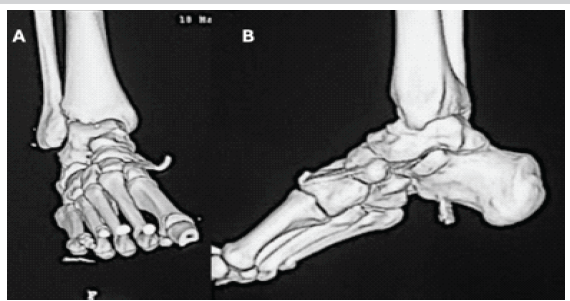
Figure 4: Post-operative computed tomography scan with 3D images after reduction.
The patient was started on limb elevation and ice packs to promote soft tissue healing and swelling reduction. A personalized, structured physiotherapy regimen was tailored for this patient. The patient was placed in a posterior splint with strict non-weight bearing for 4 weeks, following which the K-wires were removed. This was followed by gradual progression to ankle range-of-motion (ROM) exercises and weight-bearing as tolerated. The rehabilitation protocol was divided into four separate stages (Table 1).
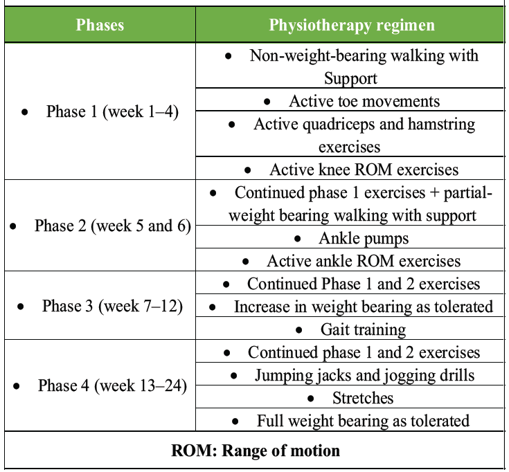
Table 1: The physiotherapy regimen followed for the patient
At 6-month follow-up, the patient was painlessly mobilizing with full ankle ROM (Fig. 5). Follow-up radiographs showed no obvious radiological abnormalities or signs of instability (Fig. 6).
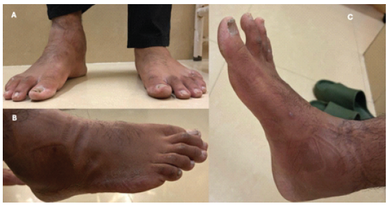
Figure 5: Clinical range of motion at 6 months showed good ankle range of motion with no pain or discomfort on plantar or dorsiflexion.
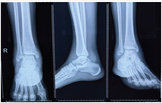
Figure 6: Follow-up radiographs at 6 months; the subtalar and ankle joints show no evidence of subluxation with maintenance of the ankle Shenton line.
Subtalar dislocation is a rare injury (1% of all dislocations) and involves derangement of both the talonavicular and talocalcaneal joints [4]. It is classified by Broca based on the direction of the rest of the foot relative to the talus and is divided into (1) the medial dislocation, (2) the lateral, and (3) the posterior. Malaigne and Burger later described the anterior displacement [5]. Medial dislocation, also known as “acquired clubfoot,” is the most common, with a 70–85% rate of incidence, and is the result of forceful inversion of the forefoot with the ankle in plantar flexion [6]. The talus pivots on the sustentaculum tali, and if the strong talocalcaneal and talonavicular ligaments rupture, the talus remains at the original position, and medial subtalar joint dislocation occurs [2,7]. The lateral dislocation is the second most common type, also known as “acquired flatfoot,” and accounts for 15–35% of all subtalar dislocations [8]. Subtalar dislocations are injuries commonly resulting from high-energy trauma and frequently involve young active males. It is associated with fractures of the talus, metatarsals, or the malleoli in many cases [9]. Few studies of isolated closed subtalar dislocations have been reported in the general literature, with most patients treated by closed reduction under sedation or anesthesia followed by immobilization for variable periods of time [6]. Results have been described as intermediate to good in various studies, with some studies finding no difference in outcome between medial and lateral dislocations [10,11]. In non-reducible cases, it was found to be imperative that open reduction be carried out. In rare cases of medial dislocations, the talar head was found to be button-holed through the extensor retinaculum or the extensor digitorum brevis, preventing reduction. Urgent reduction is needed in subtalar dislocations to prevent risk of damage to surrounding neurovascular structures, along with the articular surfaces of the subtalar, talonavicular, and talocalcaneal joints [12]. The incidence of associated injuries has been reported to be as high as 60%, emphasizing the importance of a CT scan for thorough evaluation. Moreover, a pre-existing status of pes planus may predispose the foot to such injuries by placing the foot in overpronation and valgus positioning of the heel. The associated muscular imbalance and tendon dysfunction, when combined with the weakness of the calcaneo-navicular ligament, can make the joint more vulnerable. However, the literature on such predisposing factors is lacking and requires further biomechanical or clinical investigation. Good outcomes depend on three factors: (1) Soft tissue injury to the surrounding tissue envelope, (2) early immediate reduction, and (3) short period of post-reduction immobilization [14]. Subtalar joint stiffness in subtalar dislocations can be minimized with a reduced duration of immobilization, ranging from 4 weeks for isolated dislocations up to 6 weeks for dislocations with associated fractures. In most cases, authors consider this injury to have a good prognosis if not associated with fractures or skin necrosis. The matter of the duration of immobilization remains a subject of study [14], with studies suggesting that an earlier return to weight-bearing may be successful. The incidence of associated injuries has been reported to be as high as 60%, emphasizing the importance of a CT scan for thorough evaluation.
Isolated, closed subtalar joint dislocations are rare, and following successful closed reduction, persistent instability should be assessed and managed appropriately. Immobilization of the limb for the minimum necessary time should be the ideal to prevent stiffness, and early ROM without pain should be allowed.
Isolated medial subtalar dislocations, although uncommon, require urgent reduction, thorough evaluation for associated injuries and residual instability, and early rehabilitation to restore function and prevent long-term stiffness.
References
- 1. Giannoulis D, Papadopoulos DV, Lykissas MG, Koulouvaris P, Gkiatas I, Mavrodontidis A. Subtalar dislocation without associated fractures: Case report and review of literature. World J Orthop 2015;6:374-9. [Google Scholar] [PubMed]
- 2. Simon LC, Schulz AP, Faschingbauer M, Morlock M, Jürgens C. “ Basketball foot”–long-time prognosis after peritalar dislocation. Sportverletz Sportschaden 2008;22:31-7. [Google Scholar] [PubMed]
- 3. James D. Functional outcome after early mobilization in isolated subtalar dislocation: Case report and review of current evidence. J Orthop Traumatol Rehabil 2017;9:50-2. [Google Scholar] [PubMed]
- 4. Perugia D, Basile A, Massoni C, Gumina S, Rossi F, Ferretti A. Conservative treatment of subtalar dislocations. Int Orthop 2002;26:56-60. [Google Scholar] [PubMed]
- 5. Zimmer TJ, Johnson KA. Subtalar dislocations. Clin Orthop Relat Res 1989;238:190-4. [Google Scholar] [PubMed]
- 6. Kinik H, Oktay O, Arıkan M, Mergen E. Medial subtalar dislocation. Int Orthop 1999;23:366-7. [Google Scholar] [PubMed]
- 7. Biz C, Ruaro A, Giai Via A, Torrent J, Papa G, Ruggieri P. Conservative management of isolated medial subtalar joint dislocations in volleyball players: A report of three cases and literature review. J Sports Med Phys Fitness 2019;59:1739-46. [Google Scholar] [PubMed]
- 8. Leitner B. Luxation sous-astagaliennerécente du pied avec subluxation tibiotarsienne de l’astragale. Rev Chir Orhop Reparatrice Appar Mot 1954;40:232-5. [Google Scholar] [PubMed]
- 9. Jerome JT, Varghese M, Sankaran B. Anteromedial subtalar dislocation. J Foot Ankle Surg 2007;46:52-4. [Google Scholar] [PubMed]
- 10. Merchan EC. Subtalar dislocations: Long-term follow-up of 39 cases. Injury 1992;23:97-100. [Google Scholar] [PubMed]
- 11. DeLee JC, Curtis R. Subtalar dislocation of the foot. J Bone Joint Surg Am 1982;64:433-7. [Google Scholar] [PubMed]
- 12. Bali K, Kumar V, Bhagwat K, Rawall S. Closed posterior subtalar dislocation without any associated fracture: A case report and review of the literature. Foot Ankle Surg 2011;17:e40-2. [Google Scholar] [PubMed]
- 13. Bibbo C, Lin SS, Abidi N, Berberian W, Grossman M, Gebauer G, et al. Missed and associated injuries after subtalar dislocation: The role of CT. Foot Ankle Int 2001;22:324-8. [Google Scholar] [PubMed]
- 14. Lasanianos NG, Lyras DN, Mouzopoulos G, Tsutseos N, Garnavos C. Early mobilization after uncomplicated medial subtalar dislocation provides successful functional results. J Orthop Traumatol 2011;12:37-43. [Google Scholar] [PubMed]





Related Articles in Journal of Orthopaedic Case Reports
 September 1, 2026 Traumatic Volar Dislocation of a Hemi-Scaphoid-Lunate Complex across the Carpal Ligament: A Case Report
 August 1, 2026 Complex Irreducible Dislocation of the Fourth Toe Proximal Interphalangeal Joint in a 5-year-old Boy Fixed by Soft Anchor: A Case Report
 May 1, 2026 “No Traction” Technique: A Novel, Simple, Easy, and Ergonomic Manoeuvre to Reduce Posterior Dislocation of the Hip Joint – A Case Report
 April 1, 2026 Cedell’s Fracture of the Talus Managed with Herbert Screw Fixation through Posteromedial Approach – A Case Report