

Progressive scoliosis in Crisponi syndrome can be safely managed with a multidisciplinary perioperative strategy and meticulous surgical planning, despite significant anaesthetic and systemic risks.
Mazlum Veysel Sili, Department of Orthopedics and Traumatology, Faculty of Medicine, Hacettepe University, Ankara, 06000, Turkey. E-mail: mazlumxsili@gmail.com
Abstract
Introduction: Crisponi syndrome is a rare autosomal recessive disorder caused by pathogenic variants in the CRLF1 gene and characterised by neonatal autonomic instability, feeding difficulties, recurrent hyperthermia, and persistent thermoregulatory dysfunction. Although survival has improved with advances in supportive care, musculoskeletal manifestations, particularly progressive spinal deformities, remain insufficiently described.
Case Report: We report a 16-year-old male with genetically confirmed Crisponi syndrome who developed severe progressive scoliosis. Recurrent febrile episodes limited brace compliance, and the deformity progressed to a pre-operative Cobb angle of 90.5 degrees. Pre-operative imaging demonstrated a structurally fixed, single C-shaped curve without segmentation anomalies. Following multidisciplinary evaluation addressing autonomic and anaesthetic risks, posterior spinal fusion and instrumentation from T2 to L2 were performed using pedicle screws, hooks, sublaminar bands, and multilevel chevron osteotomies. Perioperative management emphasised temperature regulation and preparation for hyperthermic crises.
Conclusion: The postoperative course was uneventful, with preserved neurological function and stable radiographic correction at the 1-month follow-up. This case demonstrates that posterior spinal fusion can be safely undertaken in patients with Crisponi syndrome when meticulous multidisciplinary planning is implemented.
Keywords: Crisponi syndrome, Scoliosis, Spinal deformity, Posterior spinal fusion, Pediatric spine surgery, Multidisciplinary management

Crisponi syndrome, also referred to as a presentation of cold-induced sweating syndrome type 1, is a rare autosomal recessive multisystemic disorder caused by pathogenic variants in the CRLF1 gene and, less frequently, in the CLCF1 gene, which encode components of a cytokine complex acting through the ciliary neurotrophic factor receptor pathway [1,2]. Clinically, Crisponi syndrome is characterised in the neonatal period by profound feeding and respiratory difficulties, facial muscle contractions, trismus, hyperthermia, and autonomic instability. Without intensive supportive care, these early manifestations can be life-threatening. Infants who survive this critical period often develop persistent thermoregulatory dysfunction and paradoxical sweating later in childhood [3]. Although the neonatal and autonomic features of Crisponi syndrome are relatively well documented, detailed descriptions of skeletal manifestations, especially progressive spinal deformities such as scoliosis, are limited in the literature. While some reports note that affected individuals may develop anomalies, including progressive kyphoscoliosis in later childhood and adolescence, the natural history and management of these deformities remain poorly characterised [4]. To date, there is a paucity of published case reports discussing the surgical management and outcomes of spinal deformity correction in patients with Crisponi syndrome, despite the significant orthopaedic challenges they may present. This case report aims to expand the clinical understanding of Crisponi syndrome by detailing the multidisciplinary assessment, surgical intervention, and early postoperative outcomes for a 16‑year‑old male with genetically confirmed Crisponi syndrome and severe scoliosis. To the best of our knowledge, while scoliosis has occasionally been noted in older children with Crisponi syndrome, this is the first published case describing its surgical correction, highlighting the perioperative challenges and successful orthopaedic outcome in this rare disorder.
A 16-year-old male patient with a genetically confirmed diagnosis of Crisponi syndrome was referred to our department due to rapidly progressing scoliosis over the past year. He was born at term via spontaneous vaginal delivery to consanguineous parents. In the neonatal period, he was admitted to the intensive care unit for 2 days due to poor feeding, hypotonia, cyanotic episodes, and generalised seizures. He received antiepileptic therapy until the age of two and required nasogastric tube feeding until the age of three. Developmental delays were noted across all domains, and he has been followed with a diagnosis of mental retardation, receiving special education until the eighth grade. He is currently attending a vocational high school with limited academic performance. Early genetic evaluation performed abroad had suggested Crisponi syndrome. The diagnosis was later confirmed through copy number variant analysis, which revealed a 971 base pair homozygous deletion at 19p13.11 involving exons 3 and 4 of the CRLF1 gene, consistent with the autosomal recessive form of the disease. Scoliosis was first identified between 6 and 12 months of age. The patient had initially been followed at an external centre, where bracing was attempted; the orthosis could only be tolerated during the winter months, as excessive sweating and recurrent febrile episodes precluded its use in warmer seasons. Follow-up at that centre was subsequently discontinued. Over the preceding year, he developed rapidly progressive spinal deformity and increasing back pain, prompting referral to our department. His earlier radiographs could not be retrieved, precluding quantification of curve progression over time. On physical examination, pre-operative standing clinical photographs demonstrated marked trunk asymmetry, right thoracic prominence, and compensatory lumbar shift without cutaneous stigmata (Fig. 1).
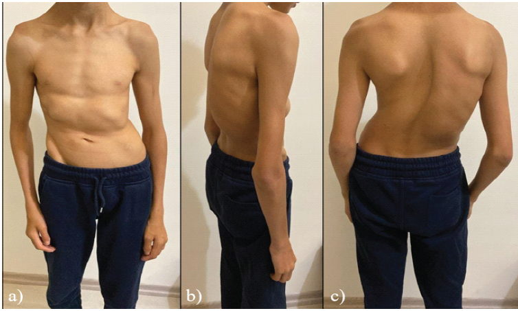
Figure 1: Pre-operative standing clinical photographs demonstrating the appearance of the spinal deformity: (a) Anterior view, (b) lateral view, and (c) posterior view.
At the time of admission, standing full-spine radiographs showed a severe single C-shaped thoracolumbar curve with its apex at T9–T10 and a standing Cobb angle of 90.5°. Pre-operative supine side-bending radiographs showed a rigid curve with essentially no correction of the Cobb angle, whereas intraoperative supine traction images under general anaesthesia confirmed partial correction of the deformity, supporting the use of corrective osteotomies (Fig. 2).
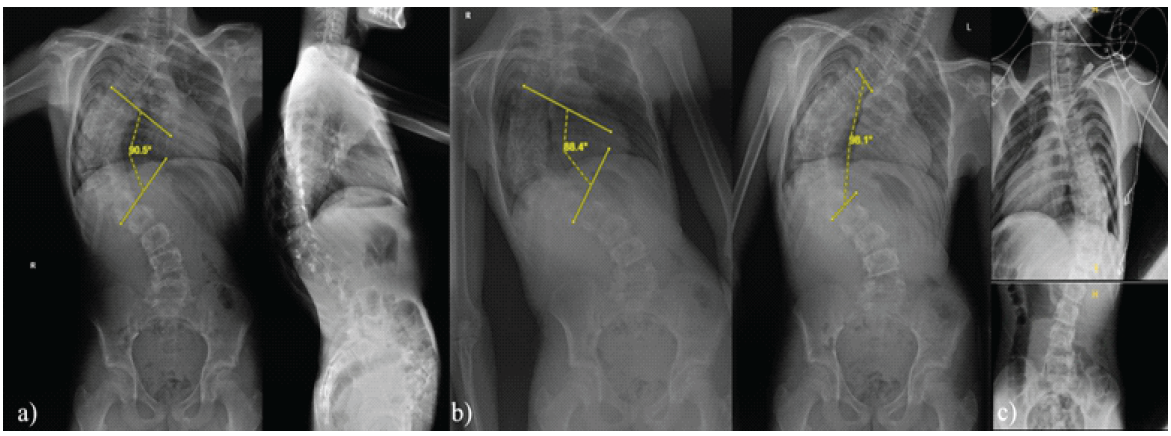
Figure 2: (a) Full-length upright scoliosis radiographs in the coronal and sagittal planes (standing anteroposterior/posteroanterior and lateral views), demonstrating a main thoracic curve with a Cobb angle of 90.5° and an apical vertebra at T10. (b) Full-length upright right and left lateral-bending radiographs obtained for assessment of coronal curve flexibility and surgical planning, with Cobb angles measuring 88.4° and 98.1°, respectively, and an apex at T10. (c) Intraoperative traction radiographs obtained under general anesthesia to assess deformity reducibility under controlled longitudinal traction. Pre-operative and intraoperative radiographic assessment of deformity and flexibility.
Computed tomography of the spine demonstrated no segmentation or fusion anomalies apart from a posterior arch defect at the S1 level. Brain magnetic resonance imaging findings were within normal limits, except for mild maxillary sinusitis. There was no evidence of brainstem pathology, hydrocephalus, or other structural abnormalities (Fig. 3).
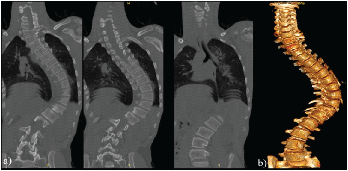
Figure 3: Coronal and three-dimensional computed tomography (CT) assessment of the spinal deformity. (a) Coronal-plane CT demonstrating a single C-shaped thoracolumbar curve with its apex at T9–T10. (b)Three-dimensional CT reconstruction facilitates multiplanar characterization of the deformity. Thin-slice CT additionally demonstrates a posterior fusion defect at S1, with no other vertebral segmentation or fusion anomalies and no osseous septum within the spinal canal.
Given the known association of Crisponi syndrome with autonomic dysregulation, the patient was evaluated preoperatively by paediatric neurology, cardiology, pulmonology, anaesthesia, and genetics. Cardiologic assessment revealed no evidence of congenital heart disease or exertional symptoms. Neurologic consultation highlighted the risk of hyperthermia, trismus, hypersalivation with aspiration, and possible rhabdomyolysis triggered by perioperative stress or low ambient temperatures. Literature-based recommendations included avoiding intraoperative hypothermia, ensuring pre-operative availability of dantrolene, and planning for intensive care monitoring post-operatively. On December 8, 2025, the patient underwent posterior spinal fusion and instrumentation from T2 to L2 under intraoperative neuromonitoring. The procedure included chevron osteotomies at T8–T9, T9–T10, T10–T11, and T11–T12 to facilitate deformity correction; placement of pedicle screws at all levels with a hook at the right T2 and a pedicle screw at the left T2; insertion of sublaminar bands at T9, T10, and T11 on the concave side of the curve; rod fixation with deformity derotation; posterior element decortication; and application of 30 cc of allograft combined with local autograft. The pedicle tracks felt osteopenic on probing; screws were therefore advanced cautiously to avoid cortical breach or pedicle fracture. Topical vancomycin powder was placed in the wound, and a Hemovac drain was inserted before layered closure (Fig. 4).
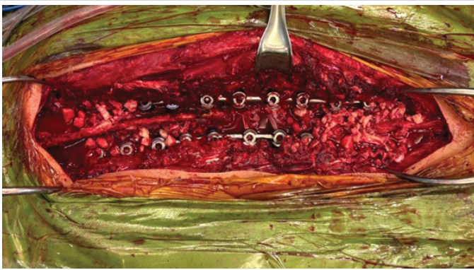
Figure 4: Intraoperative photograph obtained after completion of deformity correction and definitive posterior instrumentation. The procedure included T2–L2 posterior instrumentation with a T2 hook, multilevel chevron osteotomies at T8–9, T9–10, T10–11, and T11–12 with posterior fusion, and sublaminar band application at T9–T10–T11; the image demonstrates the final posterior construct after definitive fixation and correction.
The surgery was completed without complication, and the patient was transferred to the paediatric intensive care unit while still intubated. No signs of fever, autonomic crises, respiratory compromise, or new neurologic deficits were observed. He was extubated uneventfully, began oral intake and mobilisation within a few days, and was discharged on postoperative day 5 after standing radiographs confirmed satisfactory alignment and implant integrity. On follow-up, physical examination showed a well-healed incision, improved posture, and symmetrical shoulder levels (Fig. 5).
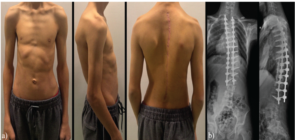
Figure 5: Post-operative clinical and radiographic outcomes. (a) Standing post-operative clinical photographs obtained in the anterior, lateral, and posterior views. (b) Standing postoperative full-length scoliosis radiographs in the coronal and sagittal planes (anteroposterior/posteroanterior and lateral views).
At 1-month follow-up, standing radiographs demonstrated preserved correction and stable instrumentation. The patient reported marked improvement in back pain, with no neurologic symptoms or implant-related complications.
Crisponi syndrome is a rare autosomal recessive disorder caused by mutations in the CRLF1 gene, typically presenting in the neonatal period with severe autonomic dysfunction, muscle rigidity, feeding difficulties, and recurrent hyperthermic episodes [5]. Although early mortality is common, improved supportive care has led to increased survival, allowing the characterisation of musculoskeletal manifestations that emerge later in life [6]. However, there remains a significant gap in the literature regarding the natural progression and surgical management of spinal deformities, particularly scoliosis, in patients with Crisponi syndrome. The case presented here is, to the best of our knowledge, the first report of a patient with Crisponi syndrome undergoing surgical correction for progressive scoliosis. While some reports have mentioned thoracic cage abnormalities such as pectus excavatum or scapular elevation in these patients, severe structural scoliosis requiring operative treatment has not been previously described in detail [7]. Our patient had been under orthopaedic follow-up at an external centre since approximately age one; however, recurrent febrile episodes – an established manifestation of autonomic instability in Crisponi syndrome – prevented brace compliance during warmer seasons, and his follow-up had lapsed over the preceding 1–2 years before referral. This is a clinically important observation, suggesting that impaired thermoregulation may contribute to rapid curve progression by limiting conservative management options [8,9]. At the time of surgical decision-making, the patient had reached a pre-operative Cobb angle of 90.5°, which is consistent with surgical thresholds reported in progressive or syndromic scoliosis [10]. Surgical intervention involved a T2–L2 posterior instrumentation, with multilevel chevron osteotomies at the curve apex and sublaminar banding at T9–T11, combined with hook-rod systems at the proximal anchor. This hybrid construct provided a balance between rigid fixation and safe deformity correction, which is especially important in syndromic patients with potential anatomical variations and neurological vulnerability [11]. Fusion was augmented with 30 cc of allograft and local autograft because the pedicle tracks felt osteopenic intraoperatively. Bone quality in this syndrome has not been systematically studied but is often compromised in syndromic scoliosis. Of critical importance in this case was perioperative anaesthetic management, given the reported risks of rhabdomyolysis, convulsions, hypersalivation, trismus, and hyperthermia-related complications such as disseminated intravascular coagulation in Crisponi syndrome [12]. Literature has highlighted that exposure to cold environments or anaesthetic stress may provoke hyperthermic crises, necessitating careful temperature control and pharmacologic preparedness. In our patient, a multidisciplinary pre-operative plan was coordinated involving paediatric neurology, anaesthesiology, pulmonology, cardiology, and genetics. Dantrolene was made readily available, and the patient was monitored postoperatively in the paediatric intensive care unit, ultimately with no complications [12]. Comparable multidisciplinary approaches have shown benefit in other forms of syndromic scoliosis, such as in Rett syndrome or Freeman-Sheldon syndrome. Nonetheless, the lack of previous surgical experience in Crisponi syndrome underscores the significance of this case, which offers a preliminary model for both recognising spinal deformity progression and safely implementing surgical intervention in this rare and complex disorder.
This case represents the first reported instance of a patient with genetically confirmed Crisponi syndrome undergoing surgical correction for progressive scoliosis. It highlights the need for awareness of potential musculoskeletal complications in long-term survivors of this rare condition. Given the unique anaesthetic and perioperative risks associated with Crisponi syndrome, a multidisciplinary approach and careful preoperative planning are essential for successful outcomes. Our experience underscores the feasibility and safety of posterior spinal fusion in such complex patients when performed under rigorous clinical oversight. A limitation of this report is the short radiographic follow-up; longer-term imaging could not be obtained because the family was unable to attend further visits for socioeconomic reasons. Future reports are warranted to better define the orthopaedic phenotype and long-term outcomes in this patient population.
Progressive scoliosis should be anticipated in long-term survivors of Crisponi syndrome, in whom the autonomic dysfunction that undermines conservative brace treatment – through heat intolerance and excessive sweating – also defines the perioperative risk during deformity correction. Early recognition of curve progression, combined with coordinated multidisciplinary planning that prioritises strict thermal control, readily available dantrolene, and intensive perioperative monitoring, allows posterior spinal fusion to be performed safely despite the syndrome’s autonomic and anaesthetic risks.
References
- 1. Buers I, Persico I, Schöning L, Nitschke Y, Di Rocco M, Loi A, et al. Crisponi/cold-induced sweating syndrome: Differential diagnosis, pathogenesis and treatment concepts. Clin Genet 2020;97:209-21. [Google Scholar] [PubMed]
- 2. Crisponi L, Crisponi G, Meloni A, Toliat MR, Nurnberg G, Usala G, et al. Crisponi syndrome is caused by mutations in the CRLF1 gene and is allelic to cold-induced sweating syndrome type 1. Am J Hum Genet 2007;80:971-81. [Google Scholar] [PubMed]
- 3. Hahn AF, Knappskog PM. Cold-induced sweating syndrome including crisponi syndrome. In: Adam MP, Bick S, Mirzaa GM, Pagon RA, Wallace SE, Amemiya A, editors. GeneReviews((R)). Seattle, WA: University of Washington; 1993. [Google Scholar] [PubMed]
- 4. Onesimo R, Sforza E, Palermo F, Giorgio V, Leoni C, Rigante D, et al. Feeding and nutritional key features of crisponi/cold-induced sweating syndrome. Genes (Basel) 2024;15:1109. [Google Scholar] [PubMed]
- 5. Piras R, Chiappe F, Torraca IL, Buers I, Usala G, Angius A, et al. Expanding the mutational spectrum of CRLF1 in Crisponi/CISS1 syndrome. Hum Mutat 2014;35:424-33. [Google Scholar] [PubMed]
- 6. Bayraktar-Tanyeri B, Hepokur M, Bayraktar S, Persico I, Crisponi L. A new case series of Crisponi syndrome in a Turkish family and review of the literature. Clin Dysmorphol 2017;26:66-72. [Google Scholar] [PubMed]
- 7. Perilli L, Dzwilewski K, Pietruszka M, Striano P, Capovilla G, Mazurkiewicz-Bełdzinska M. Early diagnostic markers in crisponi syndrome: Two cases and review. J Clin Med 2025;14:7757. [Google Scholar] [PubMed]
- 8. Negrini S, Fusco C, Minozzi S, Atanasio S, Zaina F, Romano M. Exercises reduce the progression rate of adolescent idiopathic scoliosis: Results of a comprehensive systematic review of the literature. Disabil Rehabil 2008;30:772-85. [Google Scholar] [PubMed]
- 9. Negrini S, Aulisa AG, Cerny P, De Mauroy JC, McAviney J, Mills A, et al. The classification of scoliosis braces developed by SOSORT with SRS, ISPO, and POSNA and approved by ESPRM. Eur Spine J 2022;31:980-9. [Google Scholar] [PubMed]
- 10. Delbruck H, Karl I, Hildebrand F, Hertwig MK, Pishnamaz M. Results of bracing adolescent idiopathic scoliosis in the context of clinical practice and the scoliosis research society’s criteria: 5-year observational study from a German orthopaedic university hospital. Eur J Med Res 2024;29:521. [Google Scholar] [PubMed]
- 11. Braun S, Brenneis M, Schönnagel L, Caffard T, Diaremes P. Surgical treatment of spinal deformities in pediatric orthopedic patients. Life (Basel) 2023;13:1341. [Google Scholar] [PubMed]
- 12. Allary C, Caruselli M, Fabre A, Audic F, Michel F. General anesthesia for Crisponi syndrome: Case report. Braz J Anesthesiol 2020;70:299-301. [Google Scholar] [PubMed]



Related Articles in Journal of Orthopaedic Case Reports
 January 1, 2026 Proximal Junctional Kyphosis Following Spinal Thoracic Deformity Correction in a Patient with Kabuki Syndrome: A Case Report
 December 1, 2025 Navigating Post-operative Complications: A Case Report of Cerebrospinal Fluid Leak and Pan-Resistant Enterobacter Infection Following Lumbar Fusion
 November 1, 2025 Cleft Hand and Foot Syndrome: A Report of Three Cases with Review of Literature
 July 1, 2025 Dorsal Osteoblastoma: A Rare Cause of Painful Scoliosis