

Endoscopic plantar fascia release has a steep learning curve; a K-wire as a guide will help beginners in localisation and avoid excess tissue dissection.
Dr. Hari Krishna Yadoji, Department of Orthopaedics, Care Hospital, Hitech City, Hyderabad, Telangana, India. E-mail: drharikrishna20@gmail.com
Abstract
Introduction: This case report is to emphasise the role of K-wire in localising the plantar fascia and calcaneal spur during endoscopic plantar fascia release (EPER). Plantar fasciitis is a very common problem in society. Most of the cases get good relief with physiotherapy, a change in footwear, and medication. Some cases might need steroid/platelet-rich plasma (PRP) injections. In a few refractory cases, pain remains even after extensive physiotherapy and plantar fascia injections. Such cases can benefit from plantar fascia release. Several techniques have been described for fascia release, including open, percutaneous, and endoscopic release.
Case Report: This is a 40-year-old female with severe pain in the heel while walking, particularly in the morning, for 2 years. She was treated earlier with physiotherapy, a change in footwear, steroids and a PRP injection. X-rays of the ankle show a calcaneum spur. Magnetic resonance imaging shows an 8 mm-thick plantar fascia.
Conclusion: A simple K-wire is a useful tool in a procedure like EPER, which has a steep learning curve. The use of K-wires alleviates the complications of excess soft tissue and fat pad dissection and reduces the radiographic assistance.
Keywords: Plantar fasciitis, endoscopy, K-wire.

Plantar fasciitis is a common condition of heel pain with an incidence of up to 10% in a lifetime [1]. Plantar fasciitis is the most common cause of heel pain, accounting for 80% of cases of heel pain [2]. This condition tends to affect people between 40 and 60 years, showing a higher prevalence in women [1]. Risk factors associated are obesity, prolonged weight-bearing, and reduced ankle dorsiflexion, and it can occur bilaterally in one-third of cases [3,4]. Although plantar fascia pathology is not completely understood, it seems to be a non-inflammatory structural breakdown of fascia secondary to myxoid degeneration with microtears within the fascia, collagen necrosis, and angiofibroblastic hyperplasia [5]. Conservative management is considered the gold standard in the treatment of plantar fasciitis, which includes non-steroidal anti-inflammatory drugs, plantar fascia stretches and changes in footwear, ice, and activity modifications [6]. If these methods do not give relief, steroid/platelet-rich plasma (PRP) injections can be performed. For patients with recalcitrant plantar fasciopathy, focal extracorporeal shockwave can be used with success rates between 50% and 65% [2]. Conservative treatment gives effective relief in 90% of patients. When patients do not respond even after 6–12 months, surgical treatment is required in 5–10% of patients [7]. In this subset of patients, partial or total plantar fascia release has been the mainstay of treatment, with success rates of approximately 70–90% [7]. Different surgical treatments have been described for this purpose, including open fasciotomy or minimally invasive procedures that can be performed percutaneously or endoscopically [8]. Several clinical trials reported that endoscopic plantar fascia release (EPFR) resulted in excellent relief of pain, with patient satisfaction ranging from 60% to 80% and low complication rates [9,10]. Endoscopic fascia release has a steep learning curve. Many times, the beginners face difficulty in localising which part of the foot they are in and dissect a lot of tissue/fat pad unnecessarily. This case report is about a technique where we place a K-wire for localisation and as a guide during the surgery.
This is a 40-year-old female with severe pain in the heel while walking, particularly in the morning, for 2 years. She was treated earlier with physiotherapy, a change in footwear, steroids and a PRP injection. X-rays of the ankle show a calcaneal spur as in Fig. 1. The MRI shows an 8 mm-thick plantar fascia as in Fig. 2.
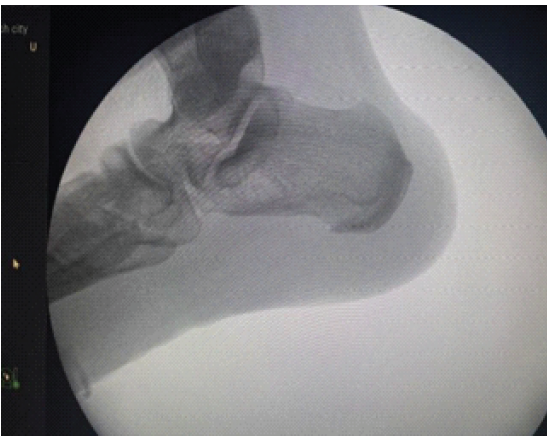
Figure 1: Radiograph showing calcaneum spur.
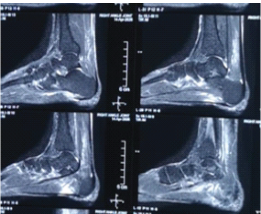
Figure 2: Magnetic resonance imaging showing thick plantar fascia.
The procedure is performed with the patient under spinal anaesthesia, and a tourniquet is applied. The patient’s leg is put in a figure of four as in Fig. 3, and a point 5 mm from the tangential line of the posterior edge of the medial malleolus to the plantar fascia (skin junction) is marked, which is going to be used as the primary viewing portal. The anterior medial entry was established two centimetres anterior to the posterior entry site, as in Fig. 4.
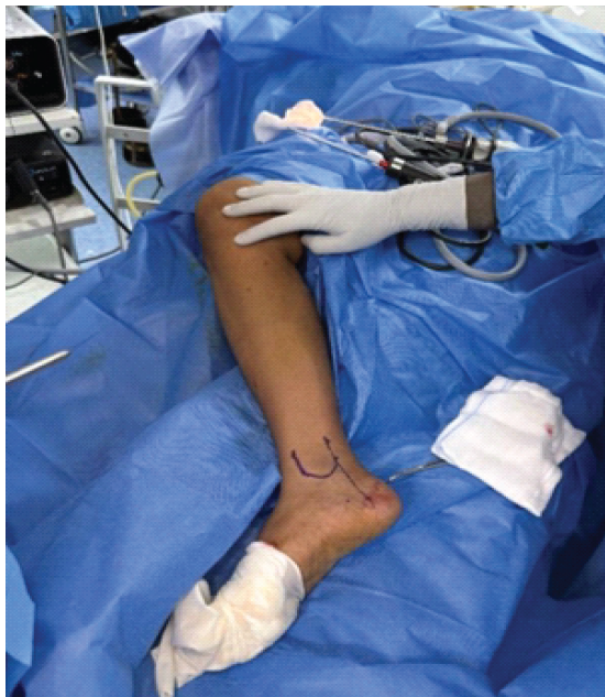
Figure 3: Position of limb (figure of four).
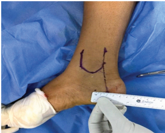
Figure 4: Medial portals.
The nick and spread technique is used for making portals. The primary viewing portal was made, and a 4 mm 300 scope was introduced. A needle is introduced through the marked anterior portal site, and only after visualising the needle is the portal made as in Fig. 5. For beginners, it is always better to make portals under vision, as it decreases the chances of damaging important structures. A K-wire is put at the spur from the plantar side under C-arm guidance and left in situ as in Fig. 6.
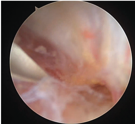
Figure 5: 2nd portal after confirming with k wire.
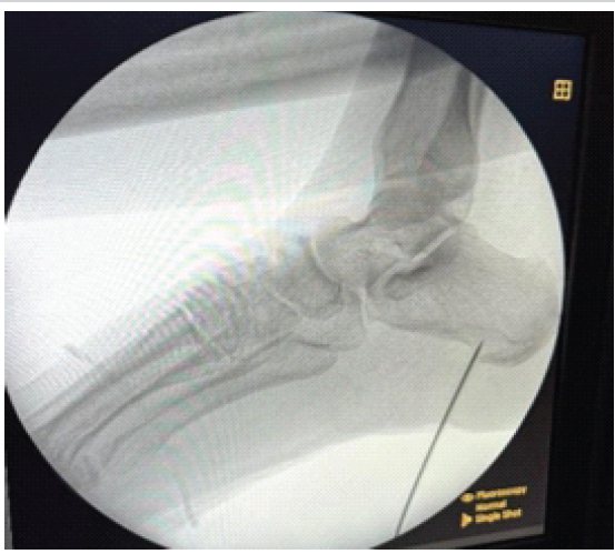
Figure 6: C arm showing K wire at spur site.
Following the trajectory of the K-wire, a shaver and radiofrequency (RF) ablator are used alternatively to remove the excess fat tissue and localise the K-wire as in Fig. 7. Once we localise the K-wire, work along the K-wire until you reach its tip. Now remove the K-wire and expose the spur and excise with a burr under C-arm guidance as in Fig. 8. The plantar fascia is released with RF as in Fig. 9. Confirm spur excision under the C-arm as in Fig. 10. Portals are closed. Partial weight-bearing with crutches is allowed immediately. Full weight-bearing is allowed after wound healing, and stretching exercises are recommended.
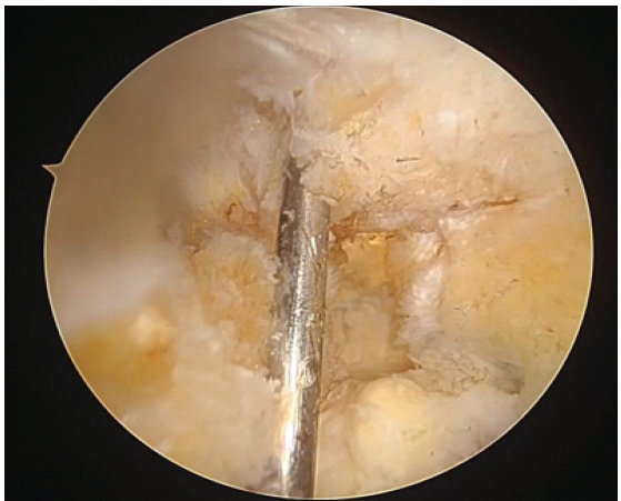
Figure 7: K wire tip localized.
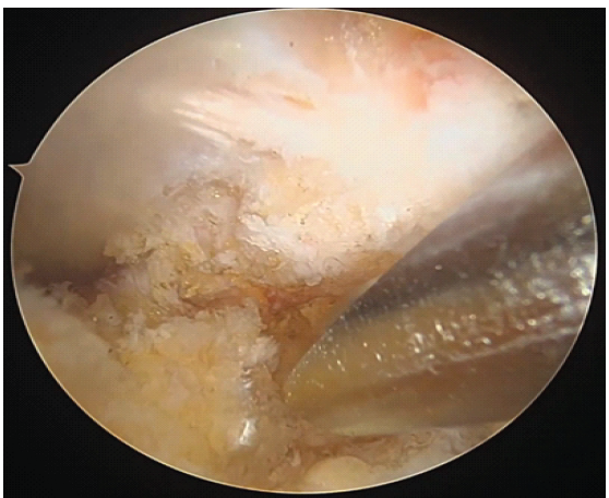
Figure 8: Burr at the spur site (*).
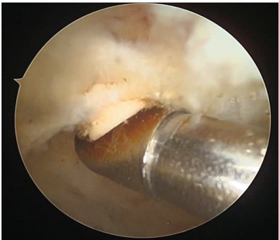
Figure 9: Plantar fascia release with ablator.
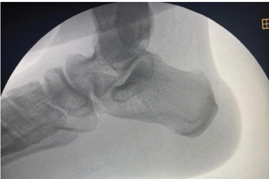
Figure 10: C arm picture after spur excision.
EPER is a minimally invasive technique that decreases complications related to open surgery, such as plantar heel hypertrophic scar formation, infection, or wound dehiscence. As a result, a shorter recovery time and early post-operative mobilisation therapy have been reported [11]. Nevertheless, the procedure has a steep learning curve. With experience, plantar fascia exposure and excision of the spur become easy with minimal dissection, but in the initial cases, one might dissect too much soft tissue or fat pad without knowing where exactly we are anatomically. Using a K-wire as a guide is the solution to these hurdles. The moment we identify the k wire, all we have to do is follow the k wire and remove tissue as required; no more blind dissection is done. The use of radiographic assistance also decreases once the k wire is identified. In particular, the identification of the spur is the most difficult part. One must release the entire fascia to visualise and excise the spur, which can lead to complications. With the help of the K-wire, once we reach the tip, remove the K-wire and dissect that area from posterior to anterior (calcaneum undersurface), minimising the damage to the plantar fascia.
A simple K-wire is a useful tool in a procedure like EPER, which has a steep learning curve. The use of K-wires alleviates the complications of excess soft tissue and fat pad dissection and reduces the radiographic assistance.
Endoscopic plantar fascia release is a great solution to long-standing and refractory plantar fasciitis. It has a steep learning curve. Use of a K-wire for guidance is a simple and easily reproducible technique, especially for beginners.
References
- 1. MacInnes A, Roberts SC, Kimpton J, Pillai A. Long-term outcome of open plantar fascia release. Foot Ankle Int 2016;37:17-23. [Google Scholar] [PubMed]
- 2. Gollwitzer H, Saxena A, DiDomenico LA, Galli L, Bouché RT, Caminear DS, et al. Clinically relevant effectiveness of focused extracorporeal shock wave therapy in the treatment of chronic plantar fasciitis: A randomized, controlled multicenter study. J Bone Joint Surg Am 2015;97:701-8. [Google Scholar] [PubMed]
- 3. Buchbinder R. Clinical practice. Plantar fasciitis. N Engl J Med 2004;350:2159-66. [Google Scholar] [PubMed]
- 4. Riddle DL, Pulisic M, Pidcoe P, Johnson RE. Risk factors for plantar fasciitis: A matched case-control study. J Bone Joint Surg Am 2003;85:872-7. [Google Scholar] [PubMed]
- 5. Lemont H, Ammirati KM, Usen N. Plantar fasciitis: A degenerative process (fasciosis) without inflammation. J Am Podiatr Med Assoc 2003;93:234-7. [Google Scholar] [PubMed]
- 6. Oliva F, Piccirilli E, Tarantino U, Maffulli N. Percutaneous release of the plantar fascia. New surgical procedure. Muscles Ligaments Tendons J 2017;7:338-40. [Google Scholar] [PubMed]
- 7. Sahu RL. Percutaneous planter fasciitis release under local anesthesia: A prospective study. Chin J Traumatol 2017;20:87-9. [Google Scholar] [PubMed]
- 8. Malahias MA, Cantiller EB, Kadu VV, Müller S. The clinical outcome of endoscopic plantar fascia release: A current concept review. Foot Ankle Surg 2020;26:19-24. [Google Scholar] [PubMed]
- 9. Ohuchi H, Ichikawa K, Shinga K, Hattori S, Yamada S, Takahashi K. Ultrasound-assisted endoscopic partial plantar fascia release. Arthrosc Tech 2013;2:e227-30. [Google Scholar] [PubMed]
- 10. Othman AM, Hegazy IH. Endoscopic plantar fasciotomy versus injection of platelet-rich plasma for resistant plantar fasciopathy. J Orthop 2015;12:S176-81. [Google Scholar] [PubMed]
- 11. Fallat LM, Cox JT, Chahal R, Morrison P, Kish J. A retrospective comparison of percutaneous plantar fasciotomy and open plantar fasciotomy with heel spur resection. J Foot Ankle Surg 2013;52:288-90. [Google Scholar] [PubMed]




Related Articles in Journal of Orthopaedic Case Reports
 August 1, 2026 Outcome Analysis of Leukocyte-Rich Platelet-Rich Plasma, Leukocyte-Poor Platelet-Rich Plasma, and Injectable Platelet-Rich Fibrin in the Management of Chronic Plantar Fasciitis: A Prospective Comparative Study
 May 1, 2026 Navigation-Assisted Endoscopic Excision of Lumbar Vertebral Osteoid Osteoma: A Case Report
 March 1, 2026 Multiple Cantilever K-Wiring Technique for Severely Comminuted Intra-articular Fragments – A Novel Surgical Technique
 December 1, 2025 Therapeutic Effect of Intralesional Corticosteroids in Comparison with Multiple Puncture Technique in Treatment of Bilateral Plantar Fasciitis