

Timely diagnosis and surgical excision can result in complete resolution of symptoms and early return to full activity.
Dr. M Venu Gopal Reddy, Department of Orthopaedic Surgery, Star Hospitals, Nanakramguda, Hyderabad - 500032, Telangana, India. E-mail: meda.venu@gmail.com
Abstract
Introduction : Ischiofemoral impingement syndrome (IFI) is a non-articular source of hip pain caused by compression of the quadratus femoris muscle in the space between the ischial tuberosity and the lesser trochanter of the femur. Among the rare structural causes of IFI, osteochondroma, a benign bone tumour, can serve and present as a space-occupying lesion, reducing the ischiofemoral space and leading to soft tissue impingement.
Case Report: We report a case of a 33-year-old male patient presenting with a six-month history of clicking and progressive left gluteal pain. Examination and imaging studies revealed a narrowed ischiofemoral space associated with an osteochondroma arising from the medial aspect of the lesser trochanter. Surgical resection of the lesion led to complete resolution of the symptoms and restoration of hip function.
Conclusion: This case highlights the need for a high index of suspicion for structural causes of IFI and underscores the importance of MRI in the diagnosis. Early surgical intervention for osteochondroma-induced IFI can result in excellent clinical outcomes.
Keywords: Ischiofemoral Impingement – IFI, Osteochondroma – OC, Quadratus Femoris Muscle – QFM.

Ischiofemoral impingement syndrome (IFI) is a non-articular source of hip pain caused by compression of the quadratus femoris muscle in the space between the ischial tuberosity and the lesser trochanter of the femur. Initially described in postoperative patients, IFI is now recognised in a wider population, including athletes and individuals with congenital or acquired anatomical variations. Among the rare structural causes of IFI, osteochondroma, a benign bone tumour, can present as a space-occupying lesion, reducing the ischiofemoral space and leading to soft tissue impingement. This report details the presentation, diagnostic workup, and surgical treatment of a 33-year-old male patient with IFI secondary to a proximal femoral osteochondroma.
- We present a case of a 33-year-old male patient, a software developer by profession, with a sedentary lifestyle and occasional recreational running with no history of trauma or previous hip surgery and systemic disease. The patient presented with a 6-month history of gradually worsening left gluteal pain and clicking sound which was dull aching in nature, worsened by prolonged sitting, walking and stair climbing. Occasionally the pain radiates to the posterior aspect of the thigh and is not associated with any neurological symptoms like tingling, numbness or weakness. Physical examination revealed antalgic gait with pain on passive extension and adduction. The IFItest was positive (pain reproduced with extension, adduction, and external rotation of the hip). The neurological examination showed normal reflexes, motor strength, and sensation in the lower limbs
Diagnostic Workup:
Plain radiographs of the pelvis with both hips showed a pedunculated bony protuberance projecting from the medial aspect of the left lesser trochanter, suspicious for osteochondroma. [Fig. 1]
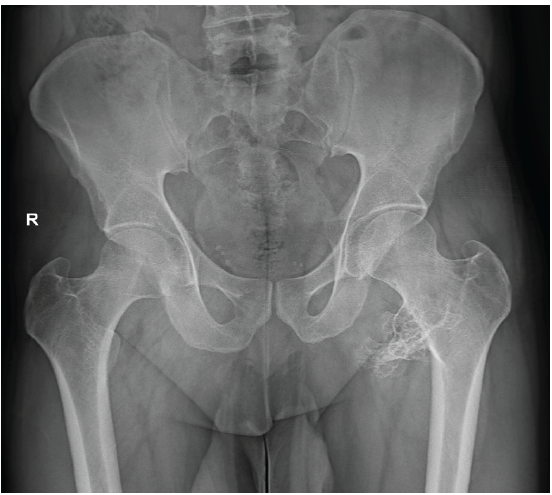
Figure 1: Pre-operative X-ray of pelvis showing protuberant growth from the medial aspect of the lesser trochanter.
Magnetic Resonance Imaging (MRI) and CT showed a significant reduction in ischiofemoral space (measured at ~10 mm), oedema and T2 signal hyperintensity in the quadratus femoris muscle. Lesion arising from the lesser trochanter with continuity of marrow and cortical bone, consistent with osteochondroma. [Figs. 2, 3] The final diagnosis was ischiofemoral impingement syndrome secondary to osteochondroma of the left lesser trochanter.
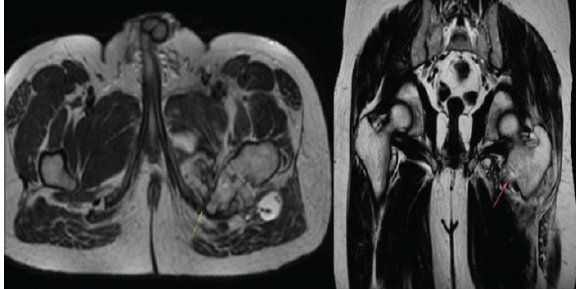
Figure 2: Axial and coronal sections of magnetic resonance imaging showing reduced ischiofemoral interval and growth from the lesser trochanteric.
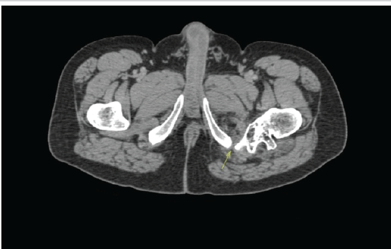
Figure 3: Computed tomography axial image showing reduced ischiofemoral interval.
Management:
Given the mechanical aetiology of the impingement, a surgical intervention was planned. Through a posterior approach to the hip, complete excision of the osteochondroma along with partial excision of the lesser trochanter with preservation of adjacent muscular and neurovascular structures was performed [Figs. 4, 5, 6].
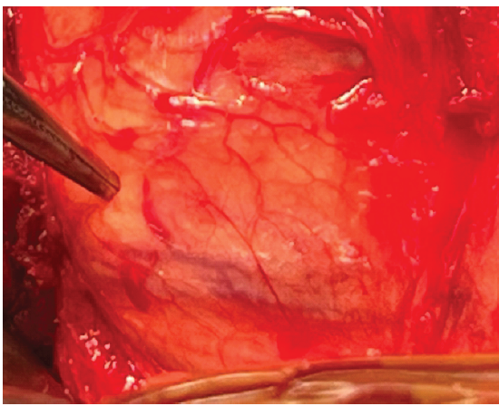
Figure 4: Intraoperative picture after exposure through posterior approach.
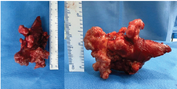
Figure 5: Intraoperative picture showing the dimensions of osteochondroma after escision.
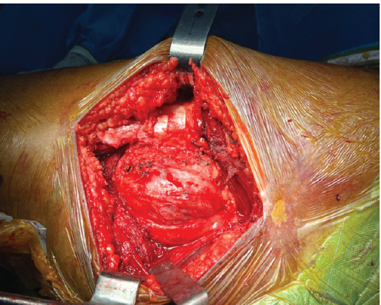
Figure 6: Intraoperative picture after excision of osteochondroma and clousure.
Intraoperative fluoroscopy confirmed complete lesion removal [Fig. 7]. The sample was sent for histopathological examination and confirmed as osteochondroma [Fig. 8]. The postoperative outcomeshowed marked reduction in gluteal pain within 2 weeks of post-op. Rehabilitation started with weight-bearing as tolerated after 6 weeks, with progressive strengthening and range-of-motion exercises starting immediately. At the 3-month follow-up there was no residual pain or functional limitations, and full hip range of motion was restored. The patient is currently two years post-op, with no new complaints.
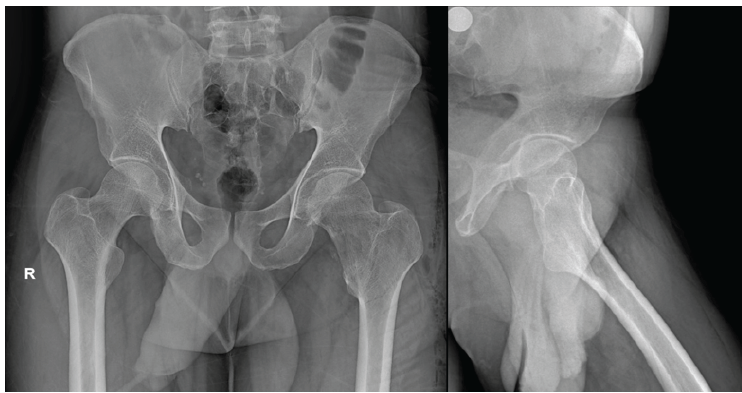
Figure 7: Post-operative X-ray of pelvis anteroposterior view and left hip lateral view after excision of osteochondroma.
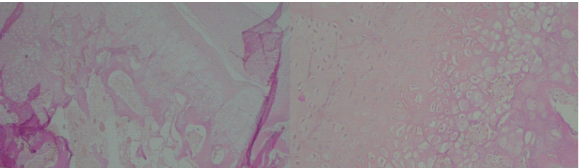
Figure 8: Histopathological examination pictures showing presence of cartilagenous cap consisting with fibrous perichondrium suggestive of osteochondroma.
Osteochondromas are benign exostoses that typically develop during skeletal development. Although often asymptomatic, they can become clinically relevant when located near anatomical structures where space is limited, such as the ischiofemoral interval. Johnson first mentioned IFI in 1977 and demonstrated the reduction in distance between the lesser trochanter and ischium in X-ray and its association with persistent hip pain in three patients. In two patients, it followed hip replacement, and in one patient, it followed a proximal femoral osteotomy. All three patients were completely pain-free after the resection of the lesser trochanter, supporting the hypothesis that abnormal contact with the ischium was responsible for the symptoms. (1) Guney et al. presented a case series about the relationship between osteochondroma and ischiofemoral impingement and IFI as one of the causes of hip pain in the early age group and the need to evaluate osteochondromas located in the lesser trochanter or ischiopubic ramus. (2) Torriani et al. described IFI in patients with abnormal signals on MRI in Quadratus femoris muscle in all aspects in 2009. (3) Kassarjian et al. described IFI as a typical impingement involving the QFM. It is the most important external rotator of the hip, which also helps in adduction. It passes through the ischiofemoral interval and is in close proximity to the sciatic nerve during its course. The most important complaint is the hip pain, which may radiate to the buttocks, knee and inguinal regions and increases with external rotation, adduction and extension. (4). Dooley et al. described the anterior impingement test as one of the most important diagnostic tools for IFI (5). Osteochondroma is a rare cause of IFI, as described in detail in the case report by Schubert et al. in 2019, with only one case report showing IFI resulting from osteochondroma. (6) Osteochondromas are the most common benign bone tumours and constitute 10-15% of all bone tumours. They can be solitary or multiple in nature. They are more common in males, whereas IFI is more common in females. Approximately 75% of OCs are encountered before the age of 20. They are mostly observed in the lower extremities, especially in the femur. (7) However, IFI is a disease of the middle age group. The OCs and IFI age groups were different. Probably this epidemiologic information is the factor that diminishes the role of OCs as an aetiology of IFI. (8) However, the X-ray findings are limited and not specific to IFI. Patti et al. reported heterogeneity and sclerosis in the lesser trochanter and ischium as the most prominent X-ray findings. It can also provide information about the narrowing of the distance between the lesser trochanter and ischium. Ultrasonography studies have not shown any benefits in the diagnosis of IFI. CT provides clear information about bone structures and narrowing of the IF interval, but it is insufficient to show soft tissue findings such as muscle oedema and fat tissue replacement [9]. MRI is the best imaging method for diagnosing IFI. In MRI, oedema in the QFM is the most important finding suggesting IFI. The lesser trochanter and tuber ischium are normally approximately 2 cm apart. This distance allows the femur to rotate without touching the tuber ischium or hamstring tendon. IFI is best evaluated using axial T2-weighted images. The presence of oedema in the muscle, instead of the absence of muscular fibre disruption, helps distinguish IF compression from QFM tear. In the chronic period, T1-weighted MRI images are useful for evaluating quadratus femoris atrophy, hyperintense fat replacement, and muscle volume loss associated with IFI [10]. Initial treatment may include conservative therapy, which includes rest, activity restriction, anti-inflammatory non-steroidal drugs, percutaneous ultrasound therapy, and physical therapy. CT- or ultrasound-guided injections with anaesthetics and steroids can be used as a diagnostic test and for symptomatic relief [11]. Surgical treatment options include resection of the lesser trochanter and decompression of the QFM [12]. Nakano et al. described an osteochondroma located at the lesser trochanter and causing impingement of the quadratus femoris muscle as a type of extra-articular hip impingement, where the patient presents with pain in the groin and sometimes radiating to the buttock area and medial aspect of the thigh. The pain is reproduced or worsened by hip extension, adduction and external rotation. (13) L R Ramos-Pascua et al. presented that surgical treatment of solitary proximal end of femur osteochondromas is mandatory and en bloc resection can be performed by a single wide approach without the need for hip dislocation. (14)
- Aldashash et al. presented a case report of a solitary osteochondroma of the proximal femur causing sciatic nerve compression, which was surgically removed with gradual improvement in symptoms. (15)
Asymptomatic osteochondromas are usually left alone and observed. Only symptomatic patients underwent marginal to wide resection with or without fixation. In our patient, excision of the osteochondroma was performed because it was a mechanical block causing clicking and pain. Regular monitoring was performed for recurrence. A positive response to surgical resection reinforces the value of early imaging and recognition of structural causes in patients with unexplained gluteal pain.
This case exemplifies a rare but treatable cause of ischiofemoral impingement. Osteochondromas should be considered in patients with persistent deep gluteal pain and imaging evidence of a bony mass near the lesser trochanter. Timely diagnosis and surgical excision can result in complete resolution of symptoms and return to full activity.
- MRI is essential for identifying both the cause and consequences (e.g., muscle edema) of IFI
- Osteochondroma should be considered in young adults with focal bony outgrowths near the hip joints
- Surgical excision offers excellent outcomes when conservative measures fail or are unlikely to succeed because of a space-occupying lesion.
References
- 1. Johnson KA.. Impingement of the lesser trochanter on the ischial ramus after total hip arthroplasty. Report of three cases. J Bone Joint Surg Am. 1977. March;59(2):268-9. PMID: . [PubMed] [Google Scholar] [Google Scholar] [PubMed]
- 2. Bunyamin Guney, Emrah Doğan, Murat Yunus Ozdemir. Osteochondroma as a Cause of Ischiofemoral Impingement – First Case Series. ISSN 1392-0138 eISSN 2029-4174 Acta medica Lituanica. 2021. Vol 28. No 1. DOI: https://doi.org/10.15388/Amed.2021.28.1.15 [Google Scholar] [PubMed] [CrossRef]
- 3. Torriani M, Souto SC, Thomas BJ, Ouellette H, Bredella MA.. Ischiofemoral impingement syndrome: an entity with hip pain and abnormalities of the quadratus femoris muscle. AJR Am J Roentgenol. 2009. July;193(1):186-90. doi: 10.2214/AJR.08.2090. PMID: . [DOI] [PubMed] [Google Scholar] [Google Scholar] [PubMed] [CrossRef]
- 4. Kassarjian A, Tomas X, Cerezal L, Canga A, Llopis E. MRI of the quadratus femoris muscle: anatomic considerations and pathologic lesions. AJR Am J Roentgenol. 2011 Jul;197(1):170-4. doi: https://doi.org/10.2214/AJR.10.5898. PMID: 21701027. [Google Scholar] [PubMed] [CrossRef]
- 5. Dooley PJ. Femoroacetabular impingement syndrome: Nonarthritic hip pain in young adults. Can Fam Physician. 2008 Jan;54(1):42-7. PMID: 18208954; PMCID: PMC2293316. [Google Scholar] [PubMed]
- 6. Schubert T, Navez M, Galant C, Docquier PL, Acid S, Lecouvet FE. Femoral osteochondroma responsible for ischiofemoral impingement, bursitis, and secondary lipoma arborescens mimicking malignant transformation. Acta Radiol Open. 2019 Dec 11;8(12):2058460119892409. doi: https://doi.org/10.1177/2058460119892409. PMID: 31903223; PMCID: PMC6923803. [Google Scholar] [PubMed] [CrossRef]
- 7. Brien EW, Mirra JM, Luck JV Jr. Benign and malignant cartilage tumors of bone and joint: their anatomic and theoretical basis with an emphasis on radiology, pathology and clinical biology. II. Juxtacortical cartilage tumors. Skeletal Radiol. 1999 Jan;28(1):1-20. doi: https://doi.org/10.1007/s002560050466. PMID: 10068070. [Google Scholar] [PubMed] [CrossRef]
- 8. Murphey MD, Choi JJ, Kransdorf MJ, Flemming DJ, Gannon FH. Imaging of osteochondroma: variants and complications with radiologic-pathologic correlation. Radiographics. 2000 Sep-Oct;20(5):1407-34. doi: https://doi.org/10.1148/radiographics.20.5.g00se171407. PMID: 10992031. [Google Scholar] [PubMed] [CrossRef]
- 9. Patti JW, Ouellette H, Bredella MA, Torriani M.. Impingement of lesser trochanter on ischium as a potential cause for hip pain. Skeletal Radiol. 2008. October;37(10):939-41. doi: 10.1007/s00256-008-0551-3. Epub 2008. August 6. PMID: . [DOI] [PubMed] [Google Scholar] [Google Scholar] [PubMed] [CrossRef]
- 10. Kassarjian A. Signal abnormalities in the quadratus femoris muscle: tear or impingement? AJR Am J Roentgenol. 2008. June;190(6):W379; author reply W380-1. doi: 10.2214/AJR.07.3540. PMID: . [DOI] [PubMed] [Google Scholar] [Google Scholar] [PubMed] [CrossRef]
- 11. Willick SE, Lazarus M, Press JM. Quadratus femoris strain. Clin J Sport Med. 2002 Mar;12(2):130-1. doi: https://doi.org/10.1097/00042752-200203000-00012. PMID: 11953562. [Google Scholar] [PubMed] [CrossRef]
- 12. Safran M, Ryu J.. Ischiofemoral impingement of the hip: a novel approach to treatment. Knee Surg Sports Traumatol Arthrosc. 2014. April;22(4):781-5. doi: 10.1007/s00167-013-2801-8. Epub 2013. December 18. PMID: . [DOI] [PubMed] [Google Scholar] [Google Scholar] [PubMed] [CrossRef]
- 13. N. Nakano, G. Yip, V. Khanduja. Current concepts in the diagnosis and management of extra-articular hip impingement syndromes. Int Orthop, 41 (7) (2017), pp. 1321-1328, 10.1007/s00264-017-3431-4. Epub 2017 Apr 11. PMID: 28401279 [Google Scholar] [PubMed] [CrossRef]
- 14. L.R. Ramos-Pascua, S. Sánchez-Herráez, J.A. Alonso-Barrio, A. Alonso-León Solitary proximal end of femur osteochondroma. An indication and result of the en bloc resection without hip luxation. PMID: 23177939. DOI: 10.1016/j.recot.2011.08.003 [Google Scholar] [PubMed] [CrossRef]
- 15. F. Aldashash, M. Elraie. Solitary osteochondroma of the proximal femur causing sciatic nerve compression. Ann Saudi Med, 37 (2) (2017), pp. 166-169, 10.5144/0256-4947.2017.166 PMID: 28377548; PMCID: PMC6150545. [Google Scholar] [PubMed] [CrossRef]






Related Articles in Journal of Orthopaedic Case Reports
 May 10, 2024 Incidental Arthroscopic Finding of Rheumatoid Arthritis in a Post-traumatic Ligamentous Injury to Knee Joint
 March 1, 2025 Neglected Chronically Dislocated Hip in a Prader-Willi Child: A Case Report and Literature Review
 March 1, 2025 Titanium Elastic Nail System for Middle One-Third Clavicle Fractures: Impact on Functional Recovery
 February 1, 2025 Demographic Insights into One-Year Mortality Following Proximal Femur Fracture Surgery in India