

Augmentation plating achieves 100% union in aseptic long bone non-unions after failed intramedullary nailing, with nail retention preserving endosteal biology and optimising functional recovery.
Dr. Deepak Singh Patel, Department of Orthopaedics, Gandhi Medical College and Associated Hamidia Hospital, Bhopal, Madhya Pradesh, India. E-mail: deepaksingh14112@gmail.com
Abstract
Introduction: Non-union of long bone fractures after intramedullary nailing (IMN) remains a demanding clinical challenge. Augmentation plating – applying a supplementary plate over the retained nail – offers superior rotational control without the morbidity of nail extraction. This prospective study evaluated clinical, radiological, and functional outcomes of this technique for aseptic femoral and tibial non-unions.
Materials and Methods: Thirty patients with aseptic diaphyseal non-union of the femur or tibia underwent augmentation plating with selective autologous bone grafting (April 2024–September 2025). Radiological healing was assessed by the modified Radiographic Union Score for Tibia (mRUST). Functional recovery was quantified by the lower extremity functional scale (LEFS) and Wu’s criteria.
Results: All 30 patients (17 femoral, 13 tibial) achieved radiological union, representing a 100% union rate. The mean time to union was 6.23 ± 0.82 months (range 5–8 months). Femoral non-unions healed significantly faster than tibial non-unions (5.88 vs. 6.69 months; P = 0.005). The mean mRUST score improved from 5.17 ± 0.79 preoperatively to 15.33 ± 0.84 at final follow-up (P < 0.001). Mean LEFS improved from 31.77 ± 3.39 to 76.60 ± 3.77 (P < 0.001). By Wu’s criteria, 60% achieved excellent and 30% achieved good results; no patient had a poor outcome. The complication rate was 10%, comprising two superficial infections managed conservatively and one deep infection managed with debridement, antibiotics, and implant retention; fracture union was achieved in all three.
Conclusion: Augmentation plating with selective autologous bone grafting delivers 100% union rates with predictable healing timelines and near-normal functional recovery in aseptic femoral and tibial non-unions. The technique is mechanically sound, biologically preserving, and operatively efficient—making it a reliable first-choice revision strategy in this clinical setting.
Keywords: Augmentation plating, femoral non-union, tibial non-union, intramedullary nail, locking compression plate, modified radiographic union score for tibia, lower extremity functional scale, Wu’s criteria, bone grafting.

Fracture non-union is, by definition, a fracture that has not healed and shows no radiological progression over at least 3 consecutive months and is, in the opinion of the treating surgeon, unlikely to unite without further intervention [1]. Among diaphyseal fractures of the lower limb, non-union following intramedullary nailing (IMN) carries a considerable burden: Reported incidences range from 1.1% to 14% for the femur and reach 9–12% for the tibia, with substantially higher rates in open, high-energy, or multiply comminuted injuries [2,3]. The failure mechanism in these cases is most often rotational instability. An intramedullary nail, while effective in neutralizing bending and axial loads, provides limited torsional control—particularly when the nail is undersized, dynamized, or applied to a non-isthmal or segmental fracture. This residual rotational micromotion perpetuates fibrous tissue at the non-union gap and precludes the osseous bridging necessary for union. Exchange nailing addresses this inadequacy only partially: Canal reaming generates biologically active graft material, and a larger nail improves canal fill, but rotational stability remains suboptimal, and the procedure imposes the morbidity of nail extraction – a step that may be technically hazardous in established, long-standing non-unions [4,5]. Augmentation plating, first described in the late 1990s, applies a supplementary compression or locking plate over a retained intramedullary nail without removing it [6]. The construct combines the axial load-sharing of the nail with the rotational and bending control of the plate. This mechanical redundancy distributes stress across two implants, reduces fatigue risk, and creates a stable environment conducive to secondary bone healing. Crucially, endosteal vascularity is preserved by retaining the nail, and operative time and blood loss are lower than exchange nailing or conversion to plating [7]. Despite growing evidence in favor of augmentation plating, most published series are retrospective, draw from single-bone cohorts, and originate from Western or East Asian centers where the epidemiological and socioeconomic context differs substantially from India. High-energy road traffic accidents, delayed referral after failed management at peripheral facilities, nutritional compromise, and heavy occupational demands characterize the typical patient in an Indian government tertiary care center—a profile that may influence both surgical planning and outcomes. Prospective data from such settings are sparse. This prospective study was therefore undertaken to evaluate the clinical, radiological, and functional outcomes of augmentation plating with selective autologous bone grafting for aseptic femoral and tibial non-unions and to characterize the operative profile and complication pattern of the procedure in a high-volume public hospital in Central India.
Study design and participants:
This prospective observational study was conducted at the tertiary health care center from April 2024 to September 2025 (IEC approval obtained 39/IEC/2024; Declaration of Helsinki followed). Thirty patients with established aseptic non-union of the femoral or tibial diaphysis were enrolled. Written informed consent was obtained from all participants before enrolment. Inclusion criteria: Age ≥18 years; radiologically and clinically confirmed aseptic non-union (no callus progression on three consecutive monthly radiographs after ≥6 months from index surgery or after conservative management); willingness to attend scheduled follow-up. Exclusion criteria: Active infections, pathological fractures, metabolic bone disease, open physes, and inability to consent.
Pre-operative assessment and surgical technique:
Standardized pre-operative evaluation included AP and lateral radiographs, hematological screening (complete blood count, erythrocyte sedimentation rate, and C-reactive protein), and the Calori Non-union Scoring System [8]. Baseline functional status was documented with the lower extremity functional scale (LEFS) [9]. All procedures were performed on a radiolucent table under fluoroscopy. The non-union site was exposed via an approach minimizing soft-tissue disruption; fibrous tissue was excised, and sclerotic bone ends were freshened until cortical bleeding was observed. The retained nail was assessed for diameter adequacy, stability, and alignment; exchange nailing or new nail insertion was performed only when the nail was mechanically deficient. An appropriately contoured locking compression plate (LCP) or dynamic compression plate was applied with at least three bicortical screw fixations proximal and distal to the non-union. An autologous cancellous bone graft harvested from the anterior iliac crest was applied selectively—at the non-union site and beneath the plate. Some hypertrophic non-unions with clear biological activity were managed with mechanical stabilization alone.
Outcome measures and follow-up:
Patients were reviewed at 2, 4, 12, and 24 weeks and at final follow-up. Radiological healing was assessed serially by the modified Radiographic Union Score for Tibia (mRUST), a four-cortex instrument (total score 4–16) validated by Leow et al. [10]. Computed tomography was not employed routinely, given cost considerations and the validated accuracy of mRUST-based plain radiographic assessment. Functional recovery was quantified by LEFS at each visit and classified by Wu’s functional outcome criteria (excellent, good, fair, poor) at final follow-up. Time to union was defined as the duration from surgery to radiological union in months. Graded weight-bearing was introduced based on serial radiological healing: Partial weight-bearing at a mean of six weeks, progressing to full weight-bearing as cortical bridging advanced.
Statistical analysis:
Continuous variables are presented as mean ± standard deviation. Preoperative and final follow-up scores were compared by paired Student’s t-test. Femoral and tibial subgroups were compared by an independent Student’s t-test. Spearman’s rank correlation assessed relationships between the Calori score, mRUST, and LEFS. Statistical significance: P < 0.05.
Demographic and surgical characteristics:
The cohort comprised 30 patients (27 males, 3 females; mean age 39.2 ± 10.4 years; range 19–55 years): 17 femoral (56.7%) and 13 tibial (43.3%) non-unions. Road traffic accidents accounted for 73.3% of injuries; the mean duration from index fracture to revision was 25.5 ± 19.2 months (range 12–120 months). Atrophic non-union was the predominant pattern (43.3%), followed by hypertrophic (33.3%) and oligotrophic (23.4%). The mean pre-operative Calori score was 10.5 ± 2.5. Nail retention was employed in 20 patients (66.7%), exchange nailing in 6 (20%), and new nail insertion in 4 (13.3%). LCPs were used in 25 patients (83.3%). Autologous bone grafting was performed in 27 patients (90%). Mean operative time was 126.3 ± 16.6 min; mean blood loss was 220.7 ± 37.3 mL. Demographic and surgical details are summarized in Table 1.
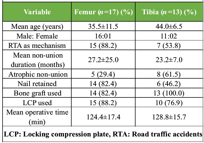
Table 1: Demographic and surgical summary
Radiological outcomes:
Union was achieved in all 30 patients (100%). The mRUST score rose progressively from 5.17 ± 0.79 preoperatively to 15.33 ± 0.84 at final follow-up (paired t-test: t = 9.14, df = 29, P < 0.001) (Table 2).
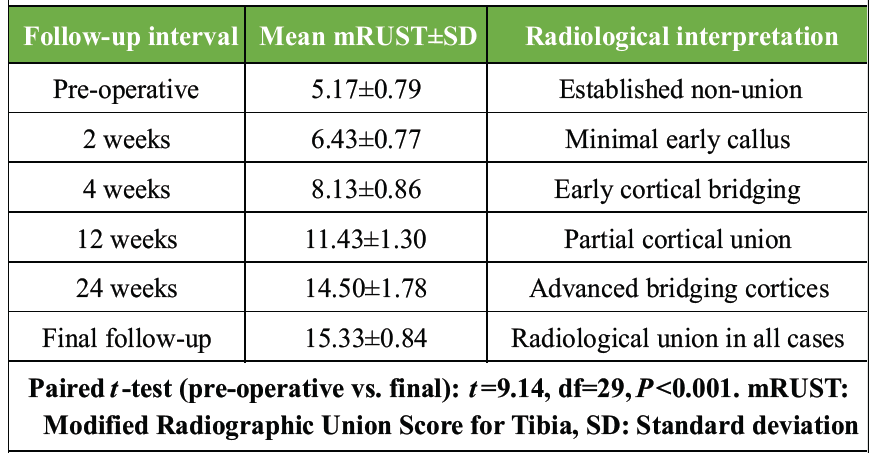
Table 2: Serial mRUST scores at follow-up intervals
The femoral group achieved a significantly higher final mRUST than the tibial group (15.71 ± 0.47 vs. 14.85 ± 0.99; P = 0.004). Mean time to union was 6.23 ± 0.82 months overall; femoral non-unions healed significantly faster (5.88 ± 0.70 vs. 6.69 ± 0.75 months; P = 0.005). Patients with retained nails had the shortest union time (6.0 ± 0.7 months) versus exchange nailing (6.7 ± 0.8 months) and new nail insertion (7.2 ± 0.9 months). Radiological outcomes are summarized in Table 2. A representative case illustrating serial radiological progression is shown in Fig. 1.
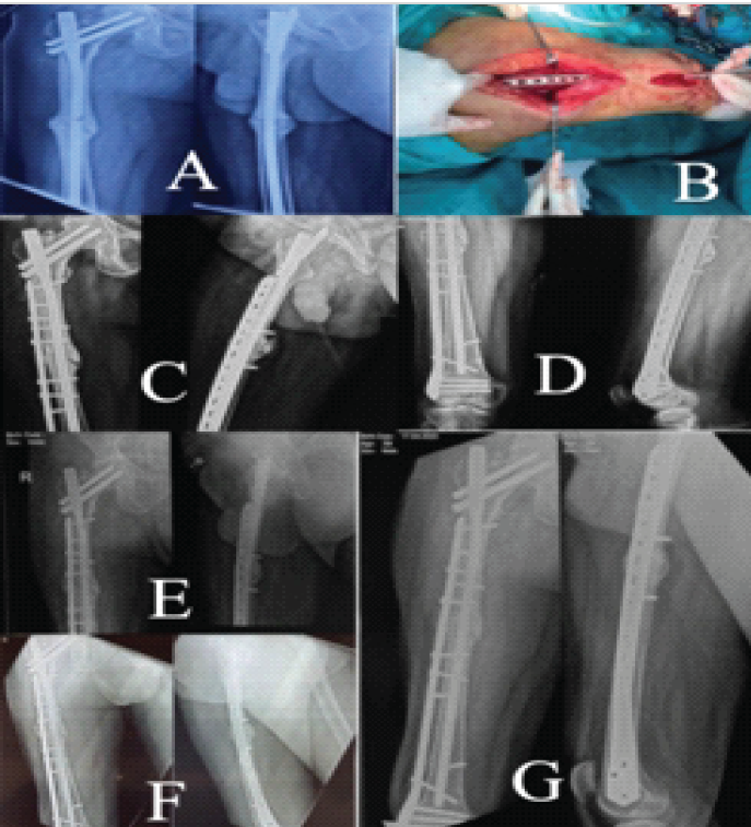
Figure 1: X-rays showing pre-operative (a), intraoperative clinical image (b), immediate post-operative (c), serial follow-up at 2 weeks (d), 4 weeks (e), 3 months (f), and 6 months (g).
Functional outcomes:
Preoperative LEFS scores indicated severe functional limitation (mean 31.77 ± 3.39). LEFS improved progressively at each follow-up visit, reaching 76.60 ± 3.77 at the final follow-up—representing near-normal lower limb function—with the improvement highly significant (paired t-test: t = 14.62, df = 29, P < 0.001). The mean LEFS improvement was 44.83 ± 4.04 points. Serial LEFS scores are presented in Table 3. Clinical images demonstrating functional recovery are presented in Fig. 2.
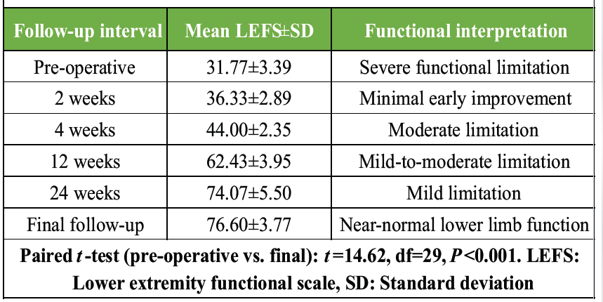
Table 3: Serial LEFS scores at follow-up intervals
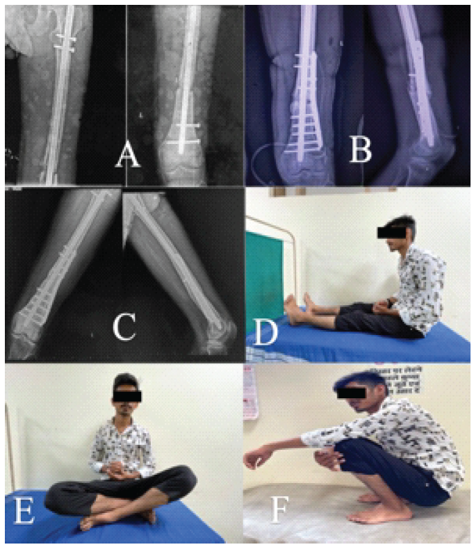
Figure 2: X-rays showing pre-operative (a), immediate post-operative (b), follow-up at 6 months (c), clinical images of functional positions (d-f) at 6 months.
By Wu’s criteria, 18 patients (60%) achieved excellent outcomes, 9 (30%) good, 3 (10%) fair, and none poor—an excellent-to-good rate of 90%. The femoral group recorded significantly higher final LEFS scores than the tibial group (78.24 ± 1.60 vs. 74.46 ± 4.70; P = 0.004), with a correspondingly higher excellent-to-good rate (94.1% vs. 84.6%). The distribution of Wu’s grades and final LEFS by anatomical site is presented in Table 4, and overall comparisons between groups are in Table 5.
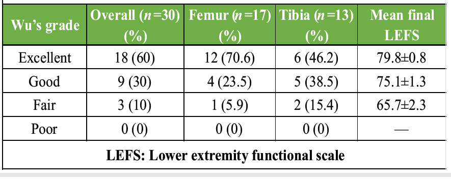
Table 4: Functional outcomes by Wu’s criteria
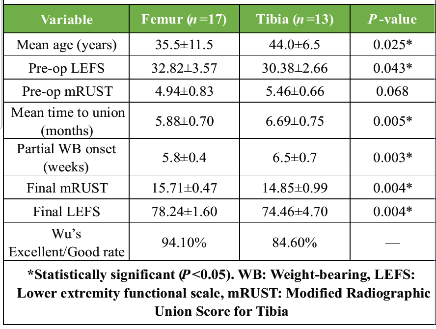
Table 5: Femur versus tibia comparison – primary outcomes
Weight-bearing:
Partial weight-bearing was initiated at a mean of 6.1 ± 0.6 weeks overall. Femoral patients commenced partial loading significantly earlier than tibial patients (5.8 ± 0.4 weeks vs. 6.5 ± 0.7 weeks; P = 0.003). Full weight-bearing was achieved at a mean of 12.3 ± 0.8 weeks, with no statistically significant difference between the two groups (P = 0.364).
Complications:
Complications occurred in 3 of 30 patients (10%). Two patients developed superficial surgical site infections, both resolving fully with wound care and culture-directed oral antibiotics. One patient developed a deep infection requiring formal debridement and implant retention under parenteral antibiotic cover (Debridement, Antibiotics, Irrigation, and Retention protocol); fracture union was nonetheless achieved, and the implant was electively removed after confirmed union at 8 months. There were no refractures, no neurovascular injuries, and no implant failures in the absence of infection. The clinical and radiological course of the deep infection case—including sequential imaging from pre-operative through post-implant removal and final functional recovery—is illustrated in Fig. 3.
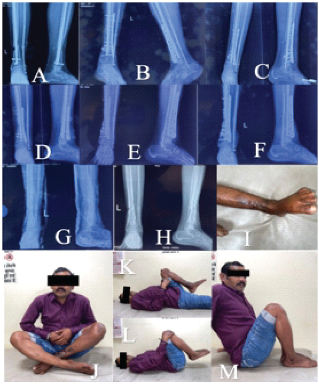
Figure 3: X-rays showing pre-operative (a), immediate post-operative (b), serial follow-up at 2 weeks (c), 4 weeks (d), 3 months (e), 8 months (f), post-implant removal (g), 1 month post-implant removal (h), clinical images of healed surgical site scar post-implant removal (i), functional positions at 9 months (j-m).
Correlation analysis:
The Calori non-union score showed only a mild, non-significant positive correlation with time to union (r = 0.35, P = 0.062) and a negligible negative correlation with final LEFS (r = −0.12, P = 0.538). In contrast, final mRUST and final LEFS scores were strongly correlated (r = 0.81, P < 0.001), confirming that radiological union translates directly to functional recovery in this cohort.
The central finding of this study is clear: Augmentation plating achieved 100% union in 30 patients with aseptic femoral and tibial non-unions, a mean time to union of approximately 6 months, and near-normal functional recovery. The strong correlation between final mRUST and final LEFS (r = 0.81, P < 0.001) confirms that radiological healing in this context translates directly to restored lower limb function—a relationship not always demonstrated in non-union studies. The functional improvement is clinically substantial: A mean LEFS gain of 44.83 points (from severe limitation to near-normal) is well above the LEFS minimal detectable change of 9 points, and the 90% excellent-to-good rate by Wu’s criteria compares favorably with the best published series—Wu’s original 1996 report (85% excellent-to-good); Birjandinejad et al. (100% union, 6.8 months); Lin et al. (100% union, 5.8 months); and Vaishya et al. (100% union, 6.25 months) [11,12,13,14]. The significantly better functional outcomes in femoral versus tibial non-unions reflect the femur’s richer muscular envelope and vascularity versus the tibia’s compromised extraosseous blood supply—a site-specific pattern consistent across the literature [12,13]. Mechanically, augmentation plating addresses the principal failure mode of IMN non-union—rotational instability—by creating a dual-implant construct in which the nail continues to share axial loads while the plate controls the rotational plane. This has been demonstrated biomechanically by Park et al. [15] and Walcher et al. [16] and has been confirmed clinically by Luo et al.’s meta-analysis of 232 patients, which showed that augmentation plating outperformed exchange nailing on union rate, time to union, blood loss, and operative time [7]. Our own operative profile (mean 126 min, 221 mL blood loss) aligns with published augmentation plating series. Nail retention (66.7% of our cohort) was associated with the shortest union time (6.0 months), consistent with Wang et al. [17], who demonstrated that retaining the nail outperforms nail removal with augmentation on all primary outcome measures. Biologically, nail retention preserves the endosteal vascularity reconstituted around the implant over months to years of implantation. Our selective grafting approach—withholding grafts from biologically active hypertrophic non-unions—achieved 100% union, in keeping with Wang et al. [18] and Metwaly and Younis [19], who demonstrated that mechanical stabilization alone suffices in biologically competent non-unions. Although atrophic, hypertrophic, and oligotrophic non-unions were not separately powered for comparative subgroup analysis, the uniform 100% union rate across all pattern types supports the versatility of augmentation plating regardless of non-union biology. The 10% complication rate reflects the realities of revision surgery in a resource-limited tertiary setting. Notably, all three infected patients achieved union. In the deep infection case, the mechanical redundancy of the nail-plate construct allowed aggressive debridement without destabilizing the fracture—a distinct advantage over single-implant systems [20]. Limitations include the single-center design, moderate sample size, absence of a control group, and follow-up limited to radiological union without long-term implant-related data. The heterogeneous cohort (femur and tibia; three non-union types; two nail-management strategies) reflects real-world practice but complicates subgroup analysis. The single-center design at a government tertiary institution in Central India—where the patient profile is characterized by high-energy road traffic injuries, delayed referral, nutritional compromise, and heavy occupational demands—limits external validity; findings may not be directly applicable to settings with different demographics, implant availability, or surgical infrastructure, and multicenter prospective validation is required to confirm generalizability. The sample of 30 patients, while consistent with several published augmentation plating series [11,12,13,14], reduces statistical power for subgroup analyses and increases the risk of Type II error in secondary comparisons; the 100% union rate should therefore be interpreted cautiously pending confirmation in larger cohorts. The observational design without randomization precludes direct causal comparison with alternative revision strategies—including exchange nailing alone or plate fixation after nail removal—and is subject to inherent selection bias in the allocation of bone grafting and nail-management strategy, both of which were guided by intraoperative findings and surgeon judgment rather than a pre-specified randomized protocol. Variability in nail-management approach and selective bone grafting reflects individualized intraoperative decision-making. While this mirrors real-world practice and is supported by published evidence [18,19], it limits strict protocol reproducibility and the generalizability of a uniform surgical recommendation. Follow-up was directed primarily at radiological union and short-term functional recovery; long-term endpoints—including implant loosening, elective implant removal rates, refracture incidence, and functional durability beyond the study period—were not assessed and represent important areas for future investigation. Functional assessment using LEFS and Wu’s criteria, while validated, does not capture broader health-related quality of life (e.g., return-to-work data) or the economic burden of the procedure—including implant costs, operative resource utilization, and productivity losses—all of which would be valuable in a predominantly working-age, occupationally active cohort treated in a resource-limited public health setting. Patients with infected non-unions, pathological fractures, and metabolic bone disorders were deliberately excluded to define outcomes in a homogeneous aseptic population; the findings are therefore not applicable to these more complex clinical scenarios. Finally, male patients comprised 90% of the cohort, reflecting the regional epidemiology of high-energy road traffic injuries in Central India; generalizability to female patients and elderly osteoporotic populations—who may exhibit different healing biology, implant anchorage, and functional recovery trajectories—remains to be established. Collectively, these findings should be regarded as hypothesis-generating evidence from a prospectively conducted observational series, with the 100% union rate representing an encouraging signal that warrants confirmation in larger, multicenter, and ideally randomized studies.
Augmentation plating with selective autologous bone grafting achieves 100% union rates, a predictable 5–8 month healing timeline, and 90% excellent-to-good functional outcomes in aseptic femoral and tibial non-unions after failed IMN. The strong correlation between radiological and functional recovery (r = 0.81, P < 0.001) underscores the clinical significance of achieving solid union. Nail retention is the preferred approach, preserving endosteal biology and reducing operative morbidity. The technique should be considered the first-choice revision strategy for appropriately selected patients with aseptic long bone non-union after failed intramedullary fixation.
Augmentation plating with nail retention is a mechanically superior and biologically preserving revision strategy for aseptic femoral and tibial non-unions after failed IMN, achieving 100% union without the morbidity of nail extraction. Functional recovery closely mirrors radiological healing—a strong mRUST–LEFS correlation (r = 0.81, P < 0.001) confirms that solid union reliably translates to near-normal lower limb function, with 90% excellent-to-good outcomes by Wu’s criteria. Selective bone grafting guided by intraoperative biological assessment is sufficient; routine grafting in hypertrophic non-unions adds avoidable donor-site morbidity. Femoral non-unions heal faster and recover better functional scores than tibial non-unions, and this should be factored into patient counseling and rehabilitation planning. Even in the presence of surgical site infection, the mechanical redundancy of the nail-plate construct permits aggressive infection control without destabilizing the fracture, allowing concurrent union—a distinct advantage over single-implant revision strategies.
References
- 1. 1 Mills LA, Aitken SA, Simpson AH. The risk of non-union per fracture: Current myths and revised figures from a population of over 4 million adults. Acta Orthop 2017;88:434-9. [Google Scholar] [PubMed]
- 2. 2 Zura R, Xiong Z, Einhorn T, Watson JT, Ostrum RF, Prayson MJ, et al. Epidemiology of fracture nonunion in 18 human bones. JAMA Surg 2016;151:e162775. [Google Scholar] [PubMed]
- 3. 3 Rupp M, Biehl C, Budak M, Thormann U, Heiss C, Alt V. Diaphyseal long bone nonunions – types, aetiology, economics, and treatment recommendations. Int Orthop 2018;42:247-58. [Google Scholar] [PubMed]
- 4. 4 Weresh MJ, Hakanson R, Stover MD, Sims SH, Kellam JF, Bosse MJ. Failure of exchange reamed intramedullary nails for ununited femoral shaft fractures. J Orthop Trauma 2000;14:335-8. [Google Scholar] [PubMed]
- 5. 5 Brinker MR, O’Connor DP. Exchange nailing of ununited fractures. J Bone Joint Surg Am 2003;85:165-73. [Google Scholar] [PubMed]
- 6. 6 Ueng SW, Chao EK, Lee SS, Shih CH. Augmentative plate fixation for the management of femoral nonunion after intramedullary nailing. J Trauma 1997;43:640-4. [Google Scholar] [PubMed]
- 7. 7 Luo H, Su Y, Ding L, Xiao H, Wu M, Xue F. Exchange nailing versus augmentative plating in the treatment of femoral shaft nonunion after intramedullary nailing: A meta-analysis. EFORT Open Rev 2019;4:513-8. [Google Scholar] [PubMed]
- 8. 8 Calori GM, Phillips M, Jeetle S, Tagliabue L, Giannoudis PV. Classification of non-union: Need for a new scoring system? Injury 2008;39 Suppl 2:S59-63. [Google Scholar] [PubMed]
- 9. 9 Binkley JM, Stratford PW, Lott SA, Riddle DL. The lower extremity functional scale (LEFS): Scale development, measurement properties, and clinical application. North American orthopaedic rehabilitation research network. Phys Ther 1999;79:371-83. [Google Scholar] [PubMed]
- 10. 10 Leow JM, Clement ND, Tawonsawatruk T, Simpson CJ, Simpson AH. The radiographic union scale in tibial (RUST) fractures: Reliability of the outcome measure at an independent centre. Bone Joint Res 2016;5:116-21. [Google Scholar] [PubMed]
- 11. 11 Wu CC. Augmentative plate fixation for femoral shaft nonunion after intramedullary nailing. J Trauma 1996;40:640-4. [Google Scholar] [PubMed]
- 12. 12 Birjandinejad A, Ebrahimzadeh MH, Ahmadzadeh-Chabock H. Plate augmentation for femoral and tibial nonunions. Arch Orthop Trauma Surg 2009;129:1199-204. [Google Scholar] [PubMed]
- 13. 13 Lin CJ, Chiang CC, Wu PK, Chen CF, Huang CK, Su AW, et al. Effectiveness of plate augmentation for femoral shaft nonunion after nailing. J Chin Med Assoc 2012;75:396-401. [Google Scholar] [PubMed]
- 14. 14 Vaishya R, Agarwal AK, Gupta N, Vijay V. Plate augmentation with retention of intramedullary nail is effective for resistant femoral shaft non-union. J Orthop 2016;13:242-5. [Google Scholar] [PubMed]
- 15. 15 Park K, Kim K, Choi YS. Comparison of mechanical rigidity between plate augmentation leaving the nail in situ and interlocking nail using cadaveric fracture model of the femur. Int Orthop 2011;35:581-5. [Google Scholar] [PubMed]
- 16. 16 Walcher MG, Day RE, Gesslein M, Bail HJ, Kuster MS. Augmentative plating versus exchange intramedullary nailing for the treatment of aseptic non-unions of the femoral shaft – a biomechanical study in a SawboneTM model. J Pers Med 2023;13:650. [Google Scholar] [PubMed]
- 17. 17 Chaofeng W, Hao G, Changjun H, Liang S, Congming Z, Teng M. Retaining intramedullary nail with plate fixation outperforms nail removal and double-plate fixation in femoral shaft nonunion. Eur J Med Res 2026;31:179. [Google Scholar] [PubMed]
- 18. 18 Wang JW, Weng LH, Lee MS. Augmentation plating with or without bone graft for femoral nonunion. Injury 2014;45:1909-13. [Google Scholar] [PubMed]
- 19. 19 Metwaly RG, Younis AS. Augmentation plate without bone graft in the management of distal tibial diaphyseal non-union. Injury 2023;54:111057. [Google Scholar] [PubMed]
- 20. 20 Mohamed MA, Noaman HH, Soroor YO, Elsayed M. Plate augmentation and bone grafting in treatment of femoral shaft nonunion initially fixed by intramedullary nail. SICOT J 2022;8:19. [Google Scholar] [PubMed]






Related Articles in Journal of Orthopaedic Case Reports
 October 1, 2026 Pertrochanteric Femur Fracture in a Post-Polio Patient treated with a Pediatric Hip Plate: A Case Report
 September 1, 2026 Salvage of Complex Bimalleolar Ankle Fracture Non-union after Failed Tibiotalocalcaneal and Charnley Arthrodesis using Fibular Strut Grafting and Ilizarov Fixation: A Case Report
 August 1, 2026 The “Terrible Tetrad” of the Elbow: A Case Series of Two Patients with a Rare Quadruple-Component Injury and a Proposed New Classification Term
 August 1, 2026 Management of Infected Non-union of Long Bones Using Antibiotic Cement-Coated Intramedullary Nails: A Prospective Case Series