

Post-operative C5 palsy can occur despite technically successful cervical decompression and stable intraoperative neuromonitoring, making meticulous post-operative neurological assessment and early rehabilitation essential for favorable outcomes.
Dr. Sandesh Subhash Agrawal, Department of Orthopedics and Spine Surgery, Shree Narayana Hospital, Raipur, Chhattisgarh, India. E-mail: preciouss333@gmail.com;
Abstract
Introduction: Post-operative C5 palsy remains an unpredictable neurological complication following posterior cervical decompression for cervical compressive myelopathy. This study aimed to analyze its incidence, clinical characteristics, management, and neurological recovery.
Materials and Methods: This retrospective case series included 9 patients (4.5%) who developed post-operative C5 palsy among 199 posterior cervical decompression procedures performed between January 2023 and January 2026. Demographic, radiological, operative, and neurological data were reviewed. All patients underwent posterior cervical decompression with instrumented stabilization under continuous intraoperative neuromonitoring using motor evoked potentials and somatosensory evoked potentials (SSEPs). Neurological recovery was assessed using serial Medical Research Council muscle strength grading.
Results: The mean age was 56.0 ± 12.4 years, with 7 males (77.8%). Severe multilevel cervical stenosis involving predominantly the C3–C7 levels was present in most patients. Stable intraoperative MEP and SSEP signals were observed in all cases. Right-sided C5 palsy occurred in 5 patients (55.6%), and neurological recovery was achieved in 8 patients (88.9%) with conservative management, with a mean recovery duration of 6.2 ± 2.1 weeks. One patient with severe multilevel stenosis and reduced preoperative cervical lordosis demonstrated a persistent neurological deficit at 6 months.
Conclusion: Post-operative C5 palsy is a delayed neurological complication that usually responds well to conservative treatment. Early recognition and structured rehabilitation remain key to optimizing neurological recovery.
Keywords: Cervical compressive myelopathy, cervical decompression surgery, cervical instrumentation, neurological deficit, posterior cervical laminectomy, post-operative C5 palsy.

Post-operative C5 palsy is a recognized neurological complication following cervical decompression surgery performed for cervical compressive myelopathy, including cervical spondylotic myelopathy (CSM) and ossification of the posterior longitudinal ligament (OPLL) [1,2,3]. It is characterized by new-onset weakness of the deltoid and/or biceps muscles, often associated with shoulder pain and sensory disturbances, without significant deterioration of lower extremity myelopathic symptoms [2,4]. Although the condition is frequently transient, persistent neurological deficits causing functional impairment have also been reported [2,5]. The reported incidence varies widely in the literature, ranging from 0.4% to 30%, depending on patient characteristics, surgical approach, and diagnostic criteria [1,2,3,4,5,6,7]. Large multicenter studies have demonstrated an overall incidence between 0.58% and 6.3% following cervical spine surgery [4,5]. The complication is more commonly observed after posterior cervical decompression procedures, including laminoplasty and laminectomy with fusion, compared with anterior approaches. Patients with multilevel cervical compression and OPLL appear to be at particularly increased risk following extensive posterior decompression and fusion procedures [3,5,6]. Despite extensive investigation, the exact pathophysiology remains incompletely understood. Proposed mechanisms include direct intraoperative nerve root injury, posterior spinal cord shift causing traction injury to the relatively short C5 nerve root, ischemia–reperfusion injury, and thermal injury during decompression [6,7,8]. Among these, traction injury secondary to post-operative spinal cord drift remains the most widely accepted theory. Clinically, C5 palsy may occur immediately after surgery or develop in a delayed manner several days postoperatively. Most of the patients demonstrate gradual neurological recovery with conservative management and rehabilitation; however, residual motor weakness may persist in a subset of patients [2,4,5]. Persistent deficits can significantly impair shoulder function, activities of daily living, post-operative satisfaction, and overall quality of life. Given its relatively low incidence but important functional implications, further institutional experiences are valuable for improving understanding of the clinical profile, recovery patterns, and prognostic factors associated with post-operative C5 palsy. In the present study, we report a case series of nine patients who developed post-operative C5 palsy following cervical decompression surgery for cervical compressive myelopathy. Patient demographics, operative characteristics, timing of symptom onset, management strategies, and neurological outcomes were analyzed, with particular emphasis on recovery patterns and factors associated with persistent neurological deficit.
Study design:
A retrospective observational case series was conducted at Shree Narayana Hospital between October 2023 and October 2025 to evaluate the clinical presentation, management, and neurological outcomes of patients who developed post-operative C5 palsy following surgery for cervical compressive myelopathy. Institutional Ethics Committee Approval Number: SNH/IEC/Cert./SPINE1.4/2026 was obtained before the commencement of the study, and patient confidentiality was maintained throughout. The study adhered to the ethical principles outlined in the Declaration of Helsinki. All eligible consecutive patients who developed post-operative C5 palsy during the study were included. Patient selection: Hospital electronic medical records, operative notes, radiological investigations, and follow-up records were retrospectively reviewed. Among 199 patients who underwent posterior cervical decompression procedures for cervical compressive myelopathy during the study period, 9 patients who developed post-operative C5 palsy were included in the present study. Inclusion criteria consisted of patients undergoing posterior cervical decompression and instrumented stabilization for cervical compressive myelopathy who subsequently developed post-operative C5 palsy during the study. Post-operative C5 palsy was defined as a new-onset reduction in deltoid muscle power by at least one Medical Research Council (MRC) grade compared with the pre-operative neurological examination, with or without associated biceps weakness, sensory symptoms, or post-operative shoulder pain, in the absence of worsening lower limb neurological function. Exclusion criteria included previous cervical spine surgery, traumatic cervical injury, brachial plexus pathology, documented intraoperative direct nerve root injury, and incomplete clinical records.
Clinical and radiological evaluation:
Demographic, clinical, operative, and follow-up data were collected from institutional records. Variables analyzed included age, sex, diagnosis, operative levels, onset of post-operative C5 palsy, severity of motor weakness, associated symptoms, duration of recovery, and final neurological outcome. Neurological assessment was performed preoperatively and during serial post-operative follow-up using the MRC grading system, with specific evaluation of shoulder abduction, deltoid, and biceps muscle strength. All patients underwent pre-operative cervical radiographs and magnetic resonance imaging (MRI). Computed tomography (CT) imaging was additionally evaluated in selected patients with complex cervical pathology or OPLL. Post-operative imaging was reviewed when clinically indicated. Patients who developed new-onset post-operative upper limb weakness underwent a detailed neurological examination. Post-operative cervical MRI and/or CT was performed to exclude epidural hematoma, residual spinal cord compression, implant malposition, or other structural causes of neurological deterioration. The diagnosis of post-operative C5 palsy was established based on new-onset deltoid weakness with or without associated biceps weakness after exclusion of compressive pathology on post-operative imaging.
Surgical procedure:
All patients underwent posterior cervical decompression with instrumented stabilization under general anesthesia using standard microsurgical techniques and intraoperative neuromonitoring. The procedure consisted of a multilevel cervical laminectomy followed by posterior fixation using lateral mass or pedicle screw-rod constructs, depending on cervical anatomy and surgeon preference. Surgical levels were determined according to radiological findings and the extent of spinal cord compression. Intraoperative somatosensory evoked potentials (SSEPs) and motor evoked potentials (MEPs) were utilized in all cases for continuous neurological monitoring. Adequate spinal cord decompression and implant positioning were confirmed intraoperatively before wound closure.
Post-operative management and rehabilitation:
Patients developing post-operative C5 palsy were managed conservatively with neurological monitoring, analgesics, short-course corticosteroids, and supervised physiotherapy rehabilitation. Rehabilitation focused on passive and active-assisted shoulder mobilization, progressive strengthening exercises of the deltoid and biceps muscles, prevention of shoulder stiffness, and restoration of upper limb function. Patients were followed up at 1, 2, 3, and 6 months postoperatively, with serial neurological examinations performed during each visit to assess recovery. Complete recovery was defined as restoration of muscle power to pre-operative MRC grade, partial recovery as improvement without full restoration of baseline strength, and persistent deficit as failure to demonstrate meaningful neurological improvement at final follow-up.
Statistical analysis:
Descriptive statistical analysis was performed using Statistical Package for the Social Sciences software version 26.0 (IBM Corp., Armonk, NY, USA) for all study variables. Continuous variables were expressed as mean ± standard deviation, while categorical variables were presented as frequencies and percentages. Inferential statistical analysis was not performed.
The present retrospective case series evaluated the clinical characteristics, radiological profile, operative variables, intraoperative neuromonitoring findings, rehabilitation course, and neurological recovery patterns of nine patients who developed post-operative C5 palsy following posterior cervical decompression and instrumented stabilization for cervical compressive myelopathy. Among 199 posterior cervical decompression procedures performed during the study, 9 patients developed post-operative C5 palsy, representing an incidence of 4.5%. Individual patient characteristics, timing of neurological deterioration, laterality of palsy, recovery duration, and final functional outcomes were additionally analyzed as depicted in Table 1.
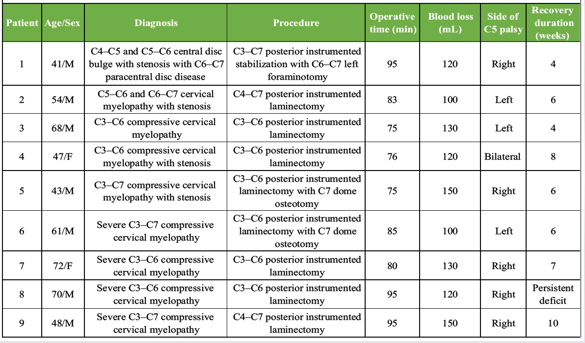
Table 1: Individual patient characteristics, operative details, and neurological recovery
Demographic and baseline clinical characteristics:
A total of 9 patients who developed post-operative C5 palsy following posterior cervical decompression and instrumented stabilization for CSM were included in this retrospective case series. All patients had symptomatic multilevel cervical compressive myelopathy with cervical canal stenosis refractory to conservative treatment. The mean age was 56.0 ± 12.4 years (range: 41–72 years), with a male predominance (7 males, 77.8%). Medical comorbidities were present in a subset of patients, with diabetes mellitus in 2 patients (22.2%), hypertension in 2 patients (22.2%), and one patient having both diabetes mellitus and hypertension. No patient had a prior history of cervical spine surgery. Radiologically, severe multilevel cervical stenosis predominantly involving the C3–C7 levels was observed in most patients. Pre-operative Nurick grades ranged from III to V, indicating moderate-to-severe cervical myelopathy, with Nurick Grade IV being the most common presentation (44.4%), followed by Grade III (33.3%) and Grade V (11.1%). The mean pre-operative cervical lordosis was 17.1° ± 3.8° (range: 12°–25°). Reduced cervical lordosis was more commonly observed in patients with severe multilevel stenosis and delayed neurolpreoperativery, as depicted in Table 2.
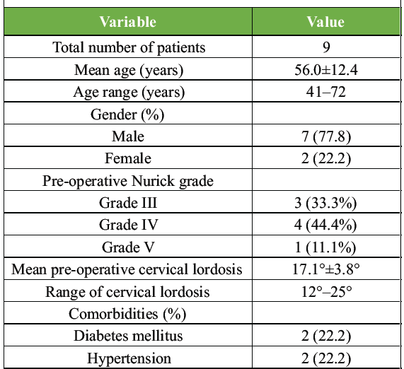
Table 2: Baseline demographic and clinical characteristics
Radiological evaluation and pre-operative cervical lordosis:
All patients underwent pre-operative radiological evaluation with MRI and standing lateral cervical radiographs. Cervical sagittal alignment was assessed using the C2–C7 Cobb angle. The mean pre-operative cervical lordosis was 17.1° ± 3.8° (range: 12°–25°). Lower pre-operative cervical lordosis values were observed in patients who demonstrated delayed or incomplete neurological recovery. The patient with the lowest preoperative cervical lordosis (12°) failed to demonstrate neurological recovery during follow-up. Multilevel cord compression with associated T2-weighted intramedullary signal changes suggestive of chronic compressive myelopathy was observed in several patients.
Operative characteristics:
The extent of decompression was tailored according to the severity and cranio-caudal extent of cervical stenosis, with multilevel posterior instrumented laminectomy involving the C3–C6 or C4–C7 levels being the most commonly performed procedure. Additional procedures included C7 dome osteotomy in 2 patients (22.2%) and unilateral C6–C7 foraminotomy in 1 patient (11.1%). Instrumented stabilization was performed in all patients to preserve cervical alignment and prevent post-operative instability or kyphotic dpreoperative mean operative duration was 84.3 ± 8.8 min (range: 75–95 min), with a mean intraoperative blood loss of 124.4 ± 18.6 mL (range: 100–150 mL). No intraoperative dural injury, vascular injury, implant malposition, or perioperative mortality was observed. Continuous multimodal intraoperative neuromonitoring using MEPs and SSEPs remained stable in all patients without significant signal deterioration or amplitude drop throughout the procedure, despite the development of post-operative C5 palsy as depicted in Table 3.
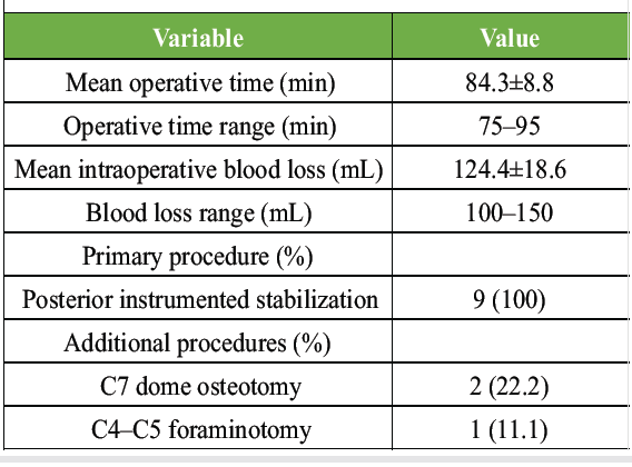
Table 3: Operative characteristics of patients with post-operative C5 palsy
Incidence and clinical pattern of post-operative C5 palsy:
Neurological deficits developed within 24 h in 6 patients and within 72 h in the remaining 3 patients. Clinically, patients presented with varying degrees of deltoid weakness and impaired shoulder abduction, with associated biceps weakness observed in selected cases. Right-sided C5 palsy was noted in 5 patients (55.6%), left-sided involvement in 3 patients (33.3%), and bilateral involvement in 1 patient (11.1%). Post-operative imaging did not demonstrate epidural hematoma, residual compression, implant malposition, or new spinal cord signal abnormality in any patient. All patients were managed conservatively using a standardized post-operative rehabilitation protocol, including intravenous corticosteroid administration, structured physiotherapy, range-of-motion exercises, strengthening rehabilitation, and electrical muscle stimulation. Patients with severe weakness required prolonged rehabilitation focusing on shoulder abduction strengthening and prevention of muscle atrophy. None of the patients required revision surgery for management of post-operative C5 palsy, as illustrated in Table 4.
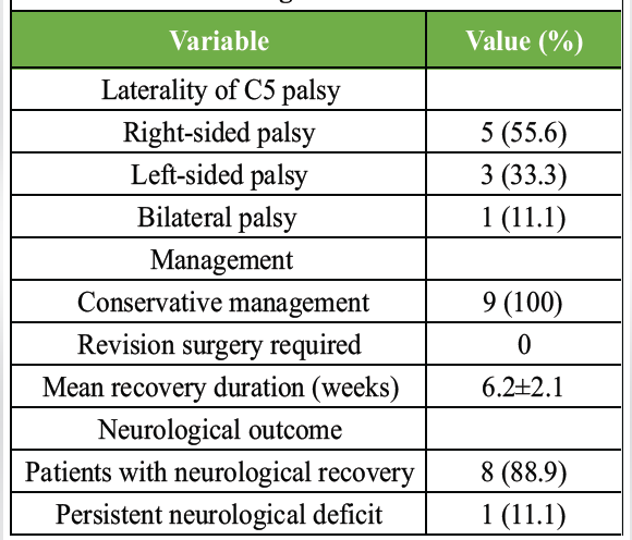
Table 4: Clinical presentation, management, and neurological outcomes
Neurological recovery and functional outcome:
All patients were followed up clinically at 1, 2, 3, and 6 months postoperatively with serial neurological assessment using the MRC grading system for shoulder abduction, deltoid, and biceps muscle power. Neurological improvement was observed in 8 patients (88.9%), with progressive recovery in shoulder abduction, deltoid strength, and elbow flexion during follow-up. The mean recovery duration was 6.2 ± 2.1 weeks (range: 4–10 weeks) as depicted in Table 5.
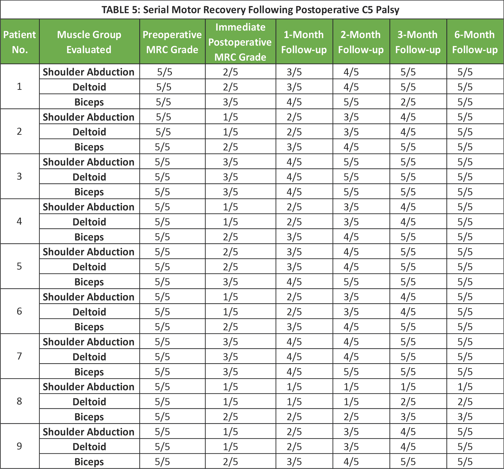 Earlier recovery was generally observed in patients with preserved cervical lordosis and less severe post-operative weakness. One patient (11.1%) with severe multilevel cervical stenosis and reduced pre-operative cervical lordosis (12°) failed to demonstrate meaningful neurological recovery despite 6 months of structured rehabilitation and physiotherapy. Overall, substantial functional improvement was achieved in the majority of patients with conservative management, as shown in Fig. 1.
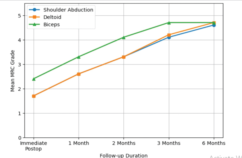
Figure 1: Trend of post-operative motor recovery based on Medical Research Council (MRC) grading of shoulder abduction, deltoid, and biceps muscle power during follow-up.
A pictorial representation of a 68-year-old male with compressive cervical myelopathy who underwent posterior cervical decompression and fusion. Pre-operative and post-operative imaging demonstrated severe cervical cord compression and satisfactory post-operative decompression, respectively (Fig. 2 and 3).
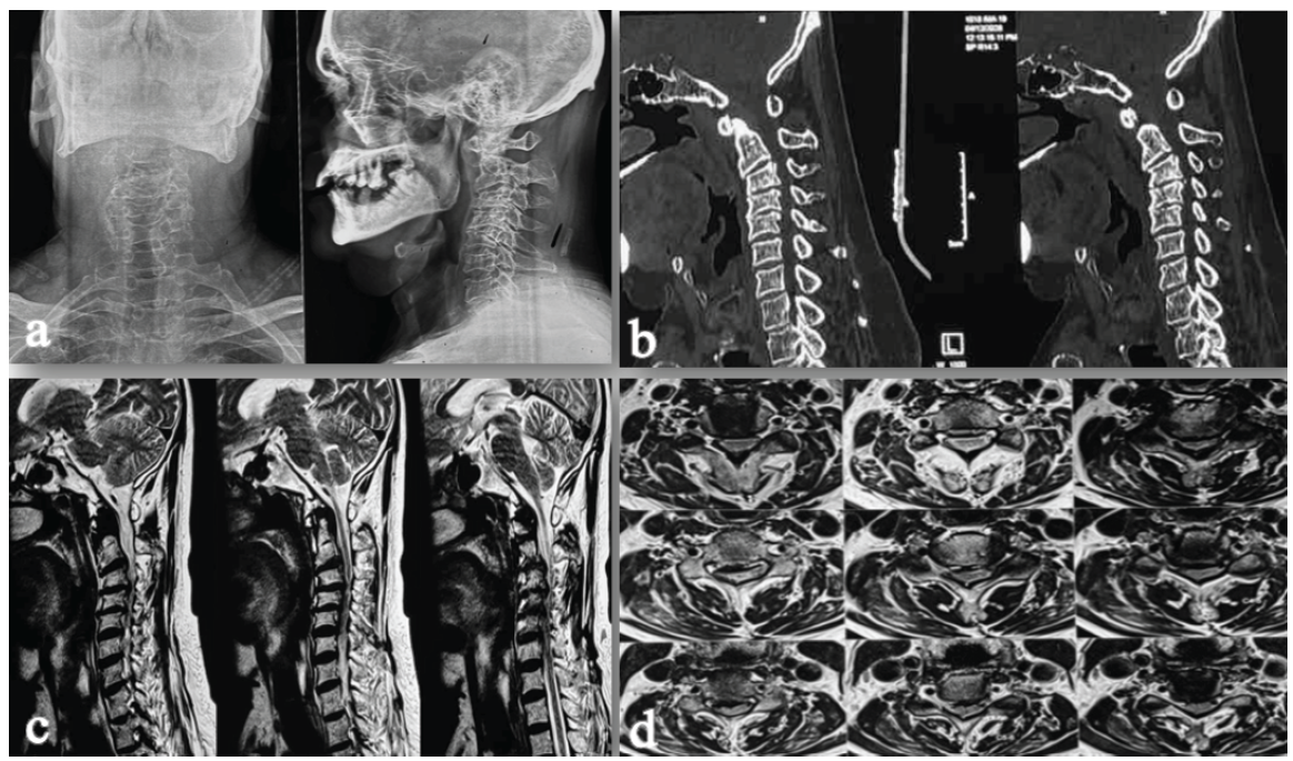
Figure 2: Pre-operative radiographic, computed tomography (CT), and magnetic resonance imaging (MRI) evaluation of the cervical spine. (a) Anteroposterior and lateral cervical spine radiographs demonstrating cervical alignment and degenerative spondylotic changes. (b) Sagittal CT images of the cervical spine showing multilevel degenerative changes with ossification/calcification contributing to spinal canal narrowing. (c) Sagittal T2-weighted MRI images demonstrating multilevel cervical spinal canal stenosis with cord compression and associated intramedullary T2 hyperintensity suggestive of compressive myelopathy. (d) Axial T2-weighted MRI images showing varying degrees of cervical spinal canal stenosis and spinal cord compression at multiple levels.
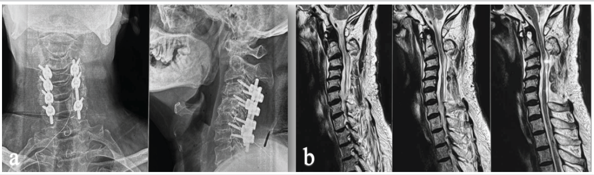
Figure 3: Post-operative imaging following C3–C6 laminectomy and posterior cervical fusion. (a) Anteroposterior and lateral radiographs demonstrating stable posterior instrumentation and maintained cervical alignment. (b) Sagittal T2-weighted magnetic resonance images demonstrating successful decompression of the cervical spinal cord with expansion of the spinal canal and improved cerebrospinal fluid signal surrounding the cord.
Post-operative C5 palsy remains a recognized neurological complication following cervical decompression surgery for cervical compressive myelopathy because of its unpredictable onset, uncertain pathophysiology, and variable neurological recovery [1,2]. It is characterized by new-onset weakness involving the deltoid and/or biceps muscles, frequently associated with shoulder pain and sensory disturbances without worsening of lower extremity neurological function [2,3]. Although the condition is often transient, persistent motor deficits resulting in functional impairment have also been reported [4,5]. The reported incidence of post-operative C5 palsy varies considerably across the literature, ranging from 0.4% to 30% depending on surgical approach, patient characteristics, and diagnostic criteria [3]. Oh et al. reported an overall incidence of 0.58% following cervical spine surgery in a multicenter epidemiological study [4], whereas Egawa et al. observed post-operative C5 palsy in approximately 6.3% of patients undergoing surgery for OPLL, particularly following posterior decompression and fusion procedures [5]. These findings suggest that posterior cervical decompression procedures and multilevel cervical pathology have been associated with a greater frequency of post-operative C5 palsy. Posterior cervical decompression surgeries have consistently demonstrated a greater association with post-operative C5 palsy compared with anterior cervical approaches [4,5,6,7]. Nassr et al. reported increased occurrence following extensive multilevel posterior decompression procedures [6], while Sargunan et al. proposed that posterior spinal cord migration following decompression may increase traction forces on the relatively short and horizontally oriented C5 nerve root [7]. In the present series, all patients developed post-operative C5 palsy following multilevel posterior decompression and instrumented stabilization, which is consistent with previously reported observations. Despite extensive investigation, the exact pathophysiological mechanism underlying post-operative C5 palsy remains incompletely understood. Proposed mechanisms include direct intraoperative nerve root injury, traction neuropathy secondary to post-operative spinal cord drift, ischemia-reperfusion injury, and thermal injury during decompression [9,10]. Among these, post-operative traction injury to the C5 nerve root remains the most widely accepted theory [3,7]. Interestingly, intraoperative neuromonitoring signals remained stable in all patients in the present series despite subsequent post-operative neurological deficits, which may support previously proposed delayed traction-related mechanisms described in the literature. Delayed neurological deterioration represents a characteristic clinical feature of this complication. Oh et al. reported that most patients developed symptoms within the first 3 post-operative days [4], whereas Egawa et al. observed symptom onset within 24 h in nearly half of the affected patients [5]. Similarly, all patients in the present study developed post-operative weakness during the early post-operative period, emphasizing the importance of meticulous neurological monitoring even in patients who are neurologically stable immediately after surgery. Clinically, post-operative C5 palsy most commonly manifests as unilateral deltoid weakness with impaired shoulder abduction. Associated biceps weakness and sensory disturbances may also occur [2,4]. In the present series, deltoid weakness represented the predominant neurological finding, with variable degrees of associated biceps involvement. Right-sided palsy was more common, while bilateral involvement was observed in one patient. Neurological recovery following post-operative C5 palsy is generally favorable, with most patients demonstrating gradual improvement following conservative management and rehabilitation [10,11]. In the current series, 8 out of 9 patients demonstrated significant neurological recovery during follow-up, with a mean recovery duration of 6.2 ± 2.1 weeks. Progressive improvement in shoulder abduction, deltoid strength, and elbow flexion was observed in most patients during serial follow-up evaluations. Patients with relatively preserved cervical lordosis demonstrated earlier neurological recovery, whereas the patient with severe multilevel stenosis and reduced pre-operative cervical lordosis (12°) failed to demonstrate meaningful neurological improvement despite 6 months of rehabilitation and follow-up. These findings suggest that cervical sagittal alignment may influence post-operative neurological recovery. Several prognostic factors associated with incomplete neurological recovery have been described, including severe initial motor weakness, older age, multimuscle involvement, prolonged recovery duration, and associated OPLL [9,11]. Persistent neurological deficits may significantly impair shoulder function, activities of daily living, rehabilitation potential, and post-operative quality of life. Consequently, early recognition and structured rehabilitation remain essential components of post-operative management. Currently, no universally accepted treatment protocol exists for post-operative C5 palsy. Conservative management remains the primary treatment strategy and generally includes corticosteroids, analgesics, physiotherapy, and rehabilitation exercises. In the present study, all patients were managed conservatively using structured physiotherapy rehabilitation, including passive and active-assisted shoulder mobilization, strengthening exercises, and electrical muscle stimulation. None of the patients required revision surgery. The exact etiology of post-operative C5 palsy remains incompletely understood and is likely multifactorial. The most widely accepted mechanism is traction injury to the relatively short C5 nerve root resulting from posterior migration of the spinal cord following decompression. Other proposed mechanisms include ischemia-reperfusion injury, direct nerve root manipulation, thermal injury during decompression, and pre-existing C4–C5 foraminal stenosis. Although no strategy eliminates the risk, meticulous surgical technique with gentle neural tissue handling, adequate but not excessive decompression, consideration of prophylactic C4–C5 foraminotomy in selected high-risk patients, and careful post-operative neurological monitoring may help reduce the incidence and facilitate early recognition of this complication. Previous studies have also suggested that prophylactic C4–C5 foraminotomy during posterior decompression may decrease post-operative nerve root tethering and reduce the incidence of post-operative C5 palsy [12]. Nevertheless, the occurrence of post-operative C5 palsy despite stable intraoperative neuromonitoring further supports the multifactorial nature of this complication [7,13,14]. The present study has several limitations. The retrospective design and relatively small sample size limit the generalizability of the findings. In addition, standardized electrophysiological and functional outcome assessments were not uniformly available because of the retrospective nature of the study. The absence of post-operative electromyography limited detailed electrophysiological characterization of the neurological deficit. In addition, no comparative control group was available, and inferential statistical analysis could not be performed because of the limited sample size. Despite these limitations, the present series provides valuable clinical insight into the presentation, radiological associations, recovery patterns, and management outcomes of post-operative C5 palsy following posterior cervical decompression surgery.
Post-operative C5 palsy remains an important neurological complication following cervical decompression surgery for cervical compressive myelopathy, particularly after multilevel posterior decompression procedures. In the present series, most patients demonstrated gradual neurological recovery with conservative management and structured rehabilitation; however, persistent motor weakness and incomplete recovery were observed in one patient despite prolonged follow-up. Early recognition, meticulous post-operative neurological monitoring, and timely initiation of rehabilitation are essential for optimizing neurological recovery and functional outcomes. The findings of the present study demonstrate generally favorable neurological recovery in most patients with conservative management while emphasizing that delayed or incomplete neurological recovery may occur in patients with severe multilevel disease and reduced cervical lordosis.
Post-operative C5 palsy is an uncommon but well-recognized complication following multilevel posterior cervical decompression and fusion. The condition typically presents as unilateral deltoid weakness with or without biceps involvement, often occurring despite stable intraoperative neuromonitoring and satisfactory post-operative imaging. Current evidence supports post-operative spinal cord drift and traction injury of the C5 nerve root as the most likely mechanism. Most of the patients demonstrate favorable neurological recovery with conservative management, including corticosteroids, physiotherapy, and structured rehabilitation. Careful pre-operative assessment of cervical alignment and C4–C5 foraminal stenosis, meticulous post-operative neurological monitoring, and early rehabilitation are essential for optimizing functional outcomes.
References
- 1. Jeon HS, Kim KN. Delayed bilateral C5 palsy following circumferential decompression and fusion in patient with cervical spondylotic myelopathy. Korean J Spine 2015;12:200-3. [Google Scholar] [PubMed]
- 2. Thompson SE, Smith ZA, Hsu WK, Nassr A, Mroz TE, Fish DE, et al. C5 palsy after cervical spine surgery: A multicenter retrospective review of 59 cases. Global Spine J 2017;7:64S-70. [Google Scholar] [PubMed]
- 3. Sakaura H, Hosono N, Mukai Y, Ishii T, Yoshikawa H: C5 palsy after decompression surgery for cervical myelopathy: Review of the literature. Spine (Phila Pa 1976) 2003;28:2447-51. [Google Scholar] [PubMed]
- 4. Oh JK, Hong JT, Kang DH, Kim SW, Kim SW, Kim YJ, et al. Epidemiology of C5 palsy after cervical spine surgery: A 21-center study. Neurospine 2019;16:558-62. [Google Scholar] [PubMed]
- 5. Egawa S, Hirai T, Sakai K, Kusano K, Tsutsui S, Matsukura Y, et al. Incidence and prognostic factors of postoperative C5 palsy after cervical OPLL surgery: A nationwide prospective multicenter study. Sci Rep 2026;16:15578. [Google Scholar] [PubMed]
- 6. Nassr A, Eck JC, Ponnappan RK, Zanoun RR, Donaldson WF 3rd, Kang JD. The incidence of C5 palsy after multilevel cervical decompression procedures: A review of 750 consecutive cases. Spine (Phila Pa 1976) 2012;37:174-8. [Google Scholar] [PubMed]
- 7. Sargunan B, Prasath V, Sudhakar K, John T. C5 palsy after cervical decompression: A 10-year single-institution study highlighting early onset, high bilaterality, and poor recovery. Cureus 2026;18:e102781. [Google Scholar] [PubMed]
- 8. Compston A. Aids to the investigation of peripheral nerve injuries. Medical research council: Nerve injuries research committee. His Majesty’s stationery office: 1942; pp. 48 (iii) and 74 figures and 7 diagrams; with aids to the examination of the peripheral nervous system. By Michael O’Brien for the guarantors of brain. Saunders Elsevier: 2010; pp. [8] 64 and 94 figures. Brain 2010;133:2838-44. [Google Scholar] [PubMed]
- 9. Hasegawa K, Homma T, Chiba Y. Upper extremity palsy following cervical decompression surgery results from a transient spinal cord lesion. Spine (Phila Pa 1976) 2007;15:197-202. [Google Scholar] [PubMed]
- 10. Bose B, Sestokas AK, Schwartz DM. Neurophysiological detection of iatrogenic C-5 nerve deficit during anterior cervical spinal surgery. J Neurosurg Spine 2007;6:381-5. [Google Scholar] [PubMed]
- 11. Miller JA, Lubelski D, Alvin MD, Benzel EC, Mroz TE. C5 palsy after posterior cervical decompression and fusion: Cost and quality-of-life implications. Spine J 2014;14:2854-60. [Google Scholar] [PubMed]
- 12. Katsumi K, Yamazaki A, Watanabe K, Ohashi M, Shoji H. Can prophylactic bilateral C4/C5 foraminotomy prevent postoperative C5 palsy after open-door laminoplasty? A prospective study. Spine (Phila Pa 1976) 2012;37:748-54. [Google Scholar] [PubMed]
- 13. Tanaka N, Nakanishi K, Fujiwara Y, Kamei N, Ochi M. Postoperative segmental C5 palsy after cervical laminoplasty may occur without intraoperative nerve injury: A prospective study with transcranial electric motor-evoked potentials. Spine (Phila Pa 1976) 2006;15:3013-7. [Google Scholar] [PubMed]
- 14. Muthu S, Kalanchiam GP, Munisamy S, Viswanathan VK. Incidence of C5 palsy in anterior cervical decompression and fusion, posterior cervical decompression and fusion and laminoplasty for degenerative cervical myelopathy: Systematic review and meta-analysis of 21,231 cases. Asian Spine J 2026;20:158-75. [Google Scholar] [PubMed]






Related Articles in Journal of Orthopaedic Case Reports
 December 1, 2024 Management of Thoracolumbar Spinal Tuberculosis by Decompression and Posterior Stabilization with Pedicle Screw Fixation
 September 8, 2018 A Rare Case of Glomus Tumor of the Thigh with Literature Review
 September 1, 2025 Massive Tendo Achilles Xanthoma Treated with en bloc Excision and Reconstruction with Flexor Hallucis Longus Tendon Transfer: A Case Report
 June 1, 2026 Hydatid Disease Masquerading as Infected Non-Union of Subtrochanteric Femur: A Rare Case Report