

TENS in humerus fractures delivers reliable bone union, excellent functional outcomes, and early mobilization with a low rate of manageable complications.
Dr. Dnyaneshwar Bhujbal, Department of Orthopaedics, Government Medical College and Hospital, Chhatrapati Sambhajinagar, Maharashtra, India. E-mail: dpbhujbal97@gmail.com
Abstract
Introduction: Humeral shaft fractures account for approximately 1–3% of all fractures and present a unique surgical challenge due to the proximity of the radial nerve. Retrograde titanium elastic nailing system (TENS) has gained increasing acceptance as a minimally invasive, load-sharing implant for managing such fractures, particularly in adolescents and young adults. This case series evaluates the functional and radiological outcomes of retrograde TENS in humeral shaft fractures.
Case Series: We present a series of 10 patients (age range 19–55 years; M:F = 7:3) treated with retrograde TENS for humeral shaft fractures between (year) and (year). Fracture patterns included transverse (5), oblique (2), spiral (2), and comminuted (1) types at various levels of the humeral shaft. All patients underwent closed or minimally open reduction and retrograde elastic nailing under fluoroscopic guidance.
Results: The mean time to clinical union was 13.8 weeks (range 10–20 weeks), and radiological union was confirmed in all patients. Functional outcomes assessed using the Constant–Murley shoulder score and Mayo Elbow Performance Score (MEPS) were excellent in 6 (60%) and good in 4 (40%) cases. Complications included one case of superficial wound infection, one of nail prominence at the elbow requiring removal, and one transient radial nerve neuropraxia that resolved completely at 8 weeks.
Conclusion: Retrograde TENS is a safe, effective, and minimally invasive option for humeral shaft fractures. It offers excellent fracture stabilization, early mobilization, and acceptable complication rates. It should be considered a viable alternative to conventional intramedullary nailing and plating, especially in younger patients.
Keywords: Titanium elastic nail, Titanium elastic nailing system, humerus shaft fracture, retrograde nailing, functional outcome, intramedullary fixation.

Fractures of the humeral shaft constitute approximately 1–3% of all skeletal fractures and nearly 20% of all humeral fractures [1]. While a significant proportion can be managed conservatively with functional bracing, surgical intervention is warranted in specific situations, including open fractures, neurovascular compromise, bilateral fractures, floating elbow injuries, polytrauma, and failure of conservative management [2]. Surgical options for humeral shaft fractures include open reduction and internal fixation (ORIF) with plates and screws, antegrade intramedullary nailing, and elastic stable intramedullary nailing. Traditional methods, although effective, are associated with complications such as shoulder impingement with antegrade nails, radial nerve palsy with plating, and significant soft-tissue dissection [3]. The titanium elastic nailing system (TENS) was originally designed by Metaizeau and Ligier for pediatric long bone fractures, but its application has been extended to adult humeral shaft fractures with promising outcomes [4]. The retrograde technique, where the nails are inserted through the olecranon fossa, offers the advantage of avoiding the rotator cuff and shoulder joint, thereby preserving shoulder function while providing adequate fracture stabilization [5]. Despite growing use, there is limited literature specifically evaluating the retrograde approach with TENS in humeral shaft fractures in the Indian population. This case series aims to evaluate the clinical, functional, and radiological outcomes of retrograde TENS in adult humeral shaft fractures managed at our institution.
The following 10 cases were managed at our department over a period of a study period. All patients provided informed consent. Ethics committee clearance was obtained from the institutional review board. Pre-operative radiographs (anteroposterior and lateral views of the humerus) were obtained in all cases. Fracture classification followed the AO/OTA classification system.
Case 1:
- Patient: 28-year-old male, presented following a road traffic accident
- Diagnosis: Closed transverse fracture of the proximal 1/3 humerus (AO 12-B1)
- Treatment: Closed reduction and retrograde TENS (3.5 mm × 2 nails) under image intensifier. Two titanium elastic nails were pre-bent and inserted through separate entry portals at the olecranon fossa under fluoroscopic guidance. Satisfactory fracture reduction and stable fixation achieved intraoperatively
- Post–operative course: Arm mobilization commenced at 2 weeks. Progressive physiotherapy of the elbow and shoulder initiated
- Outcome: Radiological union at 12 weeks. Full range of motion of the shoulder and elbow. Constant–Murley score: 92 (Excellent). No complications (Fig. 1).
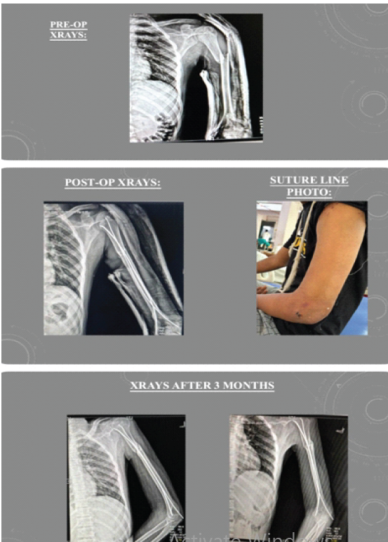
Figure 1: (a and b) Pre-operative and post-operative radiographs – Case 1.
Case 2:
- Patient: 35-year-old male, injured in a domestic fall
- Diagnosis: Closed oblique fracture, distal 1/3 humerus
- Treatment: Retrograde TENS with 3.5 mm nails. Closed reduction achieved under traction and image intensifier guidance. Two nails inserted in a divergent fashion to maximize cortical purchase
- Outcome: Union at 24 weeks. Excellent shoulder and elbow function. Constant–Murley score: 89 (Excellent). No complications (Fig. 2).
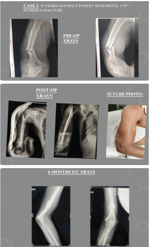
Figure 2: (a and b) Pre-operative and post-operative radiographs – Case 2.
Case 3:
- Patient: 42-year-old male, road traffic accident polytrauma
- Diagnosis: Closed comminuted mid-shaft humerus fracture (AO 12-C2)
- Treatment: Retrograde TENS using 4.0 mm nails. Fracture bridging with 2 nails; supplementary cast for 4 weeks post-fixation
- Complication: Superficial wound infection at the nail entry site managed with wound care and oral antibiotics
- Outcome: Union at 18 weeks. Good functional outcome. Mayo Elbow Performance Score (MEPS): 80 (Good). The wound healed without further intervention (Fig. 3).
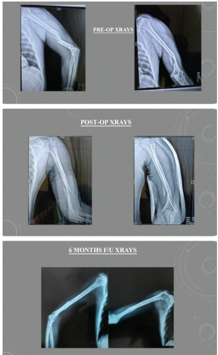
Figure 3: (a and b) Comminuted fracture pre-operative and post-operative radiographs – Case 3.
Case 4:
- Patient: 19-year-old male, sports injury (cricket)
- Diagnosis: Closed transverse fracture of midshaft humerus
- Treatment: Retrograde TENS 3.5 mm nails. Good cortical contact achieved with adequate nail spread at the fracture site
- Outcome: Fastest union in the series at 10 weeks. Excellent outcome – Constant–Murley score 95. Returned to sports at 5 months (Fig. 4).
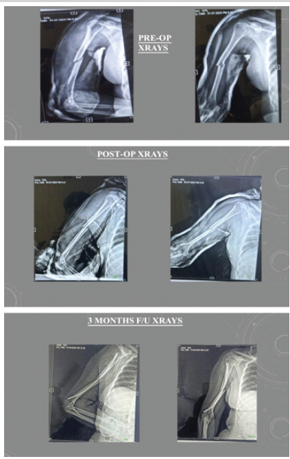
Figure 4: (a and b) Pre-operative and post-operative radiographs – Case 4.
Case 5:
- Patient: 31-year-old female, fall from height
- Diagnosis: Closed transverse mid-shaft humerus fracture
- Treatment: Retrograde TENS 3.5 mm nails
- Complication: Nail prominence at the elbow, causing discomfort; implant removed at 6 months after confirmed union
- Outcome: Union at 16 weeks. Good outcome with full motion following nail removal. Constant–Murley score: 84 (Good) (Fig. 5).
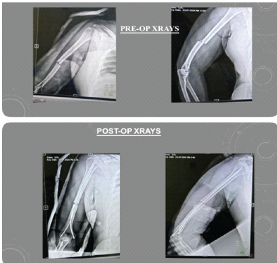
Figure 5: (a and b) Pre-operative and post-operative radiographs – Case 5.
Cases 6-10 (summary):
Cases 6 through 10 involved patients ranging from 22 to 55 years of age with various fracture patterns (transverse, oblique, spiral, and comminuted) at mid and distal shaft levels. All were treated with retrograde TENS using 3.5–4.0 mm nails. One patient (Case 7, 24-year-old male) developed transient radial nerve neurapraxia that resolved completely by 8 weeks postoperatively without surgical intervention. The remaining cases (6, 8, 9, and 10) were uneventful, with union achieved between 10 and 16 weeks. Functional outcomes were excellent in 3 cases and good in two cases based on Constant–Murley and MEPS scoring (Fig. 6).
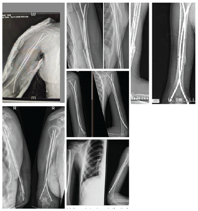
Figure 6: Representative radiographs from Cases 6–10 demonstrating fracture patterns and titanium elastic nailing system fixation.
Patient demographics:
A total of 10 patients were included in this case series. The mean age was 34.1 years (range: 19–55 years). The male-to-female ratio was 7:3. The mechanism of injury was a road traffic accident in 5 patients (50%), a fall from height or domestic fall in 3 patients (30%), and a sports injury in 1 patient (10%), with one patient sustaining polytrauma (Case 3). All fractures were closed injuries. The right humerus was involved in 6 cases and the left in 4 cases. The mean follow-up duration was 12 months (range: 10–18 months).
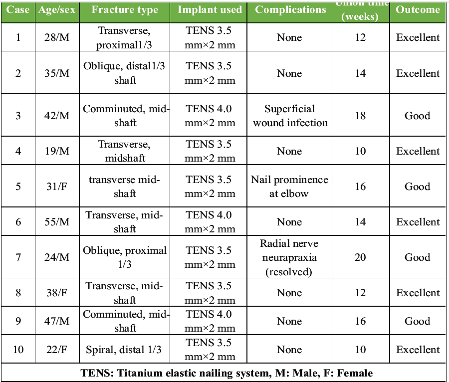
Table 1: Patient demographics, fracture characteristics, treatment details, and functional outcomes
Fracture characteristics:
Fracture patterns included transverse in 5 patients (50%), oblique in 2 patients (20%), spiral in 1 patient (10%), and comminuted in 2 patients (20%). Based on anatomical location, two fractures were in the proximal 1/3 (20%), 6 in the mid-shaft (60%), and 2 in the distal 1/3 (20%). AO/OTA classification was available for the first 3 cases: 12-B1 (Case 1) and 12-C2 (Case 3). The nail diameter used was 3.5 mm in 7 cases and 4.0 mm in 3 cases (comminuted fractures requiring a larger diameter for stability).
Radiological outcomes:
Radiological union was achieved in all 10 patients (100%). The mean time to radiological union was 13.8 weeks (range: 10–24 weeks). Simple transverse fractures (Cases 1, 4, 6, and 8) demonstrated the fastest union (10–14 weeks), while comminuted fractures (Cases 3, 9) required longer union times (16–18 weeks). The oblique distal 1/3 fracture in Case 2 had the longest union time of 24 weeks, attributed to the inherent instability of the oblique pattern at the distal shaft. Callus formation was exuberant in all cases, consistent with the biological load-sharing principle of elastic nailing. No cases of malunion, implant failure, or non-union were observed (Table 1).
Functional outcomes:
Functional outcomes were assessed using the Constant–Murley Shoulder Score (CMS) and the MEPS at final follow-up. Overall, 6 patients (60%) achieved excellent results, and 4 patients (40%) achieved good results. No patient was rated as fair or poor. The mean CMS was 88.3 (range: 81–95). The mean MEPS was 87.5 (range: 80–95). Full shoulder abduction and forward flexion (≥150°) were achieved in 9 of 10 patients. All patients demonstrated full or near-full elbow range of motion at final follow-up. The youngest patient (Case 4, 19-year-old male) returned to competitive cricket at 5 months, the best functional recovery in the series (Table 2).
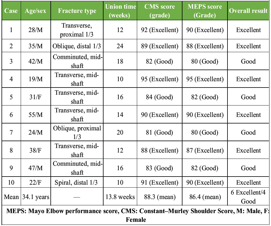
Table 2: Functional outcome scores (Constant–Murley and MEPS) at final follow-up
Complications:
Complications were encountered in 3 of 10 patients (30%). All were minor and resolved without major intervention. There were no cases of deep infection, non-union, implant breakage, or permanent neurological deficit. Table 3 provides a detailed breakdown of complications and their management.
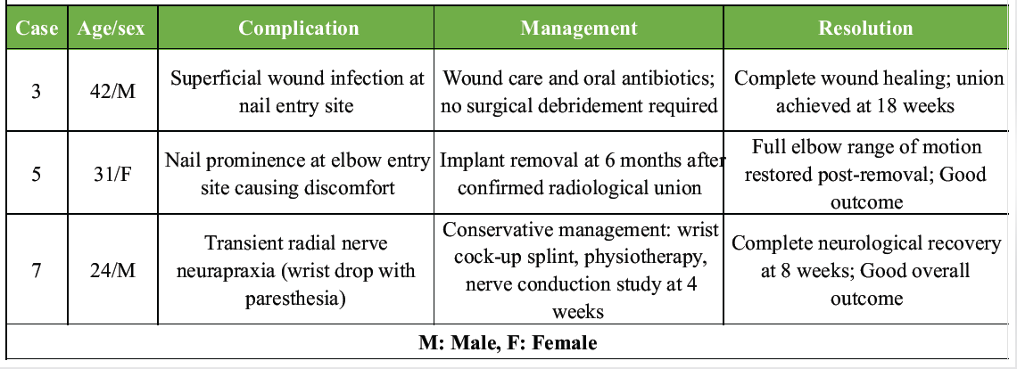
Table 3: Complications encountered and their management
Comparison with published literature:
Our results were compared with previously published series on retrograde TENS for humeral shaft fractures. Table 4 summarizes the key comparative data
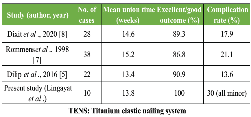
Table 4: Comparison with published literature on retrograde TENS for humeral shaft fractures
Surgical technique:
All surgeries were performed under general or regional anesthesia with the patient in a supine position with the arm on a hand table. Fluoroscopic guidance was used throughout the procedure.
Pre-bending of nails:
Two titanium elastic nails of appropriate diameter (typically 40% of the medullary canal diameter, 3.5–4.0 mm) were pre-bent with a gentle curve of approximately 30–40 degrees at a distance of 2–3 cm from the tip to ensure three-point fixation and adequate spread within the medullary canal.
Entry portals:
Two separate entry points were made approximately 2 cm proximal to the olecranon fossa, medial and lateral to the central axis, using an awl under image intensifier control. Care was taken to avoid injury to the olecranon fossa cortex and the articular surface.
Nail insertion:
Nails were inserted in a retrograde fashion (from distal to proximal) using a T-handle, guided under fluoroscopy. The fracture was reduced by closed manipulation; the nails were then advanced across the fracture site. In cases of difficult closed reduction, a small incision (approximately 2 cm) was made at the fracture site for open-assisted reduction before nail passage.
Final fluoroscopic check:
AP and lateral fluoroscopic views confirmed satisfactory fracture reduction and nail position. The nails were bent and cut flush at the entry site, leaving 0.5–1 cm outside the cortex to facilitate later removal. Wounds were closed in layers.
Post-operative protocol:
A collar-and-cuff sling was applied for 2 weeks. Pendulum exercises were started from day 2 postoperatively. Supervised physiotherapy for the shoulder and elbow range of motion commenced at week 2. Progressive resistance exercises began after radiological evidence of callus formation.
Humeral shaft fractures pose a significant therapeutic challenge. While functional bracing remains the gold standard for conservative management, achieving union in 85–90% of cases [6], surgical fixation is necessary for a subset of patients. Among operative techniques, TENS has been validated as an effective minimally invasive method. The retrograde approach was chosen in our series to avoid the known complications of the antegrade technique—particularly rotator cuff injury, shoulder impingement, and subdeltoid bursitis [7]. By entering the medullary canal through the olecranon fossa, we could preserve shoulder anatomy entirely while achieving stable fixation. Our series demonstrated a mean union time of 13.8 weeks, which is consistent with published literature. Dixit et al. reported a mean union at 14.6 weeks in a comparable series [8]. The 60% excellent and 40% good functional outcomes seen in our series align with other published reports, confirming the reliability of this technique across different fracture patterns. Comminuted fractures, as in Case 3, present a greater challenge due to the lack of cortical contact and inherent instability. In such cases, bridging fixation with supplementary external support (cast or brace) for the initial 4 weeks is recommended to prevent nail migration and loss of reduction. Our result in Case 3 was good despite this complexity, supporting the adaptability of TENS in difficult patterns. The transient radial nerve neurapraxia encountered in Case 7 was managed conservatively. This complication, while concerning, is well recognized in humeral shaft fractures and can occur with any surgical approach due to traction or contusion during reduction. Complete neurological recovery by 8 weeks is consistent with neurapraxia rather than axonotmesis [9]. Nail prominence at the elbow entry site (Case 5) is a known technical pitfall that can be minimized by ensuring adequate bending and cutting of the nail close to the cortex at the time of insertion. Patients should be counseled preoperatively about the possibility of implant removal once union is confirmed. Compared to plating, TENS offers the advantages of minimal soft-tissue dissection, absence of radial nerve exposure in most cases, preservation of fracture hematoma, and early functional rehabilitation. Compared to antegrade IM nailing, retrograde TENS avoids shoulder joint entry and rotator cuff damage [10]. The inherent elasticity of titanium allows for biological load sharing at the fracture site, promoting secondary bone healing through callus formation, which was evident radiologically in all our cases. This study has several limitations that should be acknowledged:
- Mall sample size (10 patients), limiting the statistical strength and generalizability of the findings
- Case series design without a control group, preventing direct comparison with plating, antegrade intramedullary nailing, or conservative treatment
- Single-center experience, which may not reflect outcomes in other institutions or healthcare settings
- Non-randomized study design, making the study susceptible to selection bias
- Relatively short follow-up (10–18 months), insufficient to evaluate long-term implant-related complications and functional durability
- Heterogeneous fracture patterns and locations (proximal, middle, distal third; transverse, oblique, spiral, comminuted) may have influenced outcomes but were not analyzed separately
- No standardized comparison of closed versus minimally open reduction, although both techniques were used
- Functional assessment was limited to Constant–Murley Score and MEPS; patient-reported outcome measures (e.g., DASH, QuickDASH) were not included
- No objective measurements of shoulder and elbow range of motion or muscle strength were presented
- No statistical analysis was performed to identify predictors of union, complications, or functional outcome
- Potential surgeon-related bias, as outcomes may depend on surgical expertise and familiarity with the retrograde TENS technique
- The study predominantly involved younger adults, limiting applicability to elderly osteoporotic patients who commonly sustain humeral shaft fractures
- Radiological union assessment relied on plain radiographs, without computed tomography confirmation in delayed or doubtful union cases
- Complication reporting was limited, with no detailed evaluation of implant migration, rotational malalignment, limb-length discrepancy, or patient satisfaction after implant removal
- The study cannot establish the superiority of retrograde TENS over other fixation methods, and larger multicenter prospective comparative or randomized studies are required to validate these findings and define optimal indications.
Retrograde TENS is a safe, effective, and minimally invasive surgical option for humeral shaft fractures in adults. The technique offers reliable fracture stabilization with low morbidity, preservation of shoulder function, early mobilization, and acceptable complication rates. Excellent to good functional outcomes can be expected in the majority of patients. Retrograde TENS should be considered a valuable alternative to conventional plating and intramedullary nailing, particularly in younger and active patients with mid-shaft and distal-shaft humeral fractures.
Retrograde titanium elastic nailing is a reliable, minimally invasive option for humeral shaft fractures that spares the shoulder and rotator cuff, permits early mobilization, and yields excellent-to-good functional outcomes with a low rate of major complications. It is particularly suited to younger, active patients with mid-shaft and distal-shaft fractures, provided attention is paid to adequate nail pre-bending, divergent nail spread, and flush cutting at the entry portal to minimise complications such as nail prominence and neurapraxia.
References
- 1. Tytherleigh-Strong G, Walls N, McQueen MM. The epidemiology of humeral shaft fractures. J Bone Joint Surg Br 1998;80:249-53. [Google Scholar] [PubMed]
- 2. Sarmiento A, Kinman PB, Galvin EG, Schmitt RH, Phillips JG. Functional bracing of fractures of the shaft of the humerus. J Bone Joint Surg Am 1977;59:596-601. [Google Scholar] [PubMed]
- 3. Ekholm R, Adami J, Tidermark J, Hansson K, Törnkvist H, Ponzer S. Fractures of the shaft of the humerus. An epidemiological study of 401 fractures. J Bone Joint Surg Br 2006;88:1469-73. [Google Scholar] [PubMed]
- 4. Metaizeau JP, Ligier JN. [Surgical treatment of fractures of the long bones in children. Interference between osteosynthesis and the physiological processes of consolidation. Therapeutic indications]. J Chir (Paris) 1984;121:527-37. [Google Scholar] [PubMed]
- 5. Dilip K, Devendra A, Anoop J. Retrograde elastic stable intramedullary nailing (ESIN) of humeral shaft fractures in adults: A review. J Clin Diagn Res 2016;10:RC01-3. [Google Scholar] [PubMed]
- 6. Rutgers M, Ring D. Treatment of diaphyseal fractures of the humerus using a functional brace. J Orthop Trauma 2006;20:597-601. [Google Scholar] [PubMed]
- 7. Rommens PM, Blum J, Runkel M. Retrograde nailing of humeral shaft fractures. Clin Orthop Relat Res 1998;350:26-39. [Google Scholar] [PubMed]
- 8. Dixit R, Suri HS, Rajput BS. Functional outcome of retrograde elastic nailing for humeral shaft fractures in adults: A prospective study. Int J Res Orthop 2020;6:541-45. [Google Scholar] [PubMed]
- 9. Bishop J, Ring D. Management of radial nerve palsy associated with humeral shaft fracture: A decision analysis model. J Hand Surg Am 2009;34:991-6.e1. [Google Scholar] [PubMed]
- 10. Changulani M, Jain UK, Keswani T. Comparison of the use of the humerus intramedullary nail and dynamic compression plate for the management of diaphyseal fractures of the humerus. A randomised controlled study. Int Orthop 2007;31:391-5. [Google Scholar] [PubMed]






Related Articles in Journal of Orthopaedic Case Reports
 August 1, 2025 Nailing the Outcome: A Comparative Study of Proximal Femoral Nail Anti-Rotation-II and Trochanteric Fixation Nail in Unstable Intertrochanteric Femur Fractures
 September 10, 2022 A Prospective Study on the Functional Outcome of Diaphyseal Fractures of Femur Treated with TENS Nailing in Pediatric age Group (5–15 years)
 October 1, 2026 Humeral Fractures in Metabolically Compromised Bone: Outcomes of Structural Strut Onlay Allograft with Open Reduction and Internal Fixation – A Case Series of Three Patients
 September 1, 2026 Comparative Study of Post-operative Pain and Functional Outcomes in Robotic-assisted Total Knee Replacement versus Cruciate-retaining Manual Total Knee Replacement