

In early-stage osteonecrosis of the femoral head unresponsive to bisphosphonate therapy, clinically significant chondral delamination—not evident on standard imaging—may be the true driver of persistent pain, justifying total hip arthroplasty even in the absence of radiological progression.
Dr. Sanjay Agarwala, Department of Orthopedics, P D Hinduja Hospital and Medical Research Centre, Mumbai, Maharashtra, India. E-mail: drsa2011@gmail.com
Abstract
Introduction: Osteonecrosis of the femoral head (ONFH)/avascular necrosis of the femoral head (FH) is a debilitating clinical entity. Bisphosphonates (BPS) are beneficial for conservative/medical management, especially in the early stages. There is, however, a subset of patients with early ONFH who do not respond to BPS and continue to worsen clinically despite no radiological progression. This retrospective cross-sectional study offers a possible explanation for the inexplicable cause of the severe pain in patients with early ONFH from the gross pathological changes of the retrieved FH.
Materials and Methods: A retrospective cross-sectional analysis was conducted on all patients who underwent THA for Early Ficat Arlet Stage Two by the senior surgeon following inadequate relief with BPS between August 2014 and February 2025. Gross intraoperative cartilage findings were graded as per Outerbridge Classification.
Results: 116 patients (mean age 38.1 years) met the inclusion criteria. Cartilage changes observed were: softening in 37, partial thickness fissure in 30, full-thickness fissure in 17 and exposed subchondral bone in 32 FHs.
Conclusion: Delamination of FH cartilage observed in retrieved FHs could explain the persistent pain in some patients with early ONFH unresponsive to medical therapy.
Keywords: Osteonecrosis, total hip arthroplasty, chondral lesion, delamination, avascular necrosis of femoral head, cartilage loss, femoral head sparing procedure, femoral head replacement procedure.

Osteonecrosis of the femoral head (ONFH)/avascular necrosis (AVN) of the femoral head (FH) is a debilitating pathology predominantly affecting younger adults [1]. Treatment options for ONFH include medications to manage pain and inflammation, physical therapy to improve joint mobility and strength, and in severe cases, surgical intervention to replace the damaged bone or joint [2]. Various treatment options exist for early ONFH before FH flattening/change in shape. These options include, but are not limited to, restricted weight-bearing and use of various pharmacologic agents such as lipid-lowering drugs, anticoagulants, and vasodilators [3]. The early stages (Ficat and Arlet [FA] Stages One and Two) have also been successfully managed with oral and/or intravenous bisphosphonates (BPS) [2,4,5,6,7,8,9]. Numerous FH sparing procedures (FHSP) (core decompression with or without osteoinductive substances, bone morphogenic proteins, mesenchymal stem cell implantation, growth factor-based treatment strategies, bone grafting, non-vascularised cortical grafts, free vascularised fibular grafting and osteotomies) have also been described in literature, though with highly variable outcomes [3,10,11,12,13,14,15,16,17]. FH replacement procedures, such as total hip arthroplasty (THA) or resurfacing total hip replacement, are generally reserved for late FA stages Three and Four [18]. There is, however, a subset of patients with early ONFH who continue to have persistent pain despite no radiological progression. The reasons for continued symptoms are poorly understood. Some of these patients chose to undergo THA at this early stage for pain relief and improvement in quality of life. The clinical, radiological and intraoperative correlation is yet not well-defined in the literature. This retrospective cross-sectional study explains the inexplicable cause for the severe pain in patients with early ONFH from the gross pathological changes of the retrieved FH, thereby possibly explaining the reasons for discordance between the radiological findings and clinical severity.
Study design and participants:
This was a retrospective cross-sectional study conducted at a tertiary reference centre after Internal Review Board approval (IRB/1476/AL, Feb. 2025). Medical records were reviewed for all patients with early FA stage 2 idiopathic ONFH who had undergone THA with the senior surgeon following inadequate pain relief despite receiving BPS, between August 2014 and February 2025. All other aetiologies, such as post-traumatic ONFH, were excluded. After systematically reviewing the electronic medical records (EMR), a total of 116 cases were identified and included for analysis (Fig. 1).
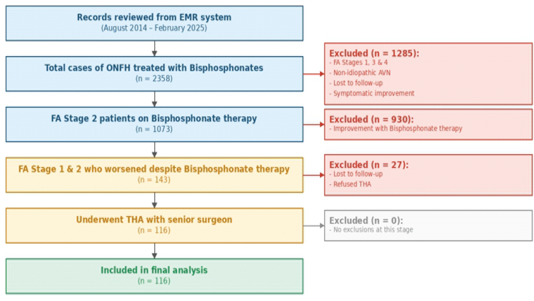
Figure 1: Flowchart illustrating the retrospective process of patient selection. THA: Total hip arthroplasty, FA: Ficat arlet, ONFH: Osteonecrosis of femoral head.
Management algorithm:
All patients with ONFH had been prescribed BPS for 3 years as per the established protocol of oral alendronate along with intravenous zoledronic acid. In addition, patients received daily supplements of calcium 500 mg and vitamin D 400 IU. Analgesics were administered as and when required. Partial weight bearing using axillary or elbow crutches was advised for the first 3 months after the initiation of bisphosphonate therapy, and weight bearing was allowed as tolerated thereafter. Patients were followed up at 3 months after initiation of therapy and then annually. Clinical and radiological progression were noted at each visit. Radiological assessment was performed with plain radiographs of both hips in anteroposterior and lateral views at every visit. FHSP, as well as THA, with their associated advantages and disadvantages, were explained to those who had persistent or worsened pain despite adequate conservative management. Patients who chose FHSP were excluded from this study. The excised heads of all patients undergoing THA were photographed and classified according to the Outerbridge classification [19].
Data collection:
Data were extracted retrospectively from the EMR system and entered into a predesigned spreadsheet. Patient demography, symptoms, radiological findings, and operative details were systematically recorded. Those patients with incomplete records were excluded from analysis. The gross findings of FH cartilage were classified as per the Outerbridge classification [19]:
- Grade 0: Normal articular cartilage
- Grade 1: Softening, blistering, or swelling of the cartilage
- Grade 2: Partial-thickness fissure and clefts <1 cm in diameter
- Grade 3: Full-thickness fissures, to the subchondral bone >1 cm in diameter
- Grade 4: Exposed subchondral bone.
Outcome measures:
The gross intraoperative findings were matched with the radiological findings, providing a possible rationale for the persisting pain and failure of BPS therapy in this retrospective cohort.
Patient demographics (Table 1):
The retrospective review included a total of 116 patients with a mean age of 38.1 years. The cohort was predominantly male, consisting of 88 men and 28 women. Regarding the side of the procedure, 51 cases involved the right side, while 65 involved the left. Notably, all 116 patients were classified as being in FA Stage 2, with no patients in Stage 1.
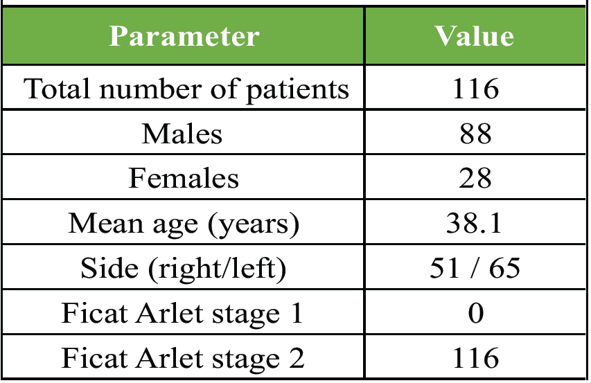
Table 1: Patient demography
Intra-operative findings (Table 2):
During the intraoperative assessment of the FHs using the Outerbridge grading system, no cases were identified as Grade 0 (normal). The most frequent finding was Grade 1 (softening or blistering), which was observed in 37 FHs. Grade 2 findings, characterised by partial thickness fissures or clefts, were present in 30 cases. Grade 3 findings, involving full-thickness fissures reaching the subchondral bone, were seen in 17 instances. Finally, 32 FHs demonstrated Grade 4 findings, indicating exposed subchondral bone.
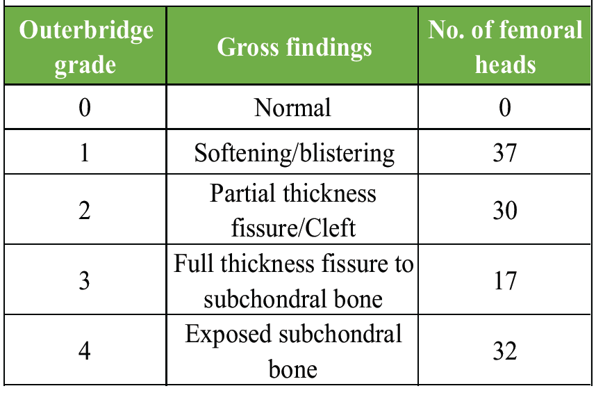
Table 2: Intraoperative findings
ONFH is a condition that predominantly affects young adults [18]. It results in a debilitating state with a varied response to conservative management. Clinical improvement with medical/conservative therapies is higher in early FA stages [4,7,9]. Conservative therapies aim to ensure a relatively pain-free quality of life and delay the onset of flattening of the FH and secondary osteoarthrosis. However, none of them adequately address cartilage changes/delamination. Some patients with early ONFH continue to have persistent pain and disability. There is scant literature on the reasons behind the failure of conservative therapy. It has been postulated that bone oedema is one of the main causes for pain in ONFH, especially in the pre-collapse stages [5,6,7]. BPS has proven to be beneficial by resolving this oedema underlying the cartilage [20]. Browne and Branch have shown that subchondral oedema must be considered as a flag for detailed evaluation of the overlying articular surface cartilage [19]. Although magnetic resonance imaging (MRI) is the gold standard in diagnosing ONFH, it has variable sensitivity and specificity in identifying cartilaginous lesions [21,22,23]. MRI remains the ideal non-invasive investigation, with arthroscopy being the ideal diagnostic and/or therapeutic intervention [24]. This retrospective cross-sectional study evaluated the morphological changes in FH cartilage from the retrieved heads of patients who had undergone THA for persistent pain in early ONFH. A variety of cartilaginous changes were found, ranging from softening to complete loss of cartilage with exposed subchondral bone, based on the Outerbridge classification (Figs. 2, 3, 4, 5).
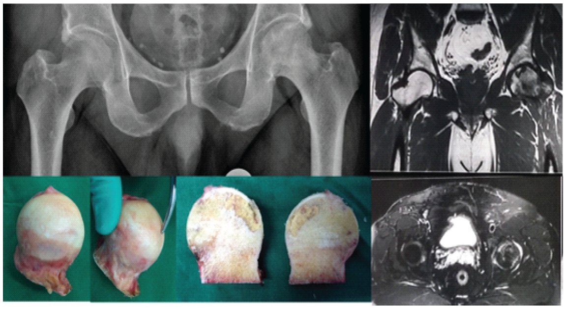
Figure 2: Softening of cartilage – left hip avascular necrosis (Outerbridge grade 1).
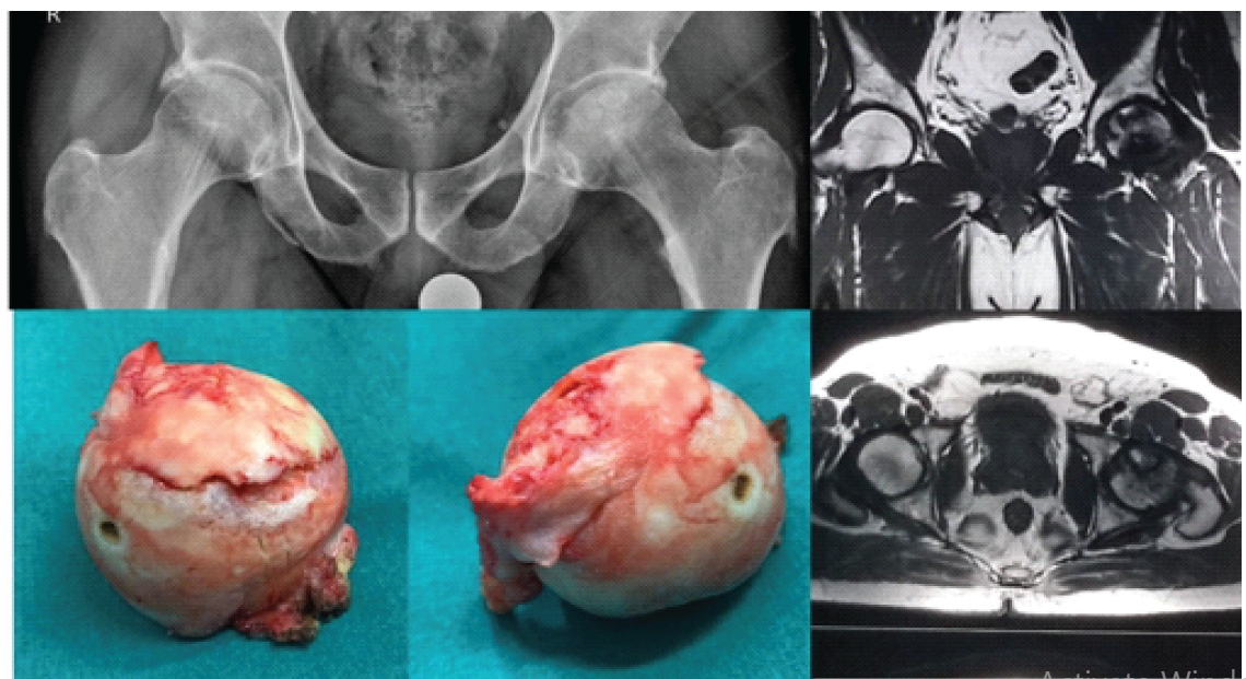
Figure 3: Partial fissure – left hip avascular necrosis (Outerbridge grade 2).
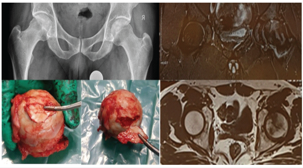
Figure 4: Complete fissure – left hip avascular necrosis (Outerbridge grade 3).
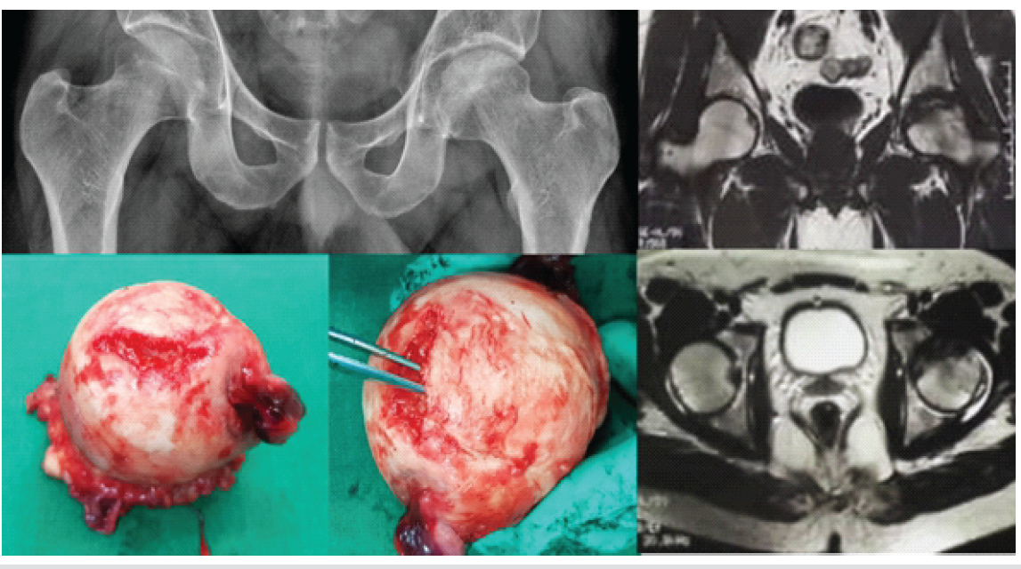
Figure 5: Exposed subchondral bone – left hip avascular necrosis (Outerbridge grade 4).
Similar gross cartilage changes were described by Babhulkar, describing severe cartilage loss with advanced stages [25]. Cartilage loss has been noted in Stage 2 in our series, which could be secondary to the underlying non-collapsed ischaemic subchondral bone. Lands et al. described a case report of THA following delamination in a case of ONFH [26]. The patient underwent THA after failure of conservative management. They postulated that articular cartilage delamination could be a cause of early-stage ONFH of the hip joint. In the early stages of AVN, the articular cartilage could start to separate from the underlying bone, known as delamination. This could cause pain and stiffness in the hip joint and can eventually lead to joint collapse and the development of osteoarthritis [26]. Although there are numerous reports on conservative management for such chondral lesions, ranging from arthroscopic debridement to mosaicplasty to microfracture, THA still remains the gold standard for such lesions [18,24,27]. Difficult surgical technique, surgeon experience, and variable outcomes are some of the reasons behind secondary THA after conservative surgeries for chondral defects [26,27]. In addition, there are reports of ON following chondral resurfacing procedures [28]. Moreover, many of the cartilaginous lesions in ONFH may be missed even on MRI, the diagnostic modality of choice [19]. Unrecognised and untreated chondral injuries can have a poor prognosis, with possible outcomes including worsening of cartilaginous injury due to AVN [29]. It can hasten progression from pre-collapse to end-stage degenerative changes [26]. It is debatable whether a chondral resurfacing procedure would help in a progressive pathology like ONFH, where the changes are occurring in the underlying bone. In a recent review, Du et al. [30] described numerous surgical options for repairing cartilage defects in the FH, such as microfracture, cartilage replantation, ACI/MAC, and autograft/allograft transplantation. Most of these techniques were originally established for the management of the knee joint. Their application for FH pathologies is limited, which can be explained by the fact that the articular surface of the FH exhibits greater curvature, deeper location, and more complex blood supply, as compared to the knee joint. Clinical evidence available on these procedures for the hip is limited to single case reports or case series, and the clinical outcomes are not comparable to those in the knee. Larger clinical studies with additional efficient solutions or options are desirable to treat cartilage defects in the FH. The limitations of this study include its retrospective design, relatively small number of cases, and the inherent possibility of selection bias. Excluding those lost to follow-up after failure of BPS therapy and including only those who eventually underwent THA means the conclusions may not apply to all patients with persistent pain despite adequate BPS therapy. Future studies with detailed MRI cartilage mapping with intra-operative correlation could provide additional information and insight. A randomised controlled trial between chondral resurfacing procedures and THA for such cases would represent an ideal study design.
Medical therapy, such as BPS, continues to be an important treatment strategy for early ONFH before a change in the shape of the FH. There is, however, a subset of patients who have persistent pain with no obvious cause in view of the seemingly normal radiology. This retrospective cross-sectional study highlights that cartilage softening/delamination in this complex ball-and-socket weight-bearing joint could possibly be the cause of the persistent incapacitating pain. THA, an established and proven surgical intervention, provides a viable option to overcome this cartilage damage.
In patients with early-stage ONFH who do not experience pain relief from bisphosphonate therapy and show no radiological progression, persistent severe pain may be due to painful chondral delamination, including cartilage softening, fissures, or loss. These cartilage changes often remain undetected on standard imaging, explaining the clinical-radiological discordance. Recognition of this pathology supports THA as an effective treatment option to alleviate pain and improve quality of life in this patient subgroup.
References
- 1. Tripathy SK, Goyal T, Sen RK. Management of femoral head osteonecrosis: Current concepts. Indian J Orthop 2015;49:28-45. [Google Scholar] [PubMed]
- 2. Agarwala S, Jain D, Joshi VR, Sule A. Efficacy of alendronate, a bisphosphonate, in the treatment of AVN of the hip. Rheumatology (Oxford) 2005;44:352-9. [Google Scholar] [PubMed]
- 3. Sen RK. Management of avascular necrosis of femoral head at pre-collapse stage. Indian J Orthop 2009;43:6-16. [Google Scholar] [PubMed]
- 4. Li D, Yang Z, Wei Z, Kang P. Efficacy of bisphosphonates in the treatment of femoral head osteonecrosis: A PRISMA-compliant meta-analysis of animal studies and clinical trials. Sci Rep 2018;8:1450. [Google Scholar] [PubMed]
- 5. Agarwala S, Vijayvargiya M. Bisphosphonate combination therapy for non-femoral avascular necrosis. J Orthop Surg Res 2019;14:112. [Google Scholar] [PubMed]
- 6. Agarwala S, Shah SB. Ten-year follow-up of avascular necrosis of femoral head treated with alendronate for 3 years. J Arthroplasty 2011;26:1128-34. [Google Scholar] [PubMed]
- 7. Agarwala S, Shah S, Joshi VR. The use of alendronate in the treatment of avascular necrosis of the femoral head: follow-up to eight years. J Bone Joint Surg Br 2009;91:1013-8. [Google Scholar] [PubMed]
- 8. Agarwala S, Sule A, Pai BU, Joshi VR. Study of alendronate in avascular necrosis of bone. J Assoc Physicians India 2001;49:949-50. [Google Scholar] [PubMed]
- 9. Prashanthkumar T, Chandan AE. A study on functional outcome of conservative management of stage 2 and 3 avascular necrosis of femoral head. Int J Contemp Med Res 2017;4:77-83. [Google Scholar] [PubMed]
- 10. Ünal MB, Cansu E, Parmaksizoğlu F, Cift H, Gürcan S. Treatment of osteonecrosis of the femoral head with free vascularized fibular grafting: Results of 7.6-year follow-up. Acta Orthop Traumatol Turc 2016;50:501-6. [Google Scholar] [PubMed]
- 11. Feng Y, Wang S, Jin D, Sheng J, Chen S, Cheng X, et al. Free vascularised fibular grafting with OsteoSet®2 demineralised bone matrix versus autograft for large osteonecrotic lesions of the femoral head. Int Orthop 2011;35:475-81. [Google Scholar] [PubMed]
- 12. Zhang L, Fan Y, Zhang Y, Chen X, Liu Y. Comparison of sartorius muscle-pedicle and circumflex iliac deep bone flap grafts in the treatment of early non-traumatic osteonecrosis of femoral head in young adults. Acta Orthop Traumatol Turc 2019;53:255-9. [Google Scholar] [PubMed]
- 13. Malizos KN, Quarles LD, Dailiana ZH, Rizk WS, Seaber AV, Urbaniak JR. Analysis of failures after vascularized fibular grafting in femoral head necrosis. Orthop Clin North Am 2004;35:305-14. [Google Scholar] [PubMed]
- 14. Tetik C, Başar H, Bezer M, Erol B, Ağir I, Esemenli T, et al. Comparison of early results of vascularized and non-vascularized fibular grafting in the treatment of osteonecrosis of the femoral head. Acta Orthop Traumatol Turc 2011;45:326-34. [Google Scholar] [PubMed]
- 15. Korompilias AV, Lykissas MG, Beris AE, Urbaniak JR, Soucacos PN. Vascularised fibular graft in the management of femoral head osteonecrosis: Twenty years later. J Bone Joint Surg Br 2009;91:287-93. [Google Scholar] [PubMed]
- 16. Gasbarra E, Perrone FL, Baldi J, Bilotta V, Moretti A, Tarantino U. Conservative surgery for the treatment of osteonecrosis of the femoral head: current options. Clin Cases Miner Bone Metab 2015;12 Suppl 1:43. [Google Scholar] [PubMed]
- 17. Atilla B, Bakırcıoğlu S, Shope AJ, Parvizi J. Joint-preserving procedures for osteonecrosis of the femoral head. EFORT Open Rev 2019;4:647-58. [Google Scholar] [PubMed]
- 18. Moya-Angeler J, Gianakos AL, Villa JC, Ni A, Lane JM. Current concepts on osteonecrosis of the femoral head. World J Orthop 2015;6:590-601. [Google Scholar] [PubMed]
- 19. Browne JE, Branch TP. Surgical alternatives for treatment of articular cartilage lesions. J Am Acad Orthop Surg 2000;8:180-9. [Google Scholar] [PubMed]
- 20. Baier C, Schaumburger J, Götz J, Heers G, Schmidt T, Grifka J, et al. Bisphosphonates or prostacyclin in the treatment of bone-marrow oedema syndrome of the knee and foot. Rheumatol Int 2013;33:1397-402. [Google Scholar] [PubMed]
- 21. Schmid MR, Nötzli HP, Zanetti M, Wyss TF, Hodler J. Cartilage lesions in the hip: Diagnostic effectiveness of MR arthrography. Radiology 2003;226:382-6. [Google Scholar] [PubMed]
- 22. Annabell L, Master V, Rhodes A, Moreira B, Coetzee C, Tran P. Hip pathology: The diagnostic accuracy of magnetic resonance imaging. J Orthop Surg Res 2018;13:127. [Google Scholar] [PubMed]
- 23. Mattila VM, Weckström M, Leppänen V, Kiuru M, Pihlajamäki H. Sensitivity of MRI for articular cartilage lesions of the patellae. Scand J Surg 2012;101:56-61. [Google Scholar] [PubMed]
- 24. Mardones R, Larrain C. Cartilage restoration technique of the hip. J Hip Preserv Surg 2016;3:30-6. [Google Scholar] [PubMed]
- 25. Babhulkar S. Osteonecrosis. 1st ed. Uttar Pradesh: Jaypee; 2013. p. 13-25. [Google Scholar] [PubMed]
- 26. Lands [first name initial?], Malige A, Reddy CS. Femoral head chondral delamination injury and associated osteonecrosis: A case report of treatment. J Orthop Allied Sci 2017;5:51-4. [Google Scholar] [PubMed]
- 27. Jordan MA, Van Thiel GS, Chahal J, Nho SJ. Operative treatment of chondral defects in the hip joint: A systematic review. Curr Rev Musculoskelet Med 2012;5:244-53. [Google Scholar] [PubMed]
- 28. Thompson SM, Saso A, Raval P, Jones M, Williams AM. Avascular necrosis complicating chondral resurfacing techniques. Knee Surg Sports Traumatol Arthrosc 2016;24:1660-4. [Google Scholar] [PubMed]
- 29. Kendell S, Helms C, Rampton J, Garrett W, Higgins L. MRI appearance of chondral delamination injuries of the knee. AJR Am J Roentgenol 2005;184:1486-9. [Google Scholar] [PubMed]
- 30. Du D, Hsu P, Zhu Z, Zhang C. Current surgical options and innovation for repairing articular cartilage defects in the femoral head. J Orthop Translat 2020;21:122-8. [Google Scholar] [PubMed]


Related Articles in Journal of Orthopaedic Case Reports
 October 1, 2026 Primary Total Hip Arthroplasty for a Displaced Femoral Neck Fracture in an Above-the-Knee Amputee: A Case Report
 October 1, 2026 Intrapelvic Neck Screw Migration after Trochanteric Fixation Nail Advanced Fixation of a Trochanteric Femoral Fracture: A Case Report and Proposed Treatment Algorithm
 September 1, 2026 Undiagnosed von Willebrand Disease Presenting as Delayed Hematoma after Total Hip Arthroplasty: A Case Report
 August 1, 2026 Total Hip Arthroplasty without Subtrochanteric Shortening Osteotomy in a Neglected Posterior Hip Dislocation: A Case Report