

The “Boba Tea Sign” on MRI is a novel, memorable visual analogy resembling tapioca pearls suspended in bubble tea. It allows a fast diagnosis of synovial chondromatosis, allowing early intervention and preventing joint degradation.
Dr. Shivam Chauhan, Department of Orthopaedics, All India Institute of Medical Sciences, Tatibandh, G E Road, Raipur - 492 099, Chhattisgarh, India. Phone: +91-9897318492. E-mail: chauhanshivam9555@gmail.com
Abstract
Introduction: Synovial chondromatosis is an uncommon benign metaplastic disorder characterized by the formation of multiple cartilaginous loose bodies within the synovial membrane. Delayed diagnosis may lead to progressive joint degeneration. We describe a classic case of knee synovial chondromatosis and propose a novel magnetic resonance imaging (MRI) finding, the “Boba Tea Sign.”
Case Report: A 35-year-old female presented with progressive left knee pain, swelling, locking, and restricted motion for 2 years. Radiographs demonstrated multiple calcified intra-articular loose bodies with arthritic changes. MRI revealed numerous rounded cartilaginous nodules suspended within synovial fluid, resembling tapioca pearls in bubble tea, termed the “boba tea sign." Due to advanced degenerative changes, the patient underwent total knee arthroplasty with extensive synovectomy and removal of loose bodies. Histopathology confirmed synovial chondromatosis. At 1-year follow-up, the patient had excellent functional recovery without recurrence.
Conclusion: The “Boba Tea Sign” is a novel and memorable MRI analogy that may facilitate recognition of synovial chondromatosis and aid in timely diagnosis and treatment planning.
Keywords: Synovial chondromatosis, knee, magnetic resonance imaging, Boba Tea Sign, loose bodies, total knee arthroplasty.

Synovial chondromatosis is a rare, benign, metaplastic disorder characterized by cartilaginous transformation of the synovium, leading to intra-articular loose bodies. It most commonly affects large joints, particularly the knee, and presents with progressive pain, swelling, and mechanical symptoms such as locking or clicking [1,2]. The etiology is idiopathic in primary synovial chondromatosis and may be secondary to trauma, osteoarthritis, or neuropathic arthropathy [3]. We present a classic case of synovial chondromatosis of the knee and propose a novel, descriptive magnetic resonance imaging (MRI) sign – the “Boba Tea Sign” – reflecting the appearance of numerous rounded nodules resembling tapioca pearls suspended in a fluid medium.
A 35-year-old female presented with a 2-year history of progressive left knee pain, swelling, and mechanical symptoms, including intermittent locking and clicking. There were no constitutional symptoms, prior trauma, or history of inflammatory arthritis. Her medical and surgical history was unremarkable. On examination, the left knee was swollen with diffuse tenderness (Fig. 1a). Passive movement elicited palpable crepitus. Range of motion was limited to 0–90°, with pain at terminal flexion. The contralateral knee and other joints were unremarkable.
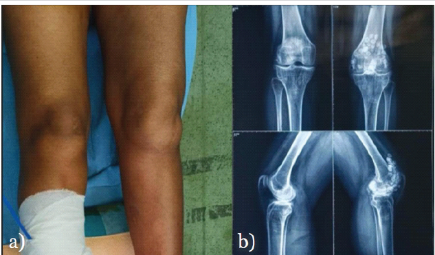
Figure 1: (a) Pre-operative clinical photograph showing swollen left knee due to multiple intra-articular loose bodies in the suprapatellar pouch and joint cavity, (b) Pre-operative anteroposterior and lateral radiographs of the left knee demonstrating multiple calcified intra-articular loose bodies with secondary arthritic changes.
Imaging and diagnosis:
Radiographs revealed multiple calcified intra-articular loose bodies in the suprapatellar pouch, intercondylar notch, and posterior capsular pouch, along with arthritic changes (Fig. 1b). MRI of the left knee showed multiple well-defined, similarly sized intra-articular bodies demonstrating variable signal intensities within joint fluid, accompanied by synovial proliferation (Fig. 2, 3). Most of them exhibited T2 hyperintensity with a peripheral T2 hypointense rim consistent with cartilaginous bodies undergoing peripheral ossification—typical findings of synovial chondromatosis [4,5].
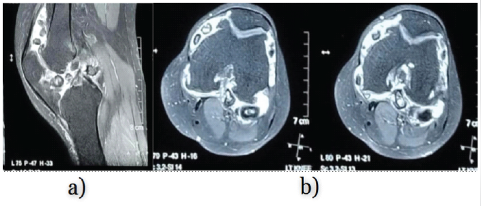
Figure 2: (a) Sagittal T2-weighted magnetic resonance imaging (MRI) slice depicting multiple intra-articular cartilaginous nodules of varying signal intensity within the joint effusion, (b) Axial T2-weighted MRI slices showing clustered, uniformly rounded cartilaginous nodules within the joint cavity and suprapatellar pouch.
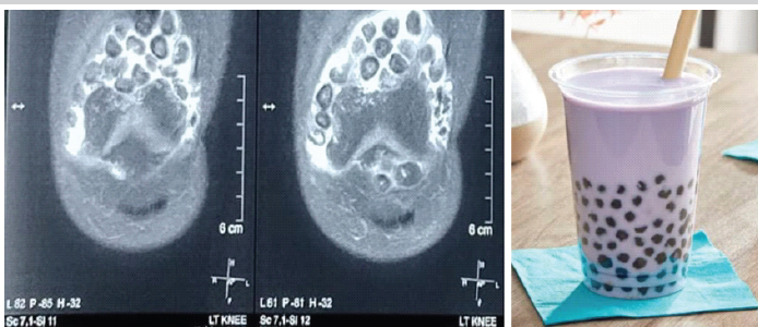
Figure 3: Coronal T2-weighted magnetic resonance imaging slices demonstrating the characteristic “Boba Tea Sign” – multiple discrete, rounded nodules suspended in synovial fluid resembling tapioca pearls in bubble tea.
The Boba Tea Sign:
On MRI, the appearance of multiple uniformly rounded, discrete nodules suspended in synovial fluid resembled the visual pattern of tapioca pearls in a bubble tea drink (Fig. 3). This unique configuration led us to coin the term “Boba Tea Sign,” a memorable analogy that may aid radiologists and clinicians in early recognition of synovial chondromatosis.
Surgical management:
In view of persistent disabling symptoms and secondary joint degeneration, the patient underwent cemented total knee arthroplasty via a medial parapatellar approach. A posterior-stabilized knee was used with patellar resurfacing. An all-polyethylene tibial component was used due to financial constraints for the patient. Extensive synovectomy was performed, and all intra-articular loose bodies were removed (Fig. 4). Histopathology confirmed synovial chondromatosis. Post-operative radiograph revealed complete removal of loose bodies and a well-aligned prosthesis (Fig. 5). The post-operative period was uneventful. Early physiotherapy and weight-bearing were initiated, with excellent recovery. At the 1-year follow-up, the patient reported significant improvement in pain and function with a full, painless range of motion (0–120°). Follow-up radiographs confirmed a well-fixed prosthesis with no residual or recurrent loose bodies (Fig. 6).
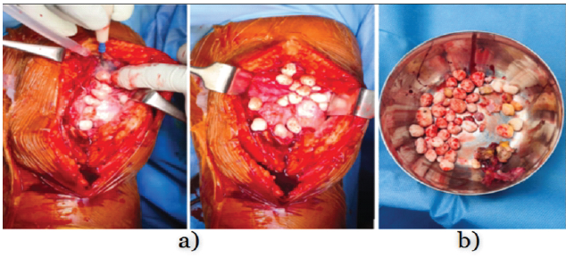
Figure 4: (a) Intraoperative photograph showing numerous cartilaginous loose bodies within the knee joint cavity after arthrotomy, (b) extracted loose bodies removed during extensive synovectomy and total knee arthroplasty.
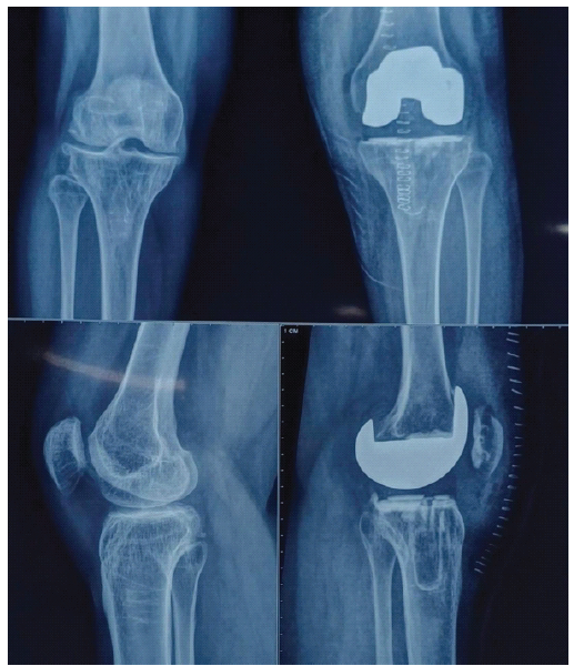
Figure 5: Immediate post-operative radiographs showing well-fixed cemented posterior-stabilized total knee prosthesis with complete removal of loose bodies and satisfactory alignment.
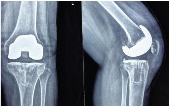
Figure 6: One-year follow-up radiographs demonstrating well-fixed total knee prosthesis with no evidence of residual or recurrent loose bodies or implant loosening.
Synovial chondromatosis is a progressive condition that may remain undiagnosed for years due to its indolent course. It is most often monoarticular and involves the knee in 60–70% of cases [6]. MRI is the imaging modality of choice, as it detects both calcified and non-calcified cartilaginous nodules and evaluates synovial proliferation [4]. The “Boba Tea Sign” on MRI provides a compelling visual analogy for the characteristic imaging findings in synovial chondromatosis. A similar concept—the “rice bodies” seen in tubercular arthritis—also exemplifies how analogical radiologic signs aid diagnostic recall [7]. To our knowledge, this is the first formal proposal of the “Boba Tea Sign” in musculoskeletal radiology. Early recognition and surgical intervention are key to preventing joint destruction and secondary osteoarthritis. Arthroscopic approaches, including synovectomy with removal of loose bodies, have demonstrated favorable outcomes in selected cases without advanced joint degeneration [8,9]. In cases with advanced degeneration, total joint replacement remains the definitive solution, with good long-term outcomes [10].
We propose the “Boba Tea Sign” as a novel, illustrative MRI finding in synovial chondromatosis, referring to the appearance of multiple rounded intra-articular nodules mimicking boba pearls suspended in fluid. This descriptive sign enhances radiologic pattern recognition and contributes to earlier diagnosis and treatment planning.
The “Boba Tea Sign” provides a distinct and easily identifiable MRI sign for diagnosing synovial chondromatosis. This allows for early diagnosis by radiologists and clinicians, allowing early and timely intervention – guiding appropriate management – including synovectomy and, in advanced degenerative cases, total joint arthroplasty, preventing further joint degeneration and improving patient outcomes
References
- 1. Davis RI, Hamilton A, Biggart JD. Primary synovial chondromatosis: A clinicopathologic review and assessment of malignant potential. Hum Pathol 1998;29:683-8. [Google Scholar] [PubMed]
- 2. Maurice H, Crone M, Watt I. Synovial chondromatosis. J Bone Joint Surg Br 1988;70:807-11. [Google Scholar] [PubMed]
- 3. Milgram JW. Synovial osteochondromatosis: A histopathological study of thirty cases. J Bone Joint Surg Am 1977;59:792-801. [Google Scholar] [PubMed]
- 4. Kramer J, Recht M, Deely DM, Schweitzer ME, Pathria MN, Resnick D. MR appearance of idiopathic synovial osteochondromatosis. J Comput Assist Tomogr 1993;17:772-6. [Google Scholar] [PubMed]
- 5. Murphey MD, Vidal JA, Fanburg-Smith JC, Gajewski DA. Imaging of synovial chondromatosis with radiologic-pathologic correlation. Radiographics 2007;27:1465-88. [Google Scholar] [PubMed]
- 6. Lim SJ, Chung HW, Choi YL, Moon YW, Park YS. Operative treatment of primary synovial ostechondromatosis of the hip. J Bone Joint Surg Am 2006;88:2456-64. [Google Scholar] [PubMed]
- 7. Tuli SM. General principles of osteoarticular tuberculosis. Clin Orthop Relat Res 2002;398:11-9. [Google Scholar] [PubMed]
- 8. Coolican MR, Dandy DJ. Arthroscopic management of synovial chondromatosis of the knee. Findings and results in 18 cases. J Bone Joint Surg Br 1989;71:498-500. [Google Scholar] [PubMed]
- 9. Ogilvie-Harris DJ, Saleh K. Generalized synovial chondromatosis of the knee: A comparison of removal of the loose bodies alone with arthroscopic synovectomy. Arthroscopy 1994;10:166-70. [Google Scholar] [PubMed]
- 10. Veras del Monte LM, Garcia FL, Perez-Prieto D, et al. Total knee arthroplasty in advanced primary synovial chondromatosis: Case report and literature review. J Orthop Case Rep 2019;9:70-3. [Google Scholar] [PubMed]





Related Articles in Journal of Orthopaedic Case Reports
 October 1, 2025 Non-calcified Synovial Chondromatosis Masquerading as a Posterior Knee Cyst: A Diagnostic Challenge
 August 1, 2026 Intra-articular Ganglionic Cysts of Knee: A Cause for Anterior Knee Pain
 June 1, 2026 Secondary Synovial Chondromatosis of the Shoulder Associated with Rotator Cuff Tear: Diagnostic Challenge and Arthroscopic Management – A Case Report
 April 1, 2026 Bucket Handle Tear of the Medial Meniscus with a New Magnetic Resonance Imaging Sign: The “Head in the Sand Sign” – A Case Report