

Large spinal neurofibromas, though uncommon, can present with progressive back pain and neurological deficits. MRI is crucial for accurately delineating the tumor and planning surgical intervention. Complete surgical excision combined with spinal stabilization can achieve excellent functional recovery and restore mobility. Early diagnosis and timely surgery are key to preventing permanent neurological impairment.
Dr. Abdullah Al Mahmud, Department of Orthopaedic Surgery, Ibn Sina Medical College Hospital, Dhaka, Bangladesh. E-mail: draalmahmud@gmail.com
Abstract
Introduction: Spinal neurofibromas are rare neurogenic tumors that can cause progressive neurological deficits due to spinal cord compression. Early diagnosis and timely surgical intervention are crucial to prevent permanent neurological impairment and restore functional mobility.
Case Report: A 38-year-old male presented to Ibn Sina Medical College Hospital with persistent mid-back pain and progressive weakness in both lower limbs, which eventually caused difficulty in independent ambulation. Neurological examination revealed decreased motor strength in the lower limbs (Medical Research Council [MRC] Grade – 4/5), sensory deficit below the D6 dermatome, and exaggerated deep tendon reflexes. Magnetic resonance imaging (MRI) of the thoracic spine demonstrated a large, well-defined, dumbbell-shaped paraspinal mass at the thoracic spine levels 5 and 6 (D5–D6) vertebral level, extending through the intervertebral foramen into the spinal canal with significant spinal cord compression. Based on clinical and radiological findings, a diagnosis of thoracic spinal neurofibroma (dumbbell type) was made. The patient underwent complete surgical excision of the tumor with spinal stabilization under general anesthesia. Intraoperatively, the tumor was encapsulated and extended from the spine to the subcutaneous tissue and right chest wall, destroying the D5–D6 vertebral body, lamina, pedicles, and part of the 5th and 6th ribs. Histopathology confirmed the diagnosis of neurofibroma. Postoperatively, the patient’s neurological function gradually improved, and follow-up imaging confirmed complete tumor resection with no residual compression. At 1-month follow-up, he was mobilizing independently, with significant pain relief and no surgical complications. At 4 years follow-up, the patient achieved independent ambulation, and neurological function had returned to normal.
Conclusion: Surgical excision with spinal stabilization is an effective treatment for thoracic dumbbell-shaped neurofibromas. Early diagnosis and intervention can lead to excellent neurological recovery, restoration of mobility, and improved quality of life.
Keywords: Surgical outcome, giant dorsal spinal neurofibroma, back pain.

Spinal neurofibromas are benign tumors arising from the peripheral nerve sheath and are commonly associated with neurofibromatosis type 1 (NF1) [1]. These tumors can occur at multiple spinal levels and often present with a variety of clinical manifestations depending on their size and location [2]. The natural history of spinal neurofibromas is variable, and understanding their clinical and genetic features is essential for appropriate management and prognosis [3]. In patients with NF2, spinal tumors are also frequently observed, with magnetic resonance imaging (MRI) studies demonstrating a high prevalence of multiple and diverse lesions along the spinal axis [4].
Spinal neurofibromas, characterized by intraspinal and paraspinal components connected through the neural foramen, present unique surgical challenges due to their proximity to critical neural and vascular structures [5]. These tumors often cause spinal cord compression, leading to pain, motor weakness, and sensory deficits, which necessitate careful pre-operative planning [6]. Surgical resection remains the mainstay of treatment to relieve compression and prevent neurological deterioration, but the extent of resection must be balanced against the risk of neurological injury [7]. Factors such as tumor location, size, and association with neurofibromatosis significantly influence surgical strategy and clinical outcomes [8]. Despite advances in microsurgical techniques, complications such as neurological deficits, recurrence, and spinal instability are important considerations during management [5,7].
Surgical excision remains the primary treatment modality for spinal neurofibromas, aiming to relieve neural compression and prevent progression of neurological deficits [9]. The extent of tumor resection is a key factor influencing long-term outcomes, with complete removal associated with lower recurrence rates and improved functional recovery [10,11]. Radiosurgery has also been explored as an adjunct or alternative for selected benign intradural spinal tumors, demonstrating favorable tumor control in cases not amenable to complete surgical excision [9]. Recent studies have highlighted the importance of individualized treatment planning, taking into account tumor type, location, size, and patient comorbidities to optimize clinical outcomes and minimize complications [12,13]. Given the rarity of large thoracic dumbbell-shaped spinal neurofibromas and the significant surgical challenges they present, reporting individual cases is valuable to expand understanding of their clinical presentation, radiological characteristics, and optimal management strategies. This case report aims to describe the successful surgical management of a large spinal neurofibroma, highlighting the role of early diagnosis, meticulous pre-operative planning, complete tumor excision, and spinal stabilization in achieving favorable neurological and functional outcomes.
A 38-year-old male was admitted to Ibn Sina Medical College Hospital with persistent back pain and progressive weakness of both lower limbs, which had worsened over several weeks, leading to difficulty with independent ambulation. Neurological examination revealed decreased motor power in the lower limbs (MRC Grade – 4/5), sensory deficit below the D6 dermatome, and exaggerated deep tendon reflexes (Modified McCormick Scale – IV).
Imaging studies were performed to evaluate the lesion. Fig. 1 illustrates the tumor along with a café au lait spot, a visible cutaneous marker associated with the lesion. Fig. 2 shows the computed tomography (CT) scan findings, demonstrating the bony changes and paraspinal extension of the mass. Fig. 3 presents the MRI findings, highlighting the large, well-defined, dumbbell-shaped mass at the D5–D6 vertebral level, extending through the intervertebral foramen into the spinal canal and causing significant spinal cord compression. Fig. 4 shows the X-ray findings, providing an overall structural overview of the thoracic spine and the lesion’s location.
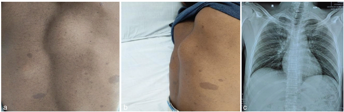
Figure 1: (a) Tumor is visible with a café au lait spot of Back view, (b) Tumor is visible with café au lait spot of Side view, (c) Pre-operative Dorsal Spine Xray represents Osteolytic lesion in costovertebral region of right 7th Rib.
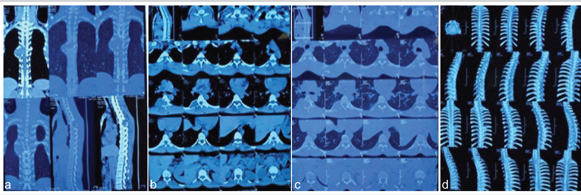
Figure 2: (a, b, c, d): Initial computed tomography (CT) scan imaging. (a) Sagital view of CT scan shows A fairly large solid density lobulated and expansile lesion (6 cm × 4 cm) seen at right posterior part of 7th rib at osteo-chondral junction. (b and c) Axial view shows lesion extends into spinal canal, para vertebral region and right D5-7neural foramen. There is remodeling of involved rib, right D7 transverse process and right part of D7 body. (d) 3D reconstruction – well-defined expansile lesion arising in the mid-thoracic region, around D5–D6 level. Extending from the spinal canal toward the posterior elements. Bony remodeling and widening of the adjacent neural foramen.
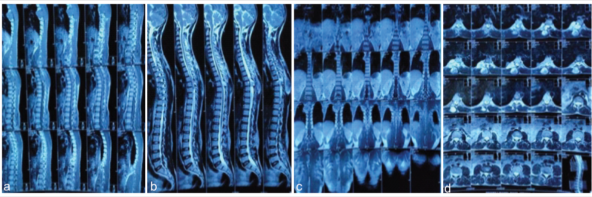
Figure 3: (a, b, c, d): Initial MRI imaging. (a, b, c) Large extra medullary intra dural lesion is noted right side and anterior aspect of spinal canal at the level of D5, D6 vertebra. Part of the lesion extends outside the spinal canal through neural foramina. The intra dural as well as extra dural extra spinal lesion measuring about 6.7 cm × 6.6 cm. (d) After contrast, heterogeneous enhancement of the lesion is noted. Heterogeneous soft tissue mass is also noted at posterior aspect of L1, L2, L3 and L4 vertebral spinal process measuring about 9.6 cm × 2.0 cm.
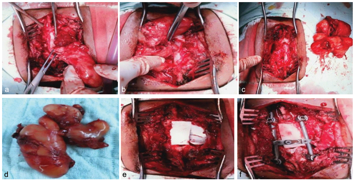
Figure 4: (a, b, c, d, e, f): Intraoperative images showing the excision of a large thoracic spinal neurofibroma. (a and b) The tumor being carefully dissected and mobilized from the paraspinal region. (c) The surgical bed after tumor removal. (d) Gross appearance of the completely excised neurofibroma. (e) Intraoperative exposure of the thoracic spinal neurofibroma at D5-7 and (f) Placement of spinal stabilization hardware following tumor resection.
Diagnosis and treatment plan
Based on clinical, radiological, and histopathological findings, the diagnosis was thoracic spinal neurofibroma (dumbbell type) at D5–D6 level with vertebral and rib destruction and cord compression.
The patient was planned for surgical intervention with the following objectives:
- Complete resection of the spinal tumor to relieve cord compression.
- Spinal stabilization to restore structural integrity following vertebral destruction.
- Post-operative rehabilitation and physiotherapy for neurological recovery.
Surgical procedure
On May 30, 2021, the patient underwent surgical removal of the spinal tumor with spinal stabilization under general anesthesia, performed by Associate Professor Dr. Abdullah Al Mahmud. A standard pre-operative time-out was performed to confirm the patient’s identity, planned procedure, operative levels, and availability of necessary imaging. The patient was then positioned prone on a radiolucent spine table with chest and iliac bolsters to allow the abdomen to hang freely. The head was kept in a neutral position on a foam headrest, with the eyes and face carefully protected. All pressure points were adequately padded, and the arms were positioned safely within <90° abduction. Fluoroscopy was arranged in anteroposterior and lateral orientation for intraoperative level localization.
After proper positioning, the surgical site was marked, and hair was clipped as required. The operative field was prepared with alcohol–chlorhexidine solution, and sterile drapes were applied. Using fluoroscopy, the levels from D4-D9 were confirmed and marked on the skin. A midline incision was made from D4-D9. Hemostasis was achieved using bipolar cautery. Subperiosteal dissection of the paraspinal muscles was carried out to expose the spinous processes, laminae, medial facets, and transverse processes from D5-D8. Self-retaining retractors were applied, and the levels were reconfirmed using fluoroscopy. During surgery, the tumor was carefully dissected from the paraspinal region, as shown in Fig. 4a. Complete excision was achieved, leaving a clear surgical bed (Fig. 4b). The gross specimen of the excised neurofibroma is shown in Fig. 4c.
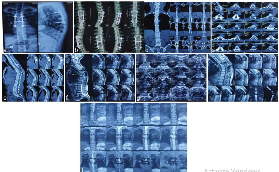
Figure 5: Postoperative findings at 4-year follow-up (a) AP and lateral radiographs and (b) 3D Reconstruction CT of Dorsal spine demonstrated satisfactory posterior pedicle screw-rod fixation across the involved thoracic vertebrae with a transverse cross-link connector. The implants were appropriately positioned with maintained spinal alignment and no radiographic evidence of implant loosening, pull-out, breakage, or loss of correction. (c,d,e) CT Scan show stable posterior instrumentation from D5 to D8 with satisfactory alignment. Mild multilevel spondylotic changes were noted, while vertebral body heights, spinal canal, and neural foramina were preserved. (f,g,h,i) MRI showed persistent D5–D8 spinal cord contusion with no significant cord compression and expected postoperative changes following D5–D8 posterior instrumentation. Mild multilevel degenerative changes were also noted.
Decompression and tumor exposure
Bony work included laminectomy from D5 to D7 levels (D5 and D7 Partially), along with medial facetectomy as required for adequate exposure. Epidural hemostasis was secured using bipolar cautery and hemostatic agents. The extradural tumor was identified and removed en bloc. The spinal canal was inspected thoroughly, and complete decompression was confirmed along the cranio-caudal extent. A bubble test was performed intraoperatively to evaluate for any parietal pleural injury.
Stabilization
Due to the extent of decompression and laminectomy, spinal stabilization was performed. Pedicle screws were inserted into D5 and D8, with two screws placed in each vertebra. Rods were contoured and secured in place, and the set screws were tightened according to the manufacturer’s specifications. A transverse cross-link connector was placed to enhance the overall stability of the construct. Fluoroscopic imaging confirmed the accuracy of screw placement and construct alignment. Adequate decompression and absence of residual compression were confirmed. After subperiosteal dissection of the paraspinal muscles and exposure of the spinous processes and laminae, the thoracic spinal neurofibroma was carefully visualized (Fig. 4a). The tumor was meticulously dissected and excised while preserving surrounding neural structures. Following tumor removal, spinal stabilization was performed using appropriate instrumentation to maintain alignment and stability (Fig. 4b).
Hemostasis, drain placement, and closure
The surgical field was irrigated thoroughly with normal saline. Final hemostasis was achieved using bipolar coagulation, bone wax, and hemostatic matrix as required. A 14 French closed-suction drain was placed in the subfascial space and brought out through a separate stab incision.
Layered closure was performed: The thoracolumbar fascia was approximated with continuous 0 Vicryl sutures, followed by deep subcutaneous closure using 0 Vicryl. Skin closure was completed with staples, and a sterile dressing was applied.
Pathology report
The histopathological examination of the excised extradural spinal tumor revealed a benign, capsulated lesion measuring 8.0 × 3.5 × 3.0 cm. microscopic sections demonstrated interlacing bundles of spindle-shaped cells arranged in a storiform pattern, with elongated nuclei and indistinct cytoplasmic borders. No features of malignancy, such as nuclear atypia, abnormal mitosis, or necrosis, were noted. These findings are consistent with neurofibroma, a benign peripheral nerve sheath tumor. Histopathological examination confirmed the diagnosis of Neurofibroma.
post-operative results and follow-up
The post-operative period was uneventful, with stable vital signs throughout hospitalization. Neurological function gradually improved, particularly motor strength in the lower limbs. Early post-operative imaging confirmed complete tumor resection with adequate spinal cord decompression and no residual compression.
At the 1-month follow-up, the patient was mobilizing independently (Modified McCormick Scale II) and reported marked reduction in pain. There was no evidence of surgical site infection, cerebrospinal fluid leakage, or early post-operative complications. The patient continued structured rehabilitation and physiotherapy.
At the 4-year follow-up, the patient demonstrated complete neurological recovery (Modified McCormick Scale I) of the lower limbs, was fully ambulatory, and had resumed his professional activities without functional limitations. Radiological evaluation revealed satisfactory spinal alignment and stable posterior instrumentation. Plain radiographs and three-dimensional CT reconstruction showed symmetrically placed thoracic pedicle screws connected by longitudinal rods with an intact transverse connector, without evidence of implant loosening, breakage, or malalignment (Fig. 5a-e). MRI confirmed preserved spinal cord morphology with sustained decompression and no evidence of residual or recurrent tumor. (Fig 5. f-i).
Spinal neurofibromas are benign tumors of the peripheral nerve sheath and can lead to progressive neurological deficits due to spinal cord or nerve root compression [12,14]. Neurofibromas with complex intraspinal and paraspinal extensions pose a unique surgical challenge because of their proximity to vital neural and vascular structures [5,6]. These tumors may present with motor weakness, sensory deficits, and pain, as observed in our patient [5]. Early diagnosis is crucial to prevent permanent neurological impairment and to plan an optimal surgical approach [14,15].
MRI is essential for pre-operative assessment, as it provides detailed information about tumor size, extent, and relationship with the spinal cord and surrounding tissues [10,12,16]. In our case, MRI clearly delineated the intraspinal and paraspinal components of the tumor and its extension through the D5–D6 intervertebral foramen, which guided surgical planning and helped anticipate intraoperative challenges [16,17]. Prior studies emphasize that accurate imaging is critical for improving surgical outcomes and minimizing complications [12,16].
Surgical excision remains the definitive treatment for symptomatic spinal neurofibromas [17]. Complete resection is associated with improved neurological recovery and decreased recurrence rates, although it can be technically challenging when tumors involve vertebral bodies, lamina, or ribs [5,6,15]. Spinal stabilization may be necessary when bony structures are resected to maintain structural integrity and prevent post-operative deformity [17]. In our patient, complete excision with stabilization resulted in excellent functional recovery and independent ambulation, consistent with outcomes reported in other series [8,10,15].
Adjunctive techniques such as intraoperative neurophysiological monitoring can enhance surgical safety by allowing real-time assessment of spinal cord function during tumor resection [18]. Tumor location, size, and degree of spinal cord compression can significantly influence clinical presentation and surgical strategy, and these factors must be considered for optimal outcomes [16,19]. In uncomplicated patients with gross total resection, follow-up of 1–5 years has been reported, and recurrence is uncommon (3–9%) and usually occurs within 4 years of surgery [20].
This case highlights several key points: Dumbbell-shaped spinal neurofibromas, although rare, can cause progressive neurological deficits; MRI is indispensable for diagnosis and surgical planning; and complete surgical excision combined with spinal stabilization can achieve excellent neurological and functional outcomes.
This case demonstrates that surgical management of spinal neurofibromas with complete excision and spinal stabilization is effective in relieving cord compression and restoring neurological function (Modified McCormick Scale from IV to I) with no evidence of recurrence in a 4-year follow-up after operation. Post-operative rehabilitation further contributes to recovery, enabling patients to regain independent mobility and return to normal daily and professional activities. Early diagnosis and intervention are essential for achieving optimal outcomes and improving quality of life.
For clinicians, early recognition of spinal neurofibromas, careful pre-operative imaging, and individualized surgical planning are key to preventing permanent neurological deficits. Meticulous operative technique and appropriate spinal stabilization strategies can significantly improve patient outcomes and quality of life.
References
- 1. Messersmith L, Krauland K. Neurofibroma. In: StatPearls. Treasure Island (FL): StatPearls Publishing; August 8, 2023. Available from: https://www.ncbi.nlm.nih.gov/books/NBK539707/ [Google Scholar] [PubMed]
- 2. Thakkar SD, Feigen U, Mautner VF. Spinal tumours in neurofibromatosis type 1: An MRI study of frequency, multiplicity and variety. Neuroradiology 1999; 41:625-9. [Google Scholar] [PubMed]
- 3. Ruggieri M, Polizzi A, Spalice A, Salpietro V, Caltabiano R, D’Orazi V, et al. The natural history of spinal neurofibromatosis: A critical review of clinical and genetic features. Clin Genet 2015; 87:401-10. [Google Scholar] [PubMed]
- 4. Mautner VF, Tatagiba M, Lindenau M, Fünsterer C, Pulst SM, Baser ME, et al. Spinal tumors in patients with neurofibromatosis type 2: MR imaging study of frequency, multiplicity, and variety. AJR Am J Roentgenol 1995; 165:951-5. [Google Scholar] [PubMed]
- 5. Crawford AH, Lykissas MG, Schorry EK, Gaines S, Jain V, Greggi T, et al. Neurofibromatosis: Etiology, commonly encountered spinal deformities, common complications and pitfalls of surgical treatment. Spine Deform 2012; 1:85-94. [Google Scholar] [PubMed]
- 6. Pollack IF, Colak A, Fitz C, Wiener E, Moreland M, Mulvihill JJ. Surgical management of spinal cord compression from plexiform neurofibromas in patients with neurofibromatosis 1. Neurosurgery 1998; 43:248-55; discussion 255-6. [Google Scholar] [PubMed]
- 7. Parsons CM, Canter RJ, Khatri VP. Surgical management of neurofibromatosis. Surg Oncol Clin N Am 2009; 18:175-96. [Google Scholar] [PubMed]
- 8. Safaee M, Parsa AT, Barbaro NM, Chou D, Mummaneni PV, Weinstein PR, et al. Association of tumor location, extent of resection, and neurofibromatosis status with clinical outcomes for 221 spinal nerve sheath tumors. Neurosurg Focus 2015;39: E5. [Google Scholar] [PubMed]
- 9. Gerszten PC, Burton SA, Ozhasoglu C, McCue KJ, Quinn AE. Radiosurgery for benign intradural spinal tumors. Neurosurgery 2008; 62:887-95, discussion 895-6. [Google Scholar] [PubMed]
- 10. Ferreira Filho AC, Blanco CM, Trindade JV, Mattos GB, Joaquim AF. Surgical outcome of spinal schwannoma and neurofibroma. Rev Assoc Méd Bras (1992) 2023;69: e20230190. [Google Scholar] [PubMed]
- 11. Seppälä MT, Haltia MJ, Sankila RJ, Jääskeläinen JE, Heiskanen O. Long-term outcome after removal of spinal neurofibroma. J Neurosurg 1995; 82:572-7. [Google Scholar] [PubMed]
- 12. Amadasu E, Panther E, Lucke-Wold B. Characterization and treatment of spinal tumors. Intensive Care Res 2022; 2:76-95. [Google Scholar] [PubMed]
- 13. Ciftdemir M, Kaya M, Selcuk E, Yalniz E. Tumors of the spine. World J Orthop 2016; 7:109-16. [Google Scholar] [PubMed]
- 14. Noureldine MH, Shimony N, Jallo GI. Benign spinal tumors. In: Human Brain and Spinal Cord Tumors: From Bench to Bedside. The Path to Bedside Management. Vol. 2. Cham: Springer International Publishing; 2023. p. 583-606. [Google Scholar] [PubMed]
- 15. Hori T, Takakura K, Sano K. Spinal neurinomas–clinical analysis of 45 surgical cases. Neurol Med Chir (Tokyo) 1984; 24:471-7. [Google Scholar] [PubMed]
- 16. Yamaguchi S, Menezes AH, Shimizu K, Woodroffe RW, Helland LC, Hitchon PW, et al. Differences and characteristics of symptoms by tumor location, size, and degree of spinal cord compression: A retrospective study on 53 surgically treated, symptomatic spinal meningiomas. J Neurosurg Spine 2020; 32:931-40. [Google Scholar] [PubMed]
- 17. Aghayev K, Vrionis F, Chamberlain MC. Adult intradural primary spinal cord tumors. J Natl Compr Canc Netw 2011; 9:434-47. [Google Scholar] [PubMed]
- 18. Lakomkin N, Mistry AM, Zuckerman SL, Ladner T, Kothari P, Lee NJ, et al. Utility of intraoperative monitoring in the resection of spinal cord tumors: An analysis by tumor location and anatomical region. Spine (Phila Pa 1976) 2018; 43:287-94. [Google Scholar] [PubMed]
- 19. Van Goethem JW, Van Den Hauwe L, Özsarlak Ö, De Schepper AM, Parizel PM. Spinal tumors. Eur J Radiol 2004; 50:159-76. [Google Scholar] [PubMed]
- 20. Gui C, Canthiya L, Zadeh G, Suppiah S. Current state of spinal nerve sheath tumor management and future advances. Neurooncol Adv 2024;6 Suppl 3: iii83-93. [Google Scholar] [PubMed]






Related Articles in Journal of Orthopaedic Case Reports
 December 1, 2024 Conservative Management of a Rare Clinical Phenomenon: Paraspinal Compartment Syndrome – A Review of Existing Literature
 November 1, 2024 The Influence of Coccygeal Dynamic Patterns in Ganglion Impar Block for the Treatment of Chronic Refractory Coccydynia
 August 1, 2026 Clinical and Functional Outcomes of Posterior Cruciate Ligament Tibial Avulsion Fractures Treated with a Mini-Open Posterior Approach and Internal Fixation: A Case Series
 October 10, 2015 Conjoint Lumbosacral Nerve Root- A Case Report