

Valgus intertrochanteric osteotomy is a cost-effective, head-preserving procedure for young patients with vertical intracapsular neck of femur fractures or selected non-unions, converting shear into compressive forces to promote union while preserving the native hip.
Dr. Faiz Ahmad Usmani, Department of Orthopaedics, Government Medical College and Hospital, Chhatrapati Sambhajinagar, Maharashtra, India. E-mail: faizusmani03@gmail.com
Abstract
Introduction: Intracapsular fractures of the femoral neck continue to represent one of the most demanding injuries encountered by orthopedic surgeons, particularly in young adults. Preservation of the femoral head remains the primary objective because arthroplasty in this age group is associated with limited implant longevity and the likelihood of multiple revision procedures. Valgus intertrochanteric osteotomy, originally described by Pauwels, alters fracture biomechanics by transforming vertical shear forces into compressive forces, thereby enhancing fracture stability and promoting union. This study evaluates the clinical and radiological outcomes of valgus intertrochanteric osteotomy in young patients presenting with Pauwels Type II and III intracapsular neck of femur fractures.
Materials and Methods: A retrospective case series comprising twenty-five patients younger than 60 years with Pauwels Type II or III intracapsular neck of femur fractures was conducted at a tertiary care center. Fresh fractures presenting within 24 h as well as delayed presentations up to 3 months were included. Patients underwent valgus intertrochanteric osteotomy using fixed-angle internal fixation according to fracture configuration and surgeon preference. Clinical records and serial radiographs were reviewed to evaluate fracture union, delayed union, fixation failure, and complications. The study was approved by the Institutional Ethics Committee, and valid written informed consent was obtained from all participants after explaining the procedure and study objectives.
Results: Among the 25 patients included in the series, fracture union was achieved in 15 patients, delayed union occurred in 4 patients, and 6 patients demonstrated fixation failure requiring further management. The overall healing rate was 76%. The procedure successfully restored mechanical alignment and preserved the native femoral head in the majority of patients.
Conclusion: Valgus intertrochanteric osteotomy continues to be an effective biological solution for selected intracapsular femoral neck fractures in young adults. Appropriate patient selection, meticulous pre-operative planning, and stable fixation remain critical determinants of successful outcomes. In appropriately selected patients, this procedure provides an economical and reproducible alternative to arthroplasty while preserving future reconstructive options.
Keywords: Neck of femur fracture, valgus osteotomy, Pauwel's fracture, head preservation, non-union, dynamic hip screw.

Intracapsular fractures of the femoral neck remain a challenging injury in orthopedic practice because of their intracapsular location and precarious vascular supply. Although arthroplasty is the preferred treatment for displaced femoral neck fractures in elderly patients, preservation of the native femoral head remains the standard goal in young adults with higher functional demands. Vertically oriented fractures are subjected predominantly to shear forces, increasing the risk of displacement, non-union, and implant failure. Valgus intertrochanteric osteotomy addresses this biomechanical disadvantage by reducing the Pauwels angle and converting shear stresses into compressive forces across the fracture site, thereby promoting union while preserving the femoral head. Over the years, this procedure has evolved with improved fixation methods, including dynamic hip screws (DHS) and angled blade plates and has demonstrated satisfactory union rates in both neglected fractures and established non-unions [1,2,3,4,5].
Despite encouraging reports, several uncertainties remain regarding the reproducibility of outcomes following valgus intertrochanteric osteotomy. Most published studies are retrospective, involve heterogeneous patient populations, or originate from specialized centers with relatively large surgical experience. Furthermore, the influence of patient selection, fracture characteristics, implant choice, and surgical accuracy on functional outcomes continues to be debated. Limited evidence is available from resource-constrained government hospitals where delayed presentation is common and access to arthroplasty may be restricted [6,7,8,9,10].
The present study was undertaken to evaluate the clinical and radiological outcomes of valgus intertrochanteric osteotomy in young patients with intracapsular neck of femur fractures treated at a tertiary care government hospital. We aimed to assess fracture union, functional recovery, and procedure-related complications, thereby providing additional evidence regarding the effectiveness of this cost-effective, head-preserving procedure in a resource-limited setting.
Study design
This case series was conducted at a tertiary care teaching hospital to evaluate the effectiveness of valgus intertrochanteric osteotomy as a head-preserving procedure in young patients with intracapsular fractures of the femoral neck. The study included ten consecutive patients treated with valgus osteotomy and internal fixation.
The study was approved by the Institutional Ethics Committee. Written informed consent was obtained from all participants after detailed explanation of the surgical procedure, possible complications, rehabilitation protocol, and the use of anonymized clinical and radiological data for academic publication.
Inclusion criteria
- Patients younger than 60 years of age.
- Pauwels Type II and Type III intracapsular neck of femur fractures.
- Fresh fractures presenting within 24 h of injury.
- Delayed fractures presenting within 3 months of injury.
Patients in whom preservation of the femoral head was considered feasible (Fig. 1).
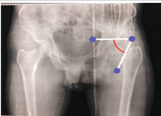
Figure 1: High Pauwel’s angle femur neck fracture.
Exclusion criteria
- Established avascular necrosis of the femoral head.
- Severe collapse of the femoral head.
- Advanced osteoarthritis of the hip.
- Grossly comminuted or non-reconstructible femoral neck fractures.
- Patients medically unfit for surgery (Fig. 2).
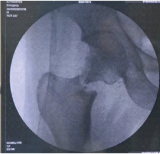
Figure 2: Severe neck comminution diagnosed on traction; surgical plan changed to hemiarthroplasty.
Pre-operative evaluation
All patients underwent detailed clinical examination and radiographic evaluation with anteroposterior pelvis and lateral hip radiographs.
Fractures were classified according to the Pauwels classification system.
The inclination of the fracture line and anticipated correction angle were assessed pre-operatively. Preoperative radiographs were used to assess fracture characteristics and determine the size of the valgus-producing wedge. Computed tomography (CT) was performed selectively to better define fracture morphology and assist surgical planning in complex cases, while magnetic resonance imaging was reserved for patients in whom avascular necrosis was suspected or radiographic findings were inconclusive. Routine laboratory investigations and pre-anesthetic fitness assessment were completed before surgery (Fig. 3).
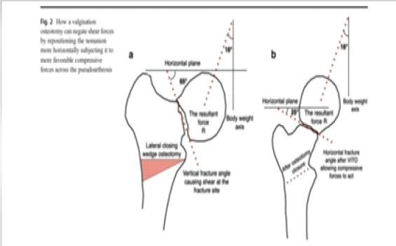
Figure 3: Biomechanics of valgus osteotomy. (21)
Surgical technique
All procedures were performed with the patient in the supine position on a traction table under fluoroscopic guidance. Reduction of the fracture was achieved whenever possible before commencing the osteotomy.
A standard lateral approach to the proximal femur was utilized. After insertion of the guide wire into the femoral neck and head along the intended implant trajectory, the planned osteotomy was marked using converging guide wires. A lateral closing wedge osteotomy, generally measuring approximately 20–25°, was created at or just distal to the level of the lesser trochanter while preserving the medial cortical hinge.
Following removal of the wedge, the distal fragment was abducted to achieve the planned valgus correction, thereby reducing the inclination of the fracture line and converting vertical shear forces into compressive forces across the fracture site. Internal fixation was performed using an appropriate fixed-angle device depending on fracture configuration and intra-operative considerations, including DHS constructs with supplementary derotation screws or angled blade plates where indicated.
Fluoroscopic assessment confirmed satisfactory fracture reduction, implant position, and correction before wound closure (Fig. 4).
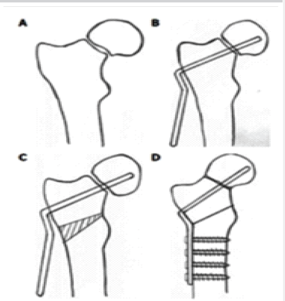
Figure 4: Demonstrative technique of valgus osteotomy.(22)
Post-operative protocol
Patients were encouraged to begin early hip and knee range-of-motion exercises under physiotherapy supervision.
Toe-touch or partial weight-bearing was advised initially, with progression according to radiographic evidence of healing and clinical assessment.
Patients were followed clinically and radiographically at regular intervals until fracture union or identification of treatment failure.
Outcome measures
Primary outcome measures included:
- Radiological fracture union.
- Time to union.
- Delayed union.
Fixation failure or non-union
Secondary outcomes included preservation of the native femoral head, maintenance of reduction, and procedure-related complications.
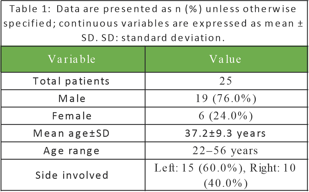
Road traffic accidents were the most common mechanism of injury, accounting for 10 cases (40.0%), followed by falls from height (12.0%). Other mechanisms included self-fall, physical assault, sports-related injuries, and slip and fall (8.0% each).
Of the 25 patients, fracture union was achieved in 15 (60%), delayed union occurred in 4 (16%), and fixation failure/non-union occurred in 6 (24%). Multivariable logistic regression demonstrated that increasing age (adjusted OR 1.08, 95% CI 1.01–1.17, P = 0.031) and high-energy mechanism of injury (adjusted OR 4.67, 95% CI 1.18–18.49, P = 0.028) were independent predictors of a poor outcome. The regression model demonstrated good calibration (Hosmer–Lemeshow P = 0.735) and acceptable discriminative ability (AUC = 0.83). (Table 1).
Patients achieving successful union demonstrated progressive pain relief, restoration of hip function, and return to activities of daily living. Radiographs revealed satisfactory maintenance of valgus correction with bridging trabeculae across the fracture site in successfully treated cases.
The procedure was technically reproducible and could be performed using widely available implants without the need for microsurgical expertise or expensive biological augmentation.
No intra-operative neurovascular complications were encountered. Careful pre-operative planning, accurate wedge calculation, preservation of the medial cortical hinge, and stable fixed-angle fixation were identified as critical technical factors influencing successful outcomes.
Although 6 cases failed to unite, these failures highlighted recognized limitations of the procedure, including inadequate reduction, persistent instability, and adverse fracture biology, emphasizing the importance of meticulous patient selection and surgical execution (Fig. 5,6,7)).
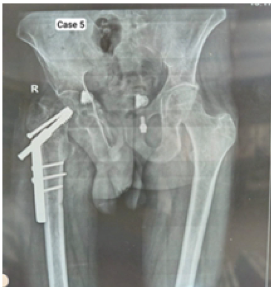
Figure 5: Implant failure in a High Pauwels angle fracture treated with valgus osteotomy and fixed primarily with dynamic hip screw.
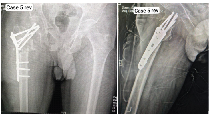
Figure 6&7: Revision surgery done, dynamic hip screws swapped for fixed-angle greater trochanter plate for proximal femur.
Management of intracapsular fractures of the femoral neck in young adults continues to represent one of the most challenging problems in trauma surgery. Unlike elderly patients, in whom arthroplasty has become the preferred treatment, every attempt should be directed towards preservation of the native femoral head in younger individuals because of their higher functional demands, superior healing potential and the finite longevity of prosthetic implants. The consequences of early arthroplasty in this age group include multiple future revision surgeries, progressive bone loss and increasing technical complexity with each subsequent reconstruction [11].
The principal biological challenge associated with intracapsular femoral neck fractures is disruption of the vascular supply to the femoral head. In addition, vertically oriented fractures generate predominantly shear forces across the fracture site, resulting in instability, fracture displacement, and increased rates of delayed union and non-union. These biomechanical factors explain why conventional fixation alone may fail in selected Pauwels Type II and Type III fractures despite satisfactory reduction [11].
Pauwels first proposed valgus intertrochanteric osteotomy in 1935 based upon biomechanical principles rather than a specific operative technique. His concept was elegant and remains relevant today. By decreasing the inclination of the fracture line, valgus correction converts detrimental shear forces into compressive forces, thereby improving the mechanical environment necessary for fracture healing. Although implant technology has evolved substantially since Pauwels’ original description, the fundamental biomechanical rationale has remained unchanged [2,11] (Fig. 8)).
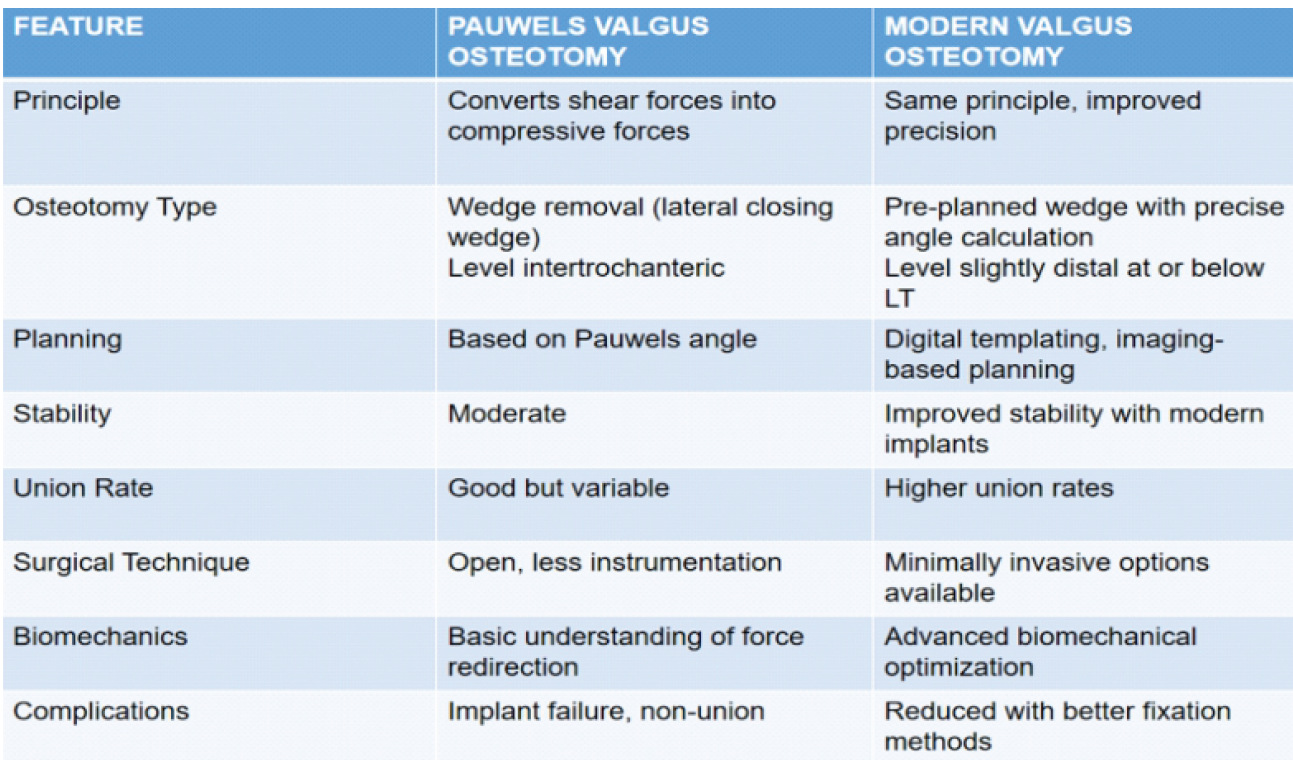
Figure 8: Differences between Pauwels and modern valgus osteotomy of proximal femur.
Our series demonstrated fracture healing in 19 of 25 patients, including four cases of delayed union, corresponding to an overall healing rate of 76%. Although the sample size remains relatively small, these findings support the continued role of valgus intertrochanteric osteotomy as an effective head-preserving procedure in carefully selected patients. The six cases of fixation failure observed in our series were associated with recognized causes of failure, including persistent instability, inadequate reduction, and compromised fixation, highlighting that meticulous surgical technique and appropriate patient selection are critical determinants of successful outcomes [6,7,8,9,12,13,14,15,16,17,18].
Historically, non-union following displaced femoral neck fractures has been reported in up to 30–50% of elderly patients. Even among younger individuals, reported rates of non-union and fixation failure remain clinically significant. The ability of valgus osteotomy to improve fracture biomechanics offers an important biological advantage over fixation alone by increasing compressive loading at the fracture interface and promoting osteogenesis under more favorable mechanical conditions [1,2,9,11].
Marti and colleagues demonstrated that valgus osteotomy could successfully salvage femoral neck non-union while maintaining the native femoral head in a substantial proportion of patients [1]. Ballmer and co-workers further refined operative planning and emphasized the importance of stable fixed-angle fixation together with accurate correction of fracture inclination [2]. These observations continue to influence contemporary surgical practice and remain applicable in present-day trauma surgery.
Yuan and collaborators highlighted the importance of careful wedge planning, recommending correction angles of approximately 20–25° in most patients. Excessive valgus correction should be avoided because it alters hip biomechanics, increases joint reaction forces, and may predispose patients to abductor insufficiency, limb-length discrepancy, and degenerative changes. The amount of correction therefore requires individualization according to fracture geometry while maintaining restoration of the mechanical axis [10,17].
Selection of the fixation device remains an important consideration in valgus intertrochanteric osteotomy. In the present series, a DHS supplemented with a cannulated cancellous (CC) derotation screw was used as the primary fixation construct in all 25 patients. This construct was chosen because it provides stable fixed-angle fixation with controlled compression across the fracture site while effectively resisting rotational forces. The DHS with an adjunctive CC screw is technically straightforward, reproducible, and widely available, making it particularly suitable for centers with limited access to more specialized implants. Although angled blade plates, dynamic condylar screws, and proximal femoral locking plates remain valuable alternatives in selected cases with challenging anatomy or fracture patterns, the uniform use of the DHS–CC screw construct in our series provided satisfactory stability and facilitated fracture healing in the majority of patients, supporting its role as a reliable implant for valgus intertrochanteric osteotomy (Fig. 9)) [3,6,7,10,13,16,18].
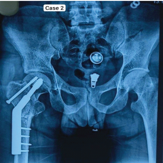
Figure 9: Dynamic hip screw with added de-rotation cannulated cancellous screw, union achieved 29 days post valgus osteotomy.
The primary implant was changed only in instances of non-union or failure.
An important advantage demonstrated by our experience is the practicality of valgus osteotomy in resource-constrained settings. The procedure does not require microsurgical expertise, vascularized bone grafting, or expensive biological substitutes. Standard trauma implants commonly available in district and tertiary hospitals can be utilized effectively while preserving the patient’s own hip joint. This is particularly relevant in developing healthcare systems where economic considerations frequently influence treatment decisions [4,5,6,14].
Despite these advantages, valgus osteotomy is not without limitations. The procedure permanently alters proximal femoral anatomy and may increase the technical difficulty of any future total hip arthroplasty. Overcorrection may lead to excessive limb lengthening, altered abductor mechanics and gait disturbance. Failure to recognize posterior comminution, femoral neck resorption or established avascular necrosis may result in persistent non-union despite technically satisfactory surgery. Careful patient selection therefore remains fundamental to achieving favorable outcomes [8,9,15,16,17,19].
Radiographic parameters including changes in Pauwels angle, neck-shaft angle, limb length discrepancy, and the degree of valgus alignment correction were not objectively measured. Quantitative assessment of these parameters would have provided a more comprehensive evaluation of the biomechanical correction achieved and its relationship with fracture healing and functional outcome [10,17].
The absence of a comparison group precludes direct evaluation of valgus intertrochanteric osteotomy against alternative head-preserving procedures or arthroplasty.
The present study has several limitations. It represents a retrospective case series from a single institution with a relatively small number of patients and lacks a comparison group treated by alternative methods. Functional outcome scores were not uniformly available for all patients, limiting objective assessment beyond radiographic healing. Nevertheless, the study reflects real-world clinical practice and demonstrates that valgus intertrochanteric osteotomy remains an effective and economical head-preserving procedure when applied to appropriately selected patients with meticulous pre-operative planning and sound surgical technique [12,13,14,15,16,17,18,20].
Valgus intertrochanteric osteotomy remains an effective biological solution for vertically oriented intracapsular neck of femur fractures and selected cases of delayed union or non-union in young adults. By converting shear stresses into compressive forces, the procedure enhances fracture healing while preserving the native femoral head. Our series demonstrated an overall healing rate of 80%, supporting the 0 continued role of this technique in appropriately selected patients. Careful patient selection, precise pre-operative planning, accurate wedge correction, and stable fixed-angle fixation remain the cornerstones of successful treatment. In resource-limited settings, valgus osteotomy continues to represent a reliable, reproducible, and cost-effective alternative to primary arthroplasty.
Valgus intertrochanteric osteotomy is an underutilized yet valuable head-preserving procedure for young patients with vertically oriented femoral neck fractures. Understanding the biomechanical principles of the operation, meticulous surgical planning, and appropriate implant selection can significantly improve fracture union while delaying or avoiding the need for hip replacement.
References
- 1. Marti RK, Schüller HM, Raaymakers EL. Intertrochanteric osteotomy for non-union of the femoral neck. J Bone Joint Surg Br 1989;71:782-7. [Google Scholar] [PubMed]
- 2. Ballmer FT, Ballmer PM, Baumgaertel F, Mast JW, Ganz R. Pauwels osteotomy for nonunions of the femoral neck. Orthop Clin North Am 1990;21:759-67. [Google Scholar] [PubMed]
- 3. Anglen JO. Intertrochanteric osteotomy for failed internal fixation of femoral neck fracture. Clin Orthop Relat Res 1997;341:175-82. [Google Scholar] [PubMed]
- 4. Kalra M, Anand S. Valgus intertrochanteric osteotomy for neglected femoral neck fractures in young adults. Int Orthop 2001;25:363-6. [Google Scholar] [PubMed]
- 5. Magu NK, Singh R, Mittal R, Garg R, Wokhlu A, Sharma AK. Osteosynthesis and primary valgus intertrochanteric osteotomy in displaced intracapsular fracture neck of femur with osteoporosis in adults. Injury 2005;36:110-22. [Google Scholar] [PubMed]
- 6. Khan AQ, Khan MS, Sherwani MK, Agarwal R. Role of valgus osteotomy and fixation with dynamic hip screw and 120° double-angle barrel plate in the management of neglected and ununited femoral neck fracture in young patients. J Orthop Traumatol 2009;10:71-8. [Google Scholar] [PubMed]
- 7. Magu NK, Rohilla R, Singh R, Tater R. Modified Pauwels’ intertrochanteric osteotomy in neglected femoral neck fracture. Clin Orthop Relat Res 2009;467:1064-73. [Google Scholar] [PubMed]
- 8. Varghese VD, Boopalan PR, Titus VT, Oommen AT, Jepegnanam TS. Indices affecting outcome of neglected femoral neck fractures after valgus intertrochanteric osteotomy. J Orthop Trauma 2014;28:410-6. [Google Scholar] [PubMed]
- 9. Deakin DE, Guy P, O’Brien PJ, Blachut PA, Lefaivre KA. Managing failed fixation: Valgus osteotomy for femoral neck nonunion. Injury 2015;46:492-6. [Google Scholar] [PubMed]
- 10. Kulkarni SG, Kulkarni GS, Babhulkar S, Kulkarni MG, Kulkarni RM. Accuracy of valgus osteotomy using dynamic hip screw. Injury 2017;48 Suppl 2:S2-7. [Google Scholar] [PubMed]
- 11. Makhni EC, Steinhaus ME, Padilla JA Contemporary management of femoral neck fractures in young adults: Fixation principles and preservation of the native hip. J Am Acad Orthop Surg 2021;29:e590-600. [Google Scholar] [PubMed]
- 12. Elgeidi A, Eissa M, El-Shafie M. Valgus intertrochanteric osteotomy for neglected femoral neck fractures in young adults: Clinical and radiological outcomes. J Orthop Surg Res 2021;16:e590-e600.. [Google Scholar] [PubMed]
- 13. Wang T, Guo W, Li B. Clinical outcomes of valgus intertrochanteric osteotomy combined with internal fixation for femoral neck fracture nonunion. Orthop Surg 2022;14:. [Google Scholar] [PubMed]
- 14. Chakrapani AS, Ragate R, Shaik A, Singh MK, Nair A, Kumar A, et al. A pilot study of functional outcomes in adult patients treated with valgus intertrochanteric osteotomy as a primary modality for intracapsular femoral neck fracture. Cureus 2024;16:e60205. [Google Scholar] [PubMed]
- 15. Seibold BT, Walker MJ, Bellaire CP, Raffa T, Dance S, Gu A, et al. Systematic review of valgus intertrochanteric osteotomy in the setting of femoral neck fractures. J Orthop 2025;67:20-6. [Google Scholar] [PubMed]
- 16. Xu H, Yang S, Zheng Y, Tian A, Wu R, Zhang X, et al. Efficacy of valgus osteotomy in treating nonunion of femoral neck fractures post-internal fixation. Front Surg 2025;12:1526384. [Google Scholar] [PubMed]
- 17. Ramasamy B, Bhowmick K, Ashok A, Livingston A, Varghese VD. Medial opening wedge valgus intertrochanteric osteotomy for femoral neck nonunion: A femoral anatomy-preserving surgical approach. Hip Int 2025;35:301-7. [Google Scholar] [PubMed]
- 18. Kumar S, Kumar P, Bedia G, Paramjit. The outcomes of valgus intertrochanteric osteotomy and lag-screw fixation in femoral neck fracture. J Contemp Clin Pract 2025;35(3). [Google Scholar] [PubMed]
- 19. Chen Y, Zhang G, Wang C, Zhu XH, Sun CG. Treatment of nonunion after femoral neck fracture with valgus intertrochanteric osteotomy: A case report with 18-year follow-up. BMC Musculoskelet Disord 2021;22:471. [Google Scholar] [PubMed]
- 20. Mia MM, Khan MS, Hossain MA, et al. Valgus osteotomy in neglected femoral neck fractures: A case report. Mugda Med Coll J 2025;8:72-5. [Google Scholar] [PubMed]
- 21. Gavaskar AS, Srinivasan P, Jeyakumar B, Raj RV.. Valgus intertrochanteric osteotomy for femur neck pseudoarthrosis: a simple solution to a complex problem. International Orthopaedics (SICOT). 2020 Apr;44(4):635-643. doi: 10.1007/s00264-019-04353-7. [Google Scholar] [PubMed] [CrossRef]
- 22. Varghese VD, Livingston A, Boopalan PR, Jepegnanam TS. Valgus osteotomy for nonunion and neglected neck of femur fractures. World J Orthop. 2016 May 18;7(5):301-7. doi: 10.5312/wjo.v7.i5.301. PMID: 27190758; PMCID: PMC4865720. [Google Scholar] [PubMed] [CrossRef]





Related Articles in Journal of Orthopaedic Case Reports
 October 1, 2026 Humeral Fractures in Metabolically Compromised Bone: Outcomes of Structural Strut Onlay Allograft with Open Reduction and Internal Fixation – A Case Series of Three Patients
 September 1, 2026 Multifactorial Non-union After First Metatarsophalangeal Arthrodesis Revised With Structural Iliac Crest Autograft: A Case Report and Comprehensive Literature Review
 September 1, 2026 Application of 3D-printed models in procedural training for dynamic hip screw fixation and intraosseous vascular access: An original article
 July 1, 2026 Megaprosthetic Total Knee Arthroplasty for Supracondylar Refracture in a Post-traumatic Osteoarthritic Knee: A Case Report