

In patients with persistent low back pain after lumbar surgery and no indication for further surgical intervention, ultrasound assessment of the thoracolumbar fascia may reveal clinically relevant abnormalities. Thoracolumbar fascial defects should be considered as a potential pain generator and may represent a target for ultrasound-guided intervention.
Dr. Selkin Yılmaz Muluk, Sağlık Bakanlığı Antalya Şehir Hastanesi Göçerler Mah. 5379 Sk. 07080, Antalya, Turkey. E-mail: selkinyilmaz@gmail.com
Abstract
Introduction: Persistent low back pain (LBP) after lumbar spine surgery is commonly attributed to recurrent stenosis, neural compression, or degenerative changes. Abnormalities of the thoracolumbar fascia (TLF) after lumbar surgery have rarely been reported as a potential source of pain. We describe a patient with persistent post-operative LBP in whom musculoskeletal ultrasound (US) demonstrated a focal fascial defect corresponding to the painful region.
Case Report: A 65-year-old man presented with LBP 2 years after lumbar surgery for spinal stenosis. Evaluation revealed no neurological deficit or indication for further surgery. US demonstrated a focal midline defect of the TLF between the L2 and L3 levels. US-guided injection of 2 mL 0.5% bupivacaine and 3 mL normal saline beneath the affected fascial plane was performed. Pain decreased at both 1-week and 1-month follow-up.
Conclusion: This case highlights a sonographically visible thoracolumbar fascial defect in a patient with persistent LBP following lumbar surgery. The favorable response to US-guided fascial plane injection suggests that post-operative fascial abnormalities may represent an underrecognized pain generator in selected patients. Studies are required to clarify their clinical significance and therapeutic implications.
Keywords: Low back pain, thoracolumbar fascia, lumbar surgery, ultrasonography, ultrasound-guided injection.

Persistent low back pain (LBP) after lumbar spine surgery remains a common clinical challenge [1]. During the evaluation of persistent post-operative pain, recurrent stenosis, nerve root compression, hardware-related complications, epidural fibrosis, and adjacent segment degeneration are among the conditions most commonly considered [1,2]. However, post-operative abnormalities of the thoracolumbar fascia (TLF) are rarely evaluated as potential pain generators. We report a patient with persistent post-operative LBP in whom ultrasound (US) examination demonstrated a focal thoracolumbar fascial defect corresponding to the painful region.
A 65-year-old man presented with chronic LBP following lumbar surgery performed 2 years earlier for lumbar spinal stenosis. Although the operation resulted in partial initial improvement, localized pain persisted postoperatively (Visual Analog Scale [VAS] 5/10) in the right paraspinal region at the L3 level. Over the subsequent 2 years, the pain remained consistently localized to the same right paraspinal region, corresponding to the site of maximal tenderness. Several months before presentation, the pain became more severe following gardening activities involving repetitive bending, increasing to 7/10 on the VAS. Previous treatments, including analgesics, muscle relaxants, and physical therapy, had provided insufficient relief.
Clinical examination
Physical examination revealed no focal neurological deficits, and there were no signs of radiculopathy. Localized tenderness was elicited exclusively over the right paravertebral area at the L3 level. Furthermore, repeat radiological assessment demonstrated no pathology requiring additional surgical intervention. A clinical photograph of the lumbar region shows the healed midline surgical scar (Fig. 1).
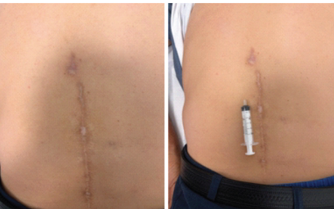
Figure 1: Post-operative lumbar incision scar. Clinical photograph demonstrating the healed midline lumbar surgical scar. The patient’s maximal pain was localized to the right paraspinal region at the L3 vertebral level.
Sonographic assessment
US examination demonstrated that thoracolumbar fascial continuity was well preserved at the cranial levels, superior to the upper limit of the incision scar (Fig. 2a). However, serial scanning from L2 through L4 revealed a focal midline thinning of the TLF, with a localized defect in fascial continuity that was most pronounced at the L3 level (Fig. 2b). Below this affected region, the fascial architecture was again intact and well preserved at the caudal levels (Fig. 2c).
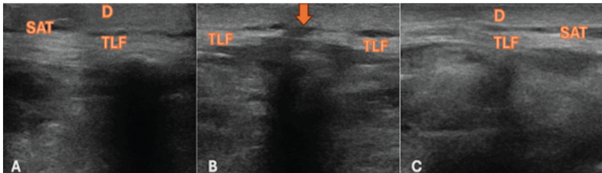
Figure 2: Ultrasound evaluation of the thoracolumbar fascia. Transverse sonograms demonstrate: (a) Preserved, thick, and continuous TLF obtained at the L1 level; (b) The L3 level demonstrating marked thinning of the thoracolumbar fascia at the midline (arrow), with the right and left fascial layers appearing separated, indicating a localized defect of normal fascial architecture; (c) Resumed thick and continuous TLF at the L5 level. TLF: Thoracolumbar fascia, D: Dermis, SAT: Subcutaneous adipose tissue.
Management and follow-up
Because no surgically relevant pathology was identified and the patient’s pain remained refractory to conservative treatments, a US-guided fascial intervention was planned. Given that the sonographic abnormality corresponded to the symptomatic area, a mixture of 2 mL of 0.5% bupivacaine and 3 mL of normal saline was prepared to be injected beneath the thoracolumbar fascial plane at the level of the defect under real-time US guidance.
Under sterile conditions, a 22-gauge needle was introduced in the transverse plane. Rather than depositing the injectate at a single point, a dynamic hydrodissection technique was used. The needle was advanced longitudinally within the fascial plane while the injection was performed, allowing progressive separation of the tissue layers, as previously described by Fusco et al. and Marrone et al. (Fig. 3) [3,4].
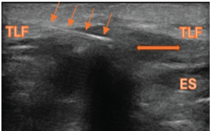
Figure 3: Ultrasound-guided fascial plane injection and hydrodissection performed at the level of L3. A diluted local anesthetic solution was injected beneath the thoracolumbar fascial plane. Using a transverse in-plane approach, the needle (arrows) was advanced during the procedure, allowing separation of the TLF from the ES muscles. The double-headed arrow demonstrates the distribution of the injectate. TLF: Thoracolumbar fascia, ES: Erector spinae.
At the 1-week follow-up, the patient’s pain intensity had decreased to a VAS score of 3/10. Fascial stretching exercises and targeted massages were recommended. By the 1-month follow-up, the pain had further resolved, reaching a VAS score of 2/10.
This case describes a patient with chronic LBP following lumbar spine surgery. In this patient, we demonstrated a focal thoracolumbar fascial defect using US and achieved successful clinical results utilizing a fascial plane injection combined with hydrodissection.
Post-operative abnormalities and persistent pain after spinal surgery are frequently attributed to epidural fibrosis, recurrent stenosis, or hardware-related complications [1,2]. While structural and mechanical alterations of the TLF, such as increased fascial thickness, reduced tissue mobility, and altered architecture, have been extensively documented in patients with non-specific chronic LBP [5], defects after lumbar surgery involving the TLF itself have rarely been described.
Although the posterior fascial layer is routinely closed during lumbar spine surgery, the post-operative healing process may result in suboptimal tissue remodeling, localized structural failure, or altered mechanical behavior [1,2,6]. In our patient, the strict absence of classical surgically relevant pathologies combined with the failure of comprehensive conservative modalities prompted us to look beyond conventional pain generators and investigate the myofascial system as the primary source of the symptoms.
The fascial system has been proposed to function as a widespread sensory organ, possessing a substantially greater density of sensory innervation than muscles, tendons, and joints [7,8]. Beyond its neurosensory properties, the TLF plays a biomechanical role in load transfer between the trunk and lower extremities, contributing significantly to lumbosacral stability [9]. Impaired fascial gliding and structural defects frequently develop following trauma, excessive mechanical loading, or surgical interventions, potentially leading to local inflammation, peripheral sensitization, and subsequent functional impairment [9]. In our patient, the persistent focal pain remained localized for 2 years and gradually increased in severity despite conservative treatment.
Musculoskeletal US offers a highly practical and dynamic method for evaluating fascial morphology and function in vivo. Beyond assessing thickness and echotexture, high-resolution US can identify focal irregularities and defects of fascial continuity [10]. It also allows for the real-time assessment of fascial mobility and gliding behavior [9,11]. In the present case, US demonstrated preserved fascial continuity at adjacent cranial and caudal levels, while revealing a focal midline fascial defect precisely at the symptomatic site of maximal pain.
Although alternative pain generators, including paraspinal muscle pathology or post-operative scar-related pain, cannot be completely excluded, the close spatial correspondence between the sonographic abnormality and maximal tenderness, together with the favorable response to targeted fascial intervention, supports the possibility that the disturbed TLF contributed to the patient’s pain. In this case, gardening activities may have aggravated symptoms arising from a pre-existing post-operative fascial abnormality rather than representing the primary cause of the fascial defect.
To address this localized pathology, we performed an US-guided fascial plane intervention. Current evidence suggests that fascia itself represents a distinct therapeutic target; the clinical benefit of such interventions extends beyond the pharmacological effects of local anesthetics, stemming significantly from the mechanical modification of the fascial planes [3,4]. Utilizing the dynamic hydrodissection technique effectively separates the bound, compromised tissue layers and promotes the mechanical release of the fascia from the anatomic structures [3,4]. Furthermore, experimental studies have demonstrated that such a hydrorelease reduces gliding resistance between fascial interfaces, offering a clear mechanical explanation for symptom improvement [11]. The local anesthetic solution not only provides an immediate nociceptive blockade of the sensitized fascial free nerve endings but also facilitates the physical defect of localized micro-adhesions and fibrous septa [3].
Following the intervention, the patient demonstrated a reduction in pain intensity, with the VAS score decreasing from 7/10 to 3/10 at 1 week. To optimize long-term tissue remodeling, prevent recurrent adhesions, and encourage proper fascial hydration, a tailored regimen of fascial stretching exercises and targeted massages was introduced. This combined therapeutic approach yielded sustained clinical improvement, with the patient’s pain further resolving to a VAS score of 2/10 at the 1-month follow-up. While case reports possess inherent limitations regarding generalizability, our findings suggest that high-resolution US evaluation of the TLF should be considered in the diagnostic workup of patients with persistent post-surgical LBP refractory to conventional care.
This case demonstrates a focal thoracolumbar fascial defect identified by US in a patient with persistent LBP following lumbar surgery. The observed improvement after US-guided fascial plane injection suggests that fascial abnormalities after lumbar surgery may represent a previously underrecognized contributor to pain in some patients. Further studies are needed to clarify their prevalence, clinical significance, and therapeutic implications.
• In persistent post-operative low back pain, not only neural and bone pathologies should be considered
• The thoracolumbar fascia can be evaluated with ultrasound
• A fascial defect may be a possible source of pain
• Ultrasound-guided fascial intervention may be a treatment option.
References
- 1. Miękisiak G. Failed back surgery syndrome: No longer a surgeon’s defeat-a narrative review. Medicina (Kaunas) 2023;59:1255. [Google Scholar] [PubMed]
- 2. Van de Minkelis J, Peene L, Cohen SP, Staats P, Al-Kaisy A, Van Boxem K, et al. 6. Persistent spinal pain syndrome type 2. Pain Pract 2024;24:919-36. [Google Scholar] [PubMed]
- 3. Fusco P, Pascarella G, Stecco C, Blanco R, Forero M, Pawa A, et al. Factors to consider for fascial plane blocks’ success in acute and chronic pain management. Minerva Anestesiol 2024;90:87-97. [Google Scholar] [PubMed]
- 4. Marrone F, Pullano C, De Cassai A, Fusco P. Ultrasound-guided fascial plane blocks in chronic pain: A narrative review. J Anesth Analg Crit Care 2024;4:71. [Google Scholar] [PubMed]
- 5. Pirri C, Pirri N, Guidolin D, Macchi V, Porzionato A, De Caro R, et al. Ultrasound imaging of thoracolumbar fascia thickness: Chronic Non-specific lower back pain versus healthy subjects; A sign of a “Frozen back”? Diagnostics (Basel) 2023;13:1436. [Google Scholar] [PubMed]
- 6. Yilmaz E, Tawfik T, O’Lynnger TM, Iwanaga J, Blecher R, Abdul-Jabbar A, et al. Wound closure after posterior multi-level lumbar spine surgery: An anatomical cadaver study and technical note. Cureus 2018;10:e3595. [Google Scholar] [PubMed]
- 7. Suarez-Rodriguez V, Fede C, Pirri C, Petrelli L, Loro-Ferrer JF, Rodriguez-Ruiz D, et al. Fascial ınnervation: A systematic review of the literature. Int J Mol Sci 2022;23:5674. [Google Scholar] [PubMed]
- 8. Mika A, Oleksy Ł, Fede C, Pirri C, Stecco C. Editorial: Fascia as a multi-purpose structure of connective tissue – dysfunction, diagnostics and treatment. Front Med (Lausanne) 2024;11:1472116. [Google Scholar] [PubMed]
- 9. Soares HR, Pinheiro AR, Crasto C, Barbosa P, Dias N, De Carvalho P. Diagnostic ultrasound assessment of deep fascia sliding mobility in vivo: A scoping review – part 1: Thoracolumbar and abdominal fasciae. J Bodyw Mov Ther 2021;27:92-102. [Google Scholar] [PubMed]
- 10. Pirri C, Pirri N, Macchi V, Porzionato A, De Caro R, Stecco C. Ultrasound imaging of thoracolumbar fascia: A systematic review. Medicina (Kaunas) 2024;60:1090. [Google Scholar] [PubMed]
- 11. Shiwaku K, Otsubo H, Suzuki D, Pirri C, Kodesyo T, Kamiya T, et al. Biomechanical effects of fascial hydrorelease: A cadaveric study. BMC Musculoskelet Disord 2025;26:306. [Google Scholar] [PubMed]





Related Articles in Journal of Orthopaedic Case Reports
 September 1, 2026 Medial Malleolar Screw Fixation Causing Flexor Hallucis Longus Impingement: A Rare Case of Checkrein Deformity
 September 1, 2026 Unilateral Biportal Endoscopic Excision of a Lumbar Discal Cyst: A Case Report and Technical Note
 June 1, 2026 Clinical and Ultrasonographic Outcomes of Percutaneous Achilles Tenotomy Using an 18-Gauge Needle in Idiopathic Clubfoot: A Prospective Study
 June 1, 2026 Influence of Menopause Onset on the Progression of Lumbar Degenerative Disease: A Clinical Correlation Study