

Conventional occipitocervical fusion with foramen magnum decompression provides safe, effective stabilization for selected patients with complex or infective craniovertebral junction pathology when reduction is not feasible.
Dr. Sandesh Subhash Agrawal, Department of Orthopedics and Spine Surgery, Shree Narayana Hospital, Raipur - 492001, Chhattisgarh, India. E-mail: preciouss333@gmail.com
Abstract
Introduction: Basilar invagination (BI) with atlantoaxial instability is a complex craniovertebral junction (CVJ) disorder that causes progressive cervicomedullary compression and disabling myelopathy. Although reduction-based techniques such as distraction–compression–extension–reduction (DCER) have expanded surgical options, conventional occipitocervical fusion remains an important stabilization strategy in selected patients with rigid deformity, advanced neurological compromise, or infective CVJ pathology.
Materials and Methods: This retrospective case series included three patients with BI and atlantoaxial instability, including one with infective C1–C2 spondylodiscitis, who underwent posterior occipitocervical fusion with foramen magnum decompression between 2024 and 2025. Clinical outcomes were assessed using the Nurick grade, Visual Analog Scale (VAS), and Oswestry Disability Index (ODI). Radiographic evaluation included the atlantodental interval (ADI), clivo-axial angle (CXA), Chamberlain’s line, and McRae’s line. Perioperative complications and neurological outcomes were recorded. All patients completed 12 months of follow-up.
Results: All patients presented with advanced myelopathy (Nurick Grade III–V) and marked craniovertebral instability (ADI, 6.2–9.0 mm; CXA, 125–132°). Posterior occipitocervical fusion with foramen magnum decompression was successfully completed in all patients without intraoperative neurological deterioration, implant-related complications, or revision surgery. Neurological status remained stable or improved throughout follow-up. The patient with infective spondylodiscitis demonstrated improvement in VAS (8–3) and ODI (64–38%), whereas all patients achieved radiographic construct stability at final follow-up.
Conclusion: Occipitocervical fusion with foramen magnum decompression provided satisfactory neurological stabilization, radiographic stability, and functional improvement in this small series of patients with complex BI and atlantoaxial instability. It remains a valuable stabilization strategy for selected patients when reduction-based techniques are not feasible.
Keywords: Basilar invagination, atlantoaxial instability, Occipitocervical fusion, craniovertebral junction, cervical myelopathy, spondylodiscitis, clivo-axial angle, Nurick grade.

Basilar invagination (BI) with atlantoaxial instability is a complex craniovertebral junction (CVJ) disorder characterized by superior migration of the odontoid process into the foramen magnum, resulting in progressive cervicomedullary compression and neurological deterioration [1,2]. Patients commonly present with cervical myelopathy, gait disturbance, neck pain, and long-tract signs secondary to chronic neural compression and instability [1,2].
The surgical management of BI has evolved from decompression-focused procedures to stabilization-based strategies aimed at restoring alignment and achieving durable fusion. Contemporary reduction techniques, such as distraction–compression–extension–reduction (DCER), have demonstrated favorable radiological and clinical outcomes in selected reducible deformities [1]. However, these techniques require specialized instrumentation and appropriate anatomical conditions, limiting their applicability in patients with rigid deformity, severe instability, poor bone quality, or infective CVJ pathology [3,4].
Conventional occipitocervical fusion provides rigid stabilization of the CVJ and remains an established treatment option for complex BI, particularly when reduction-based techniques are not feasible [3,4,5]. In infective C1–C2 spondylodiscitis, progressive osseoligamentous destruction further compromises stability, making robust posterior fixation essential for neural protection and spinal stability [6].
We present a retrospective three-patient case series of BI with atlantoaxial instability, including one patient with infective C1–C2 spondylodiscitis, managed with conventional occipitocervical fusion and foramen magnum decompression. This study highlights the clinical presentation, radiological characteristics, surgical management, and 12-month outcomes, emphasizing the continued role of conventional occipitocervical fusion as a reliable stabilization strategy in selected complex CVJ pathologies.
Study design and setting
This retrospective observational case series included three consecutive patients with BI associated with atlantoaxial (C1–C2) instability who underwent posterior occipito-cervical fusion at a single tertiary referral spine center. One patient had concomitant infective C1–C2 spondylodiscitis with CVJ instability. Patients with traumatic craniovertebral injuries, neoplastic lesions, previous CVJ surgery, or incomplete clinical records were excluded. All procedures were performed by the same experienced spine surgery team using a standardized surgical protocol. Owing to the descriptive nature of the study and the small sample size, only descriptive analysis was performed.
Clinical assessment
All patients underwent a detailed neurological evaluation with emphasis on motor weakness, gait disturbance, and long tract signs such as hyperreflexia, Hoffmann’s sign, Babinski sign, and the finger escape phenomenon. Functional disability was assessed using the Oswestry Disability Index (ODI), whereas pain severity was quantified using the Visual Analog Scale (VAS). Neurological status was further graded using the Nurick grading system to assess the severity of myelopathy and functional impairment.
Radiological assessment
Pre-operative imaging included X-ray cervical spine with dynamic views, computed tomography (CT) of the CVJ for bony anatomy and instability assessment, and magnetic resonance imaging (MRI) for evaluation of corticomedullary compression and infective pathology. Radiological parameters assessed included atlantodental interval (ADI), Chamberlain’s line, McRae’s line, and the clivo-axial angle (CXA) to quantify instability and BI severity. Immediate post-operative radiographs were reviewed to assess implant position, construct alignment, and maintenance of reduction.
Surgical technique
All patients were counseled regarding disease severity and surgical risks, including neurological deterioration, vascular injury, and potential need for post-operative ventilatory support. After informed consent, patients were intubated with care to avoid excessive neck movement, and a video laryngoscope was used when a difficult airway was anticipated.
Gardner–Wells tongs were applied for cranial stabilization. Intraoperative neuromonitoring with motor evoked potentials and somatosensory evoked potentials was used in all cases. Baseline signals were recorded in the supine position, followed by prone positioning with neutral alignment and gentle 2 kg traction. Reverse Trendelenburg positioning was used to minimize venous bleeding.
A standard posterior midline approach was used to expose the occipital bone and subaxial cervical spine. Foramen magnum decompression was performed in all cases, including removal of approximately 1 cm of occipital bone and partial posterior arch resection of C1 (up to 1.5 cm bilaterally) to decompress the cervicomedullary junction. Instrumentation was performed under fluoroscopic guidance. C2 laminar screws were used. Additional lateral mass screws were placed in subaxial cervical vertebrae depending on the fixation requirement. Occipital fixation was achieved using 8–12 mm cortical screws. Rods were contoured and assembled, followed by controlled alignment and sequential tightening. Neuromonitoring signals were checked after each critical step. Decortication of the occiput and posterior elements was performed, and autologous iliac crest bone graft was placed to promote fusion. Wound closure was performed in layers over a suction drain. Postoperatively, patients were mobilized on day 2 with a rigid cervical collar.
Patients were evaluated clinically and radiologically at 6 weeks, 6 months, and 12 months after surgery. Clinical outcomes included Nurick grade, VAS, and ODI, while radiographs were obtained to assess implant stability, construct alignment, and maintenance of alignment throughout follow-up.
Case 1
A 43-year-old male presented with progressive gait instability, ataxia, and paraesthesia involving all four limbs. Neurological examination revealed cervical myelopathy with hyperreflexia and positive long tract signs (Nurick Grade IV). CT and MRI demonstrated BI with atlantoaxial instability, an ADI of 7.5 mm, CXA of 128°, odontoid migration 6 mm above Chamberlain’s line, violation of McRae’s line, and significant cervicomedullary compression (Fig. 1). The patient underwent posterior occipitocervical fusion extending to C5 with foramen magnum decompression. At 12-month follow-up, neurological function, gait, and balance had improved without implant-related complications or neurological deterioration.
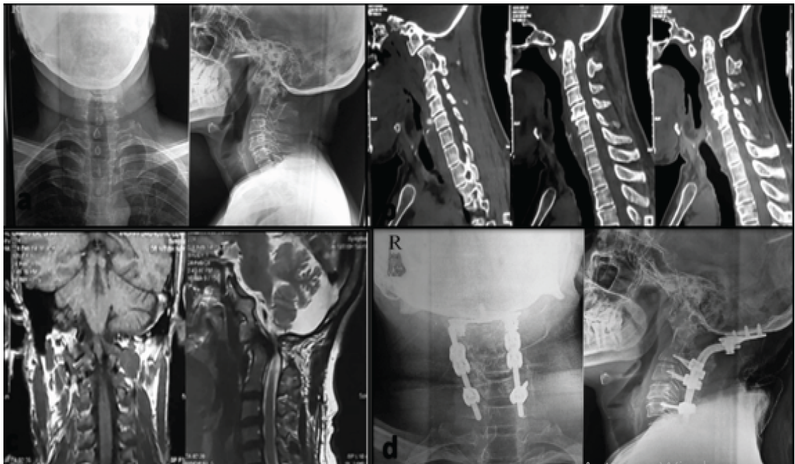
Figure 1: Pre-operative and post-operative radiological assessment of basilar invagination with atlantoaxial instability following occipitocervical fusion. (a) Pre-operative anteroposterior and lateral cervical radiographs demonstrating craniovertebral junction malalignment with basilar invagination and atlantoaxial instability. (b) Pre-operative sagittal computed tomography images showing superior migration of the odontoid process, atlantoaxial subluxation, reduction in the clivo-axial angle, and significant craniovertebral junction deformity. (c) Pre-operative magnetic resonance imaging demonstrates marked cervicomedullary compression with ventral brainstem indentation secondary to basilar invagination and C1-C2 instability. (d) Post-operative anteroposterior and lateral radiographs demonstrating satisfactory occipitocervical fixation extending from the occiput to C5 with screw-rod construct, restoration of craniovertebral alignment, and adequate stabilization following foramen magnum decompression and C1 posterior arch excision.
Case 2
A 14-year-old boy presented with neck pain, progressive gait difficulty, and loss of hand dexterity. Examination demonstrated bilateral hand weakness, hyperreflexia, positive Hoffmann’s, Babinski, and finger escape signs, and Nurick Grade III myelopathy. Imaging confirmed BI with atlantoaxial instability, an ADI of 6.2 mm, CXA of 132°, odontoid migration 4 mm above Chamberlain’s line, and mild violation of McRae’s line, producing cervicomedullary compression (Fig. 2). Posterior occipitocervical fusion with foramen magnum decompression was performed. At 12 months, the patient showed sustained neurological improvement with better gait, hand function, and independent ambulation.
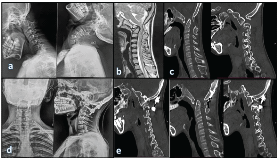
Figure 2: Dynamic radiographic, computed tomography (CT), and magnetic resonance imaging (MRI) evaluation demonstrating craniovertebral junction instability and post-operative occipitocervical stabilization. (a) Pre-operative dynamic flexion-extension cervical radiographs demonstrating significant atlantoaxial instability with abnormal craniovertebral junction mobility and reducibility assessment. (b) Pre-operative sagittal MRI showing basilar invagination with severe cervicomedullary compression and ventral indentation at the cervicomedullary junction. (c) Pre-operative sagittal CT images demonstrating superior migration of the odontoid process, atlantoaxial subluxation, and altered clivo-axial alignment consistent with craniovertebral instability. (d) Post-operative anteroposterior and lateral cervical radiographs demonstrating satisfactory occipitocervical fixation extending from the occiput to C2 with restoration of craniocervical alignment following posterior stabilization. (e) Post-operative sagittal CT images confirming appropriate placement of the occiput-to-C2 fusion construct, adequate decompression at the craniovertebral junction following foramen magnum decompression and C1 posterior arch excision, and improved cervicomedullary alignment.
Case 3
A 23-year-old male presented with severe neck pain and progressive quadriparesis. Neurological examination revealed diffuse upper motor neuron signs with Nurick Grade IV–V myelopathy. Pre-operative VAS and ODI were 8/10 and 64%, respectively. CT and MRI demonstrated infective C1–C2 spondylodiscitis with atlantoaxial instability, BI, ADI of 9.0 mm, CXA of 125°, odontoid migration 8 mm above Chamberlain’s line, violation of McRae’s line, and marked cervicomedullary compression (Fig. 3). The patient underwent posterior occipitocervical fusion to C3 with foramen magnum decompression. At 12-month follow-up, VAS improved to 3/10, ODI to 38%, and neurological status remained stable without implant-related complications or revision surgery.
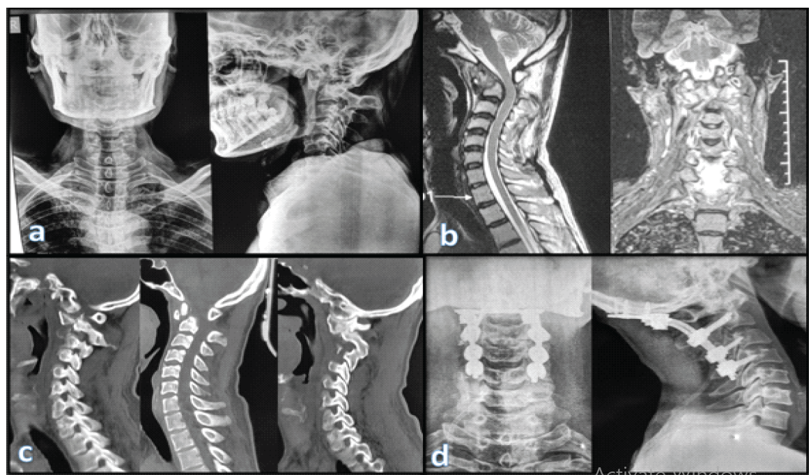
Figure 3: Radiological evaluation of infective C1-C2 spondylodiscitis with basilar invagination before and after occipitocervical fusion. (a) Pre-operative anteroposterior and lateral cervical radiographs demonstrating craniovertebral junction malalignment with atlantoaxial instability and features suggestive of basilar invagination. (b) Pre-operative sagittal and coronal magnetic resonance imaging demonstrating infective spondylodiscitis involving the C1-C2 articulation with associated soft-tissue inflammation, cervicomedullary compression, and ventral brainstem indentation. (c) Pre-operative sagittal computed tomography images demonstrating osseo ligamentous destruction at the atlantoaxial complex, superior migration of the odontoid process, increased atlantodental interval, and altered clivo-axial alignment consistent with severe craniovertebral instability. (d) Post-operative anteroposterior and lateral radiographs demonstrating satisfactory occipitocervical fixation extending from the occiput to C4 with restoration of Craniocervical alignment following posterior stabilization, foramen magnum decompression, and C1 posterior arch excision.
Follow-up outcomes
All three patients completed clinical and radiological follow-up at 6 weeks, 6 months, and 12 months. No implant-related complications, wound infection, neurological deterioration, implant loosening, or revision surgery occurred during the follow-up period. Serial radiographs demonstrated maintenance of construct alignment and implant stability in all patients. Progressive improvement in neurological function and ambulatory status was observed throughout the 12-month follow-up.
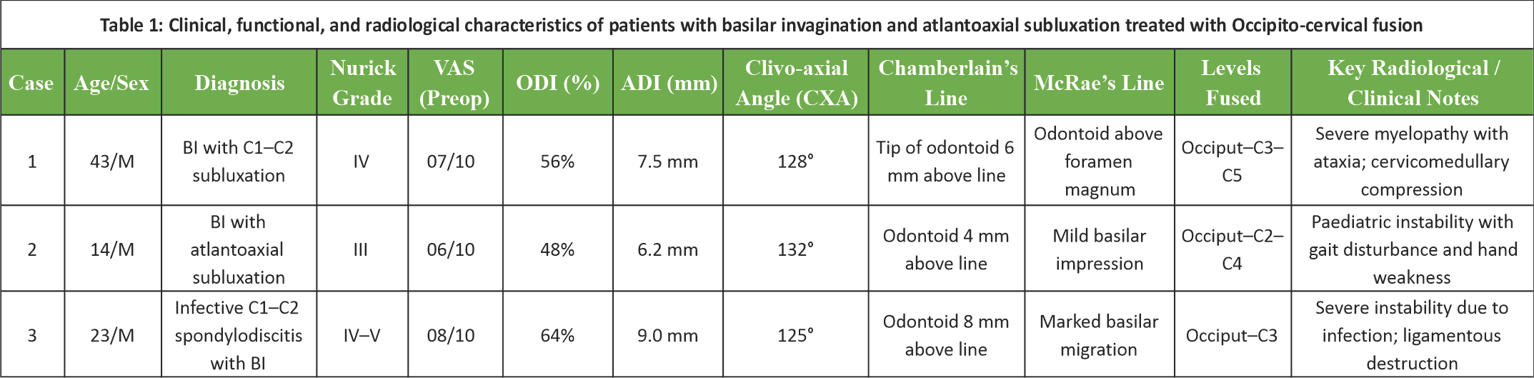
Table 1: Clinical, functional, and radiological characteristics of patients with basilar invagination and atlantoaxial subluxation treated with occipitocervical fusion.
The CVJ is one of the most biomechanically complex regions of the spine, where even minor instability can result in significant neurological compromise because of its close relationship to the brainstem and upper cervical spinal cord. BI with atlantoaxial instability is increasingly recognized as a dynamic instability-driven disorder characterized by progressive odontoid migration and cervicomedullary compression rather than a static congenital anomaly [1,2]. In the present series, all patients presented with advanced myelopathy (Nurick Grade III–V) and radiological evidence of significant CVJ instability, highlighting the progressive nature of untreated BI and the importance of timely surgical stabilization.
Modern management of BI has evolved from decompression-focused procedures to stabilization- and realignment-based strategies. Goel proposed that atlantoaxial instability is the primary pathological driver of BI and that C1–C2 fixation alone may achieve indirect reduction without direct decompression in selected patients [3,4,5]. Subsequently, reduction-based techniques such as DCER were developed to achieve controlled anatomical realignment and have demonstrated favorable radiological and clinical outcomes in carefully selected reducible deformities [1]. However, these techniques require appropriate anatomical conditions and specialized expertise, limiting their applicability in patients with rigid deformity, severe instability, or infective CVJ pathology [5,6].
However, reduction-based techniques are not feasible in all patients with BI. Rigid deformity, advanced instability, infective osseoligamentous destruction, and poor bone quality may preclude safe anatomical reduction. In our series, all patients demonstrated advanced CVJ instability, characterized by an ADI of 6.2–9 mm, CXA of 125–132°, and odontoid migration beyond Chamberlain’s and McRae’s lines, consistent with significant cervicomedullary compression [6,7]. Although DCER provides excellent deformity correction in reducible BI [8,9], conventional Occipito-cervical fusion with foramen magnum decompression achieved sustained neurological stabilization and functional improvement throughout the 12-month follow-up in our patients, supporting stabilization as an effective strategy in carefully selected complex CVJ pathologies [10].
Long-term studies have demonstrated that occipitocervical fusion provides durable stabilization and sustained neurological improvement in complex CVJ disorders [11,12]. Modern screw-rod constructs further enhance biomechanical stability and clinical outcomes [13]. The infective case in our series illustrates the value of stabilization-first surgery in C1–C2 spondylodiscitis, where progressive osseopligamentous destruction frequently precludes reduction-based procedures. Consistent with previous reports [9], rigid posterior stabilization with foramen magnum decompression resulted in sustained neurological recovery despite severe structural compromise.
Biomechanically, Occipito-cervical fusion eliminates pathological motion at the CVJ, providing immediate stability and facilitating neurological recovery [10,11,12,14]. Although upper cervical motion is sacrificed, preservation of neurological function and spinal stability remains the primary objective in advanced CVJ instability. Because post-operative CT was not routinely performed, this study focused on clinical outcomes and construct stability rather than radiological fusion [15].
This study has several limitations. The small sample size and retrospective single-center design limit statistical analysis and generalizability. Although all patients completed 12 months of follow-up, longer-term evaluation is required to assess implant longevity, adjacent segment degeneration, and radiological fusion. In addition, the absence of a comparison group precludes direct evaluation of conventional occipitocervical fusion against reduction-based techniques such as DCER.
Conventional occipitocervical fusion remains a dependable stabilization strategy for selected patients with BI and atlantoaxial instability, particularly in advanced, rigid, or infective cases. In this series, sustained neurological stabilization and functional improvement were maintained throughout the 12-month follow-up, highlighting that restoration of stability alone can achieve favorable clinical outcomes in carefully selected patients.
Occipitocervical fusion remains a safe, reliable, and effective treatment for advanced basilar invagination with atlantoaxial instability, including infective craniovertebral junction pathology. Even without complex reduction techniques, rigid stabilization can provide significant neurological recovery, pain relief, and durable functional improvement when guided by careful clinical and radiological assessment.
References
- 1. Sarat Chandra P, Bajaj J, Singh PK, Garg K, Agarwal D. Basilar invagination and atlantoaxial dislocation: Reduction, deformity correction and realignment using the DCER (distraction, compression, extension, and reduction) technique with customized instrumentation and implants. Neurospine 2019;16:231-50. [Google Scholar] [PubMed]
- 2. Goel A, Pareikh S, Sharma P. Atlantoaxial joint distraction for treatment of basilar invagination secondary to rheumatoid arthritis. Neurol India 2005;53:238-40. [Google Scholar] [PubMed]
- 3. Dave BR, Vashishtha A, Krishnan A, Mayi SC, Rai RR, Dave MB, et al. Multiple rod construct for occipito-cervical fusion: Overcoming challenges with enabling technologies. Cureus 2026;18:e107690. [Google Scholar] [PubMed]
- 4. Kukreja S, Ambekar S, Sin AH, Nanda A. Occipitocervical fusion surgery: Review of operative techniques and results. J Neurol Surg B Skull Base 2015;76:331-9. [Google Scholar] [PubMed]
- 5. Dave BR, Vashishtha A, Krishnan A, Mayi SC, Rai RR, Dave MB, et al. Occipito-cervical fusion using screw rod plate system in craniocervical pathologies: A prospective cohort analysis of long-term functional and radiological outcome with minimum two years of follow-up. Cureus 2025;17:e100489. [Google Scholar] [PubMed]
- 6. Itoh T, Tsuji H, Katoh Y, Yonezawa T, Kitagawa H. Occipito-cervical fusion reinforced by Luque’s segmental spinal instrumentation for rheumatoid diseases. Spine (Phila Pa 1976) 1988;13:1234-8. [Google Scholar] [PubMed]
- 7. Deutsch H, Haid RW Jr., Rodts GE Jr., Mummaneni PV. Occipitocervical fixation: Long-term results. Spine (Phila Pa 1976) 2005;1:530-5. [Google Scholar] [PubMed]
- 8. Fairbank JC, Couper J, Davies JB, O’Brien JP. The oswestry low back pain disability questionnaire. Physiotherapy 1980;66:271-3. [Google Scholar] [PubMed]
- 9. Huskisson EC. Measurement of pain. Lancet 1974;2:1127-31. [Google Scholar] [PubMed]
- 10. Nurick S. The pathogenesis of the spinal cord disorder associated with cervical spondylosis. Brain 1972;95:87-100. [Google Scholar] [PubMed]
- 11. He B, Yan L, Xu Z, Chang Z, Hao D. The causes and treatment strategies for the postoperative complications of occipitocervical fusion: A 316 cases retrospective analysis. Eur Spine J 2014;23:1720-4. [Google Scholar] [PubMed]
- 12. Sasso RC, Jeanneret B, Fischer K, Magerl F. Occipitocervical fusion with posterior plate and screw instrumentation. A long-term follow-up study. Spine (Phila Pa 1976) 1994;19:2364-8. [Google Scholar] [PubMed]
- 13. Liu G, Li Q, Sheng F, Xu N, Li M, Wang Y, et al. Outcomes of occipitocervical fixation using a spinous process screw in C2 as a third anchor point for occipitocervical fixation: A case presentation. BMC Musculoskelet Disord 2020;21:307. [Google Scholar] [PubMed]
- 14. Kanayama M, Cunningham BW, Weis JC, Parker LM, Kaneda K, McAfee PC. Maturation of the posterolateral spinal fusion and its effect on load-sharing of spinal instrumentation. An in vivo sheep model. J Bone Joint Surg Am 1997;79:1710-20. [Google Scholar] [PubMed]






Related Articles in Journal of Orthopaedic Case Reports
 June 22, 2021 Basilar Invagination: Surgical Treatment by Novel Anterior Implant
 September 1, 2026 Spinal Epidural Abscess Following Spinal Anesthesia in a Bacteremic Patient: A Case Report
 August 1, 2026 Post-operative C5 Palsy Following Posterior Cervical Decompression and Fusion: A Case Report
 July 1, 2026 Role of Surgical Stabilization in the Management of Post-discectomy Septic Spondylodiscitis: Two Successful Case Reports