


Introduction
Ewing sarcoma is an aggressive primary malignancy of bone that predominantly affects children and adolescents [1]. While modern multimodal therapy, combining systemic chemotherapy with local control measures, has significantly improved survival rates, management of the primary tumor remains a cornerstone of treatment [2,3]. For extremity tumors, limb salvage surgery is now the standard of care, offering oncological outcomes equivalent to amputation while preserving function and improving quality of life [4]. However, reconstruction of large skeletal defects following tumor resection in skeletally immature patients is exceptionally challenging. Options such as massive allografts are associated with high complication rates in growing children, including infection, non-union, fracture, and the need for multiple revision surgeries [5]. Endoprosthetic reconstruction is also associated complications and revision rates, including aseptic loosening, infection, and periprosthetic fracture [6]. Extracorporeal irradiation (ECI) with autograft reimplantation has emerged as a valuable biological reconstruction technique [7]. This method involves resecting the tumor-bearing bone, sterilizing it ex vivo with a high dose of radiation, and reimplanting it as a perfectly matched, non-immunogenic autograft [8,9]. It preserves the patient’s native bone stock, provides an osteoconductive scaffold, and avoids many of the limitations of alternative reconstructive methods [10,11]. This report describes the case of a 4-year-old boy with Ewing sarcoma involving the entire tibia, who was successfully treated with en bloc resection, ECI, and immediate soft-tissue reconstruction using a free anterolateral thigh (ALT) flap.
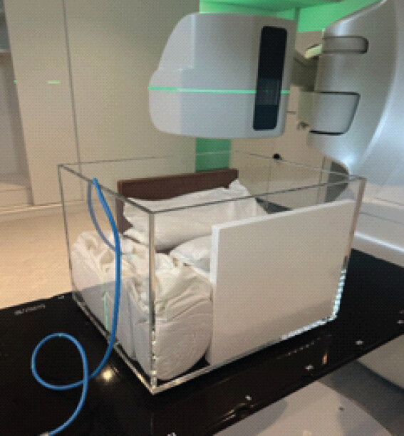
While the ECI technique is well established, its application in extreme clinical scenarios has not been widely reported. A custom phantom setup was developed by the Medical Physics team at the Radiation Oncology Department to simulate tissue-equivalent conditions for ECI. The phantom consisted of an acrylic container with an RW3 slab phantom (PTW, Freiburg, Germany) positioned at the base and along the lateral sides to provide water-equivalent backscatter and lateral scatter equilibrium. The remaining volume was tightly packed with rice bags to simulate soft-tissue density, thereby creating a homogeneous irradiation medium (Fig. 1).
The significance of this case report lies in its detailed description of the application of this advanced technique to a highly unusual and challenging presentation: a massive Ewing sarcoma occupying the majority of the tibia in a 4-year-old child. This report provides a transparent, step-by-step account of a successful and complex single-stage surgical solution.
Case Report
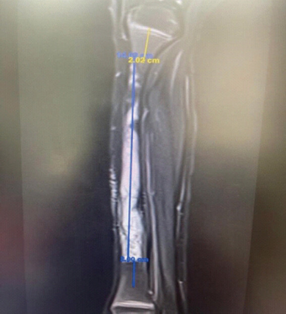
A 4-year-old boy presented with progressive pain and swelling in his right lower leg. Initial radiographic evaluation and magnetic resonance imaging (MRI) at presentation revealed an aggressive, permeative lesion involving the majority of the right tibia, sparing the growth plate as well as the proximal and distal parts. Pre-operative MRI measurements demonstrated a tumor length of 14.6 cm, occupying a significant portion of the total tibial length of 22.7 cm (Fig. 2).
A comprehensive staging workup was performed. A whole-body bone scan demonstrated intense, isolated radiotracer uptake throughout the right tibia, with no evidence of other skeletal metastases. Bone marrow biopsies were negative for metastatic disease. A computed tomography (CT) scan of the chest revealed a small, non-specific 2-mm nodule in the right lower lobe, which was deemed too small to biopsy and was scheduled for surveillance.
An open biopsy of the tibial lesion was performed at a different hospital on 22 December 2024, and histopathological analysis confirmed the diagnosis of Ewing sarcoma. The patient was subsequently started on neoadjuvant chemotherapy on 22 February 2025, following the Children’s Oncology Group AEWS0031 protocol. He demonstrated a good clinical response and completed induction chemotherapy.
The case was discussed at the multidisciplinary tumor board meeting. Given the patient’s very young age and the extensive involvement of the entire tibia, both definitive radiation therapy and surgical options for local control were considered. The high morbidity associated with irradiation of the entire tibia in a growing child – including growth arrest and an increased risk of secondary malignancy – was weighed against the complexity of surgical reconstruction. After detailed discussions with the family regarding the risks and expected outcomes of each approach, a joint decision was made to proceed with limb salvage surgery. The patient completed chemotherapy on 29 May 2025, restaging investigations showed no evidence of metastasis, and surgery was scheduled for 22 June 2025.
Surgical technique
The patient was taken to the operating room for the definitive surgical procedure.
Preparation and incision
Under general anesthesia, the patient was positioned supine. The right lower extremity and the contralateral (left) thigh were prepared and draped in a sterile fashion. A pneumatic tourniquet was applied to the right thigh, but was not inflated initially. The surgical plan involved two teams working simultaneously: One performing the tumor resection and the other harvesting the free ALT flap. A longitudinal incision was made over the anterior aspect of the right leg, extending from the tibial tubercle to the ankle joint.
En bloc tumor resection
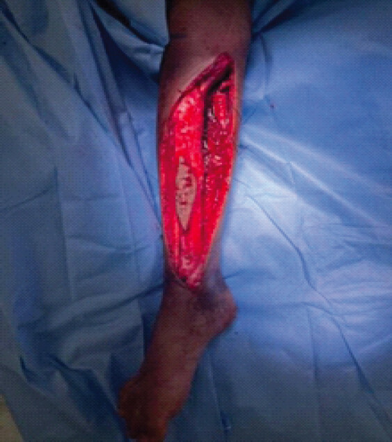
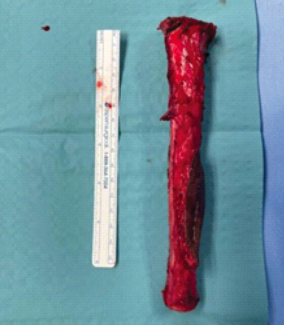
The incision was carried down to the periosteum, which was incised and reflected along with the tumor pseudocapsule. Meticulous dissection was performed to achieve wide surgical margins. The anterior tibial vessels and the deep peroneal nerve were identified and carefully preserved. Dissection proceeded circumferentially around the tibia. The biopsy tract was clearly identified and maintained in continuity with the main tumor mass (Fig. 3). Proximal and distal osteotomies were performed through the tibial epiphyses using an oscillating saw, preserving the articular cartilage of both the knee and ankle joints. The entire tibia was thus resected en bloc and removed from the sterile field for preparation (Fig. 4).
ECI
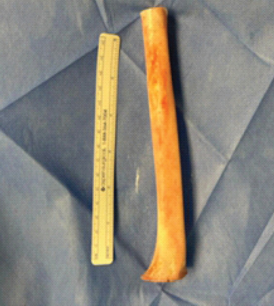
The resected tibia was transferred to an adjacent sterile preparation table. All adherent soft tissue was meticulously removed from the bone (Fig. 5). The bone was then double-bagged in sterile wrapping and immediately transported to the radiation oncology suite.
Specimen handling and immobilization
The excised bone specimen was transported in a sterile, multilayer-wrapped plastic container. On arrival at the Radiation Oncology Department, the specimen was placed in an additional sterile plastic bag and positioned within the phantom separation setup. The bone was immobilized and covered with rice bags to ensure tissue equivalence and to eliminate potential air gaps. A representative bone specimen (approximately 20 cm in length and matched in width to the excised patient bone) was centrally positioned within the phantom. A certified reference-grade dosimetry system consisting of a PTW Farmer ionization chamber (Type 30013; PTW, Freiburg, Germany) was placed adjacent to the specimen for in-phantom dose verification. The chamber was connected to a PTW UNIDOS Tango electrometer (PTW, Freiburg, Germany), enabling continuous real-time monitoring of the delivered dose. To minimize dosimetric heterogeneity, rice bags were tightly packed around the bone to eliminate any residual air gaps. The phantom was subsequently aligned on the treatment couch using the in-room laser system of a Varian TrueBeam linear accelerator. Calibration and dose uniformity checks were performed before irradiation to verify setup accuracy.
Dose calculation and delivery
ECI was delivered using a Varian TrueBeam linear accelerator. A single fraction of 50 Gy was prescribed [12]. In addition, it was delivered over approximately 20 min using 6 MV photon energy. A lateral opposed beam arrangement with a square field size of 30 × 30 cm² was selected to ensure complete coverage of the bone specimen with adequate margins. The total number of monitor units (MU) required to deliver the prescribed dose was 9,414 MU. In vivo dose verification was performed using a PTW Farmer ionization chamber (Type 30013) connected to a UNIDOS Tango electrometer. The recorded dose was within 3% of the prescribed dose, confirming the accuracy of treatment delivery. This dose has been shown to effectively sterilize malignant bone while preserving its structural integrity, thereby enabling safe reimplantation without compromising biomechanical function[13]. In addition, it has been widely used for extracorporeal sterilization of tumor-bearing bone prior to re-implantation[14].
Post-treatment handling
Following completion of irradiation, the bone specimen was removed from the phantom, re-packaged in its transport container, and immediately returned to the operating room for surgical reimplantation.
ALT flap harvest
Simultaneously, a second surgical team harvested a free ALT flap from the contralateral thigh. The perforators arising from the descending branch of the lateral circumflex femoral artery were identified using a handheld Doppler device. An appropriately sized skin paddle was designed, and the flap was elevated, incorporating the fascia. The vascular pedicle was meticulously dissected to obtain sufficient length for subsequent microvascular anastomosis.
Reimplantation and internal fixation
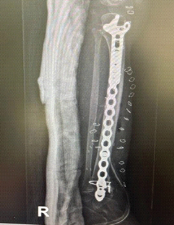
Following irradiation, the sterilized tibial segment was returned to the operating room and reimplanted in its anatomical position. Rigid internal fixation was achieved using a long titanium plate with 11 screw holes, securing the proximal and distal ends of the irradiated autograft to the preserved epiphyseal bone fragments.
Microvascular anastomosis and flap inset
The recipient vessels – the anterior tibial artery and vein – were prepared in the right leg under an operating microscope. The ALT flap was transferred to the defect, and microvascular anastomosis was performed by connecting the flap artery and vein to the recipient vessels. Adequate flap perfusion was confirmed by brisk capillary refill and visible dermal bleeding. The flap was then inset and contoured to cover the fixation plate and the anterior aspect of the reconstructed tibia, thereby providing well-vascularized soft-tissue coverage.
Closure
A closed suction drain was placed deep to the flap. The wound was closed in layers, with subcutaneous sutures and skin staples. The donor site on the thigh was partially closed primarily and partially covered with a split-thickness skin graft. A well-padded posterior splint was applied to the right leg to immobilize the knee and ankle. The patient tolerated primarily and partially covered with a split-thickness skin graft. A well-padded posterior splint was applied to the right leg to immobilize the knee and ankle. The patient tolerated the prolonged procedure well and was transferred to the pediatric intensive care unit for post-operative monitoring.
Post-operative course
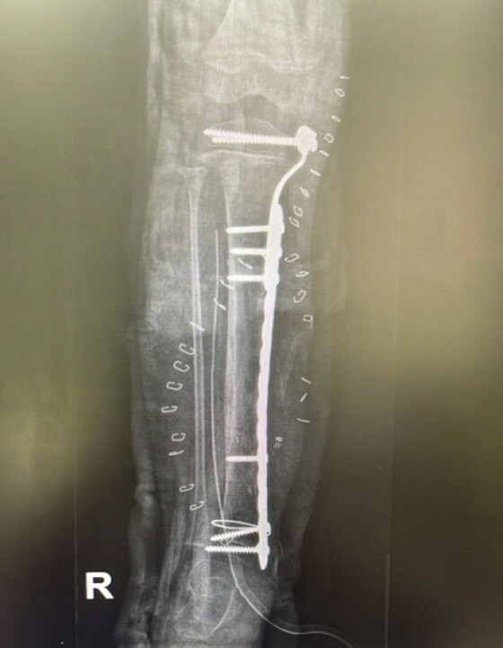
The patient’s post-operative recovery was uneventful. The free ALT flap remained viable with excellent perfusion. Subsequent histopathological analysis of the resected tibial specimen confirmed wide, negative surgical margins (tumor-free) and demonstrated extensive chemotherapy-induced necrosis. Postoperatively, the patient was immobilized in a splint and maintained on a non–weight-bearing protocol. A one-month post-operative radiograph demonstrated a stable construct, with the hardware in the appropriate position and no loss of alignment (Fig. 6). The patient subsequently resumed the planned adjuvant chemotherapy and was enrolled in a gentle physical therapy program. The patient was followed up at 2, 4, and 6 months postoperatively and showed excellent clinical progress, with satisfactory bone healing (Fig. 7). He required three sessions of wound debridement, after which definitive closure was achieved, resulting in excellent skin condition. A follow-up MRI performed on 14 December confirmed no evidence of tumor recurrence.
Discussion
This case highlights a complex yet successful single-stage solution to one of the most challenging scenarios in pediatric orthopedic oncology: Ewing sarcoma involving the entire tibia. The choice of local control in such cases remains controversial. Definitive radiotherapy would result in severe growth arrest, significant limb length discrepancy, and a high risk of chronic radiation-induced fibrosis and secondary malignancy [15]. Amputation, although oncologically sound, is associated with substantial functional and psychological consequences. Consequently, limb salvage surgery was considered the preferred approach, making the choice of reconstruction critically important.
The use of an ECI autograft offered several distinct advantages over alternative reconstructive options in this 4-year-old child. An endoprosthesis would be unsuitable due to the presence of open physes and would necessitate multiple revision surgeries for loosening and lengthening throughout childhood and adolescence [6]. A massive allograft, while a biological option, carries a high complication rate – including fracture (up to 30%), non-union, and infection – and lacks growth potential [5,16]. In contrast, the ECI autograft provides an exact anatomical fit, preserves the patient’s native bone stock, and serves as an osteoconductive scaffold that allows creeping substitution and eventual biological incorporation [11,12,17,18]. Although the irradiated bone itself is devitalized, it provides a durable and potentially permanent reconstructive solution. Previous reports of ECI for diaphyseal tibial sarcomas have demonstrated satisfactory oncological control, bony union, and good functional outcomes, supporting its use as a biological reconstruction option in this anatomical site [19].
Management was further complicated by the presence of a non-specific pulmonary nodule. Although too small to be definitively characterized, its presence in a patient with Ewing sarcoma necessitates treatment with full systemic chemotherapy, as would be administered for metastatic disease, along with close radiographic surveillance. The decision to proceed with aggressive surgical local control was guided by the principle that achieving optimal local control is paramount for overall survival, irrespective of systemic disease status .
The surgical technique addressed two major challenges simultaneously. First, the skeletal defect was bridged using an ECI autograft and stabilized with rigid internal fixation. Second, the extensive soft-tissue defect created by the tumor and its resection was reconstructed using a free vascularized ALT flap. This represents a crucial adjunct to the ECI procedure. Irradiated bone is inherently avascular and is associated with an increased susceptibility to infection and non-union [13,19]. The introduction of a large, well-vascularized tissue envelope such as the ALT flap not only provides durable soft-tissue coverage but also enhances the local biological environment, potentially reducing the risk of infection and facilitating graft incorporation and healing [20]. While the combination of ECI with vascularized bone grafts (e.g., free fibula transfer) has been shown to reduce non-union rates, the use of a vascularized soft-tissue flap is likely to confer a comparable biological advantage [21].
This case exemplifies a maximalist approach to biological limb salvage, integrating the oncological safety of wide resection, the biological benefits of an ECI autograft, and the reconstructive capacity of microsurgical free tissue transfer. Although complex and technically demanding, this strategy offers a young child the best possible opportunity for a durable, functional, and salvaged limb.
Conclusion
En bloc resection with ECI and reimplantation, combined with immediate vascularized free flap coverage, represents a viable and effective limb-salvage strategy for extensive diaphyseal Ewing sarcoma, even in very young children. This single-stage approach addresses both skeletal and soft-tissue defects, providing a durable biological reconstruction while avoiding the long-term complications associated with prosthetic replacement and allografts. This case contributes to the growing body of evidence supporting ECI as an important technique in the orthopedic oncologist’s armamentarium for complex pediatric limb salvage.
Clinical Message
Extracorporeal irradiation with reimplantation can be considered for limb salvage in young children with extensive Ewing sarcoma, particularly when conventional reconstructive options are limited. Vascularized soft-tissue coverage may further support reconstruction and healing.
Conflict of Interest:
Nil
Source of Support:
Nil
Consent:
The authors confirm that informed consent was obtained from the patient for publication of this article
References
- Kumar N, Gupta B. Global incidence of primary malignant bone tumors. Curr Orthop Pract 2016;27:530–4. [Google Scholar] | [PubMed]
- Gitelis S, Bayne CO, Frank JM, Fillingham YA, Kent PM. Surgery in malignant bone tumors. Curr Probl Cancer 2013;37:192–7. [Google Scholar] | [PubMed]
- Sluga M, Windhager R, Lang S, Heinzl H, Bielack S, Kotz R. Local and systemic control after ablative and limb sparing surgery in patients with osteosarcoma. Clin Orthop Relat Res 1999;358:120–7. [Google Scholar] | [PubMed]
- Hong AM, Millington S, Ahern V, McCowage G, Boyle R, Tattersall M. Limb preservation surgery with extracorporeal irradiation in the management of malignant bone tumor: The oncological outcomes of 101 patients. Ann Oncol 2013;24:2676–80. [Google Scholar] | [PubMed]
- Donati D, Capanna R, Campanacci D, Del Ben M, Ercolani C, Masetti C. The use of massive bone allografts for intercalary reconstruction and arthrodeses after tumor resection. A multicentric European study. Chir Organi Mov 1993;78:81–94. [Google Scholar] | [PubMed]
- Skinner JA, Cannon SR, Briggs TW, Sewell MD, Hanna SA, McGrath A. Intercalary diaphyseal endoprosthetic reconstruction for malignant tibial bone tumours. J Bone Joint Surg Br 2011;93:1111–7. [Google Scholar] | [PubMed]
- Hong A, Stevens G, Stalley P, Pendlebury S, Ahern V, Ralston A. Extracorporeal irradiation for malignant bone tumors. Int J Radiat Oncol Biol Phys 2001;50:441–7. [Google Scholar] | [PubMed]
- Spira E, Lubin E. Extracorporeal irradiation of bone tumors. A preliminary report. Isr J Med Sci 1968;4:1015–9. [Google Scholar] | [PubMed]
- Sys G, Uyttendaele D, Poffyn B, Verdonk R, Verstraete L. Extracorporeally irradiated autografts in pelvic reconstruction after malignant tumour resection. Int Orthop 2002;26:174–8. [Google Scholar] | [PubMed]
- Singh VA, Nagalingam J, Saad M, Pailoor J. Which is the best method of sterilization of tumour bone for reimplantation? A biomechanical and histopathological study. Biomed Eng Online 2010;9:48. [Google Scholar] | [PubMed]
- Davidson AW, Hong A, McCarthy SW, Stalley PD. En-bloc resection, extracorporeal irradiation, and re-implantation in limb salvage for bony malignancies. J Bone Joint Surg Br 2005;87:851–7. [Google Scholar] | [PubMed]
- Puri A, Gulia A, Jambhekar N, Laskar S. The outcome of the treatment of diaphyseal primary bone sarcoma by resection, irradiation and re-implantation of the host bone: Extracorporeal irradiation as an option for reconstruction in diaphyseal bone sarcomas. J Bone Joint Surg Br 2012;94:982–8. [Google Scholar] | [PubMed]
- Puri A, Gulia A, Agarwal M, Jambhekar N, Laskar S. Extracorporeal irradiated tumor bone: A reconstruction option in diaphyseal Ewing's sarcomas. Indian J Orthop 2010;44:390–6. [Google Scholar] | [PubMed]
- Chan LW, Imanishi J, Ngan SY, Chander S, Chu J, Thorson R. Extracorporeal irradiation and reimplantation with total hip arthroplasty for periacetabular pelvic resections: A review of 9 cases. Sarcoma 2016;2016:2549616. [Google Scholar] | [PubMed]
- Sharma DN, Rastogi S, Bakhshi S, Rath GK, Julka PK, Laviraj MA. Role of extracorporeal irradiation in malignant bone tumors. Indian J Cancer 2013;50:306–9. [Google Scholar] | [PubMed]
- Matejovsky Z Jr, Matejovsky Z, Kofranek I. Massive allografts in tumour surgery. Int Orthop 2006;30:478–83. [Google Scholar] | [PubMed]
- Kotb SZ, Mostafa MF. Recycling of extracorporeally irradiated autograft for malignant bone tumors: Long-term follow-up. Ann Plast Surg 2013;71:493–9. [Google Scholar] | [PubMed]
- Kreig AH, Lenze U, Schultze L, Gross MW, Haug M. Extracorporeal irradiation and reimplantation of tumor-bearing bone segments following diaphyseal sarcoma resection at the tibia. Anticancer Res 2019;39:2015–23. [Google Scholar] | [PubMed]
- Zhang S, Wang XQ, Wang JJ, Xu MT. En bloc resection, intraoperative extracorporeal irradiation and re-implantation of involved bone for the treatment of limb malignancies. Mol Clin Oncol 2017;7:1045–52. [Google Scholar] | [PubMed]
- Heller L, Kronowitz SJ. Lower extremity reconstruction. J Surg Oncol 2006;94:479–89. [Google Scholar] | [PubMed]
- Muramatsu K, Ihara K, Hashimoto T, Seto S, Taguchi T. Combined use of free vascularised bone graft and extracorporeally-irradiated autograft for the reconstruction of massive bone defects after resection of malignant tumour. J Plast Reconstr Aesthet Surg 2007;60:1013–8. [Google Scholar] | [PubMed]
© 2026 Journal of Orthopaedic Case Reports - Published by Indian Orthopaedic Research Group
About the Authors
-

Ali H. Alyami
-

Abaad Saad Althagafi
-

Rawan Hameed AlRashed
-

Ghufran Aljawi
-

Yasir Barakati
-

Ramh AlGarni
Related Articles in Journal of Orthopaedic Case Reports
 September 1, 2026 Proximal Humerus Ewing Sarcoma: Technical Challenges and Strategies for Bone Defect Reconstruction
 August 1, 2026 Extensive Giant Cell Tumor Involving Entire Tibia with Talar Extension and Pulmonary Metastases: Successful Limb Preservation with Denosumab and Angioembolization
 May 10, 2024 Lower-Limb Salvage Procedure using Anterolateral Thigh Free Flap and Multiple Tendon Grafts in a Road Injury Patient: A Case Report
 October 1, 2026 Soft-Tissue Closure through Acute Deformation and Gradual Correction Using Taylor Spatial Frame in a Gustilo-Anderson Type IIIB Open Tibial Pilon Fracture: A Case Report