


Introduction
Spondylolysis is a developmental defect or an acquired abnormality of the pars interarticularis, often attributed to repetitive microtrauma sustained by a neural arch [1,2]. The purported incidence of spondylolysis is approximately 6%, and 85–95% of these defects develop at the L5 level [2,3,4]. The bipedal ambulation in an erect posture predisposes the lordotic lumbar spine of the human beings to high degrees of static and dynamic stresses during routine physiological activities [5]. These abnormal forces have been implicated as the inciting events in unilateral (U/L) or bilateral (B/L) spondylolysis [6, 7].
A majority of the acute pars interarticularis fractures are successfully managed in a conservative manner [8,9]. Symptomatic pediatric patients and young adults (especially with an athletic background) with chronic stress fractures or pars defects can benefit from non-fusion surgeries such as direct pars repair [10,11,12]. Although diverse techniques for pars repair have been described with varying success rates, there is considerable ambiguity in the relative efficacy of each technique, particularly in the relatively older cohort of patients in their late adolescence or early adulthood [13,14,15]. With the advent of modern imaging modalities, such procedures are increasingly performed through minimally invasive approaches [16,17,18,19,20]. The current case report discusses an uncommon presentation of a 19-year-old boy, who was treated with a novel “triple procedure” approach involving a combination of bilateral intralaminar screws, autogenous iliac crest bone graft and temporary intersegmental pedicle screw fixation. The case is reported for the relative rarity of the presentation; and the innovative approach employed in the management of this condition.
Case Report
A 19-year-old (tall and relatively well-built) boy presented with chronic mechanical low back pain over the past 2 years. The pain was progressively worsening in the last 6 months and was typically aggravated on hyperextension postures and heavy lifting activities (VAS: varying between 3/10 and 9/10). There was no radicular symptom or sensory involvement.
He underwent an initial, short course of conservative management; following which he was advised plain radiographs of his lumbar spine. His X-rays (Fig. 1a, Fig. 1b, c) revealed a defect in the pars interarticularis of the L5 vertebra with hypoplasia of the neural arch. There was minimal instability on dynamic radiographs. Further, he was advised a computed tomography (CT) and magnetic resonance imaging (MRI) scan of his lumbar spine (Fig. 1d, Fig. 1e, f, h). While the CT scan revealed bilateral L5 pars deficiencies and posterior neural arch hypoplasia; MRI revealed L5-S1 Pfirmann Grade 1 disk space degeneration. In addition, there was no hyperintense signal in the peri-pedicular region, suggesting that the lesion was a chronic pars defect.
![Figure 1: Pre-operative images. (a) Anteroposterior view of plain lumbar radiograph demonstrating mildly hypoplastic spinous process and adjacent lamina [spina bifida occulta], (b and c) dynamic radiographs – flexion and extension lateral views showing lytic defect in the L5 pars interarticularis with minimal intervertebral instability. (d and e) Computed tomography (CT) images showing right and left parasagittal views at the level of pars interarticularis indicating a chronic lytic pars defect with sclerotic margins. (f and g) Axial CT images showing chronic lytic pars defect with sclerotic margins and significant gap at L5 vertebra. (h) Mid-sagittal section of T2-weighted image (magnetic resonance imaging) scan depicting Pfirmann Grade 1 degeneration of L5-S1 disk space with no significant spinal canal compromise.](https://jocr.co.in/wp/wp-content/uploads/131FF1-converted-1024x573.jpg)
Based on his clinico-radiological presentation, the possible treatment options were discussed. He was advised a course of conservative treatment including diverse interventions, namely anti-inflammatory medications, physiotherapeutic regimens, exercises (back and core strengthening exercises), and lifestyle modifications. Nevertheless, despite these conservative measures, his symptoms persisted. He was offered the option of diagnostic pars block. However, he did not consent to undergo the block and wished to go ahead with a definitive procedure.
The possible treatment options and their prognostic implications were discussed. Based on a detailed discussion with the patient and the family members, the plan to go ahead with direct pars repair (in the form of non-fusion surgery) was made. Considering his presentation at a relatively older age and larger physical build, the possibility of treatment failure, including non-union/ pseudoarthrosis, implant failure, iatrogenic neural arch fractures, progressive disk degeneration, and persistent instability, was discussed. In view of the poorer outlook in our patient, the “novel” triple approach involving a combined treatment strategy of intralaminar screw fixation (Buck technique), autologous iliac crest bone graft application and intersegmental pedicle screw stabilization was planned.
Surgical procedure
The procedure was performed under general anesthesia (GA), with the patient positioned prone. Using a midline incision centered over the spinolaminar junction at L5, multifidus was raised subperiosteally from the L5 and S1 spinous processes and L5 lamina. The spinous processes of L5/S1 vertebrae, L5-S1 interlaminar space, and caudal half of B/L L5 laminae were exposed (without exposing the medial facet/ facetal joint) and checked under image intensifier. Although there was some hypoplasia of the spinous processes, the reminder of the posterior arch was relatively intact in our patient. The fibrous and sclerotic tissues were excised from the lytic defect, and pseudoarthrosis zone was prepared using a high-speed burr (Midas Rex™ MR8™) until bleeding bone was exposed. The entry point was created as a notch at the caudal margin of the left L5 lamina using a 2 mm diamond burr, approximately 10 mm lateral to the base of the spinous process. A guidewire was drilled under C-arm guidance across the lytic defect with a trajectory directed towards the ipsilateral pedicle, angled approximately 30° lateral to the sagittal plane (Fig. 2a and b). The position of the guide wire was checked on C-arm imaging with anteroposterior (AP) and lateral radiographic views. After drilling over the guide wire, an appropriately sized 4.5 mm titanium cortical screw was inserted along the aforementioned trajectory across the defect (Fig. 2c, Fig. 2d, e). However, the screw was not completely tightening until this stage. Autologous bone graft was procured from the iliac crest and packed across the lytic defect (Fig. 2f). The screw was then finally tightened to achieve good purchase in the solid ipsilateral pedicular bone. The same procedure was repeated on the contralateral side.
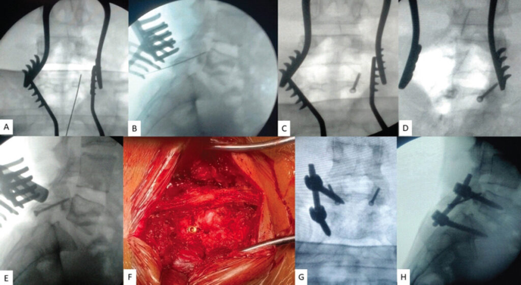
After this, the repair was further protected with temporary, intersegmental fixation using a unilateral (left-sided) pedicle screw-rod construct applied through a separate Wiltse approach. Compression was applied across the pedicle screws before final tightening of the construct was carried out (Fig. 2g and h).
Post-operative protocol
Following the surgery, the patient was allowed to ambulate on the 1st post-operative day (POD) with a lumbosacral (LS) brace. The brace was continued for a period of 6 weeks. The patient was followed up at the end of 3-, 6-, and 12-week follow-up time points, followed by 6, 12, and 2 years postoperatively. At each post-operative follow-up, the patient was examined clinically and radiologically (plain, standing X-rays of lumbar spine – AP and lateral views). After 6 weeks, he was started on a rehabilitative protocol involving back and core strengthening exercises.
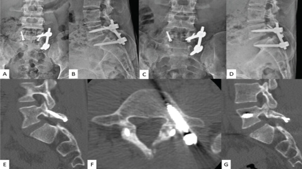
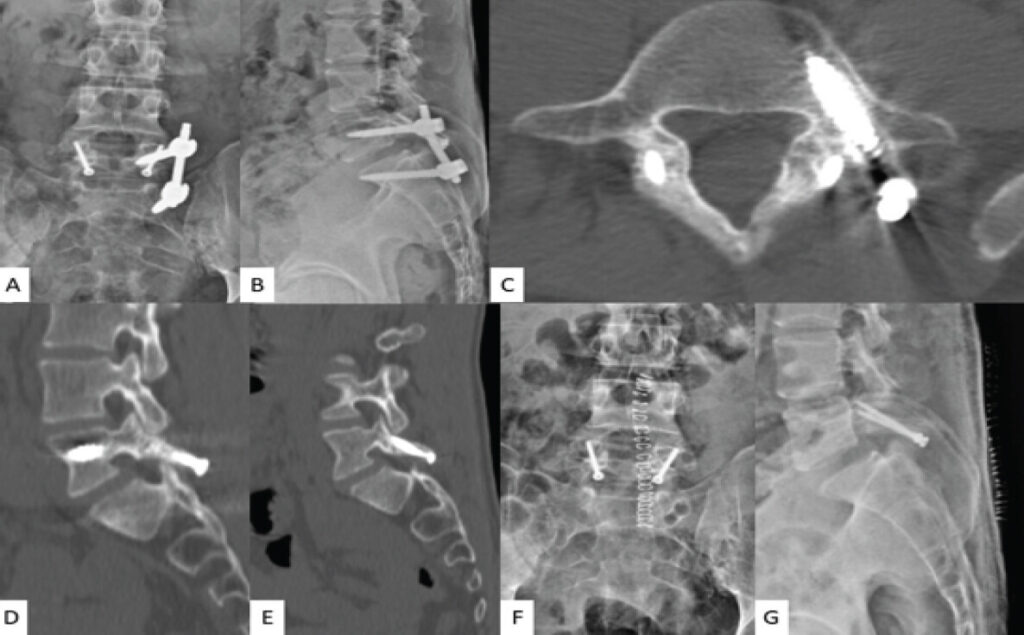
At the end of 1- and 2-year follow-up time points, CT scans of the lumbar spine were performed (Fig. 3a, Fig. 3b, c, d, e, f, g and Fig. 4a, Fig. 4b, c, d, e). There was excellent healing of the pars defect, with satisfactory osseous replacement of the deficiency in the neural arch. The patient also had satisfactory resolution of his back symptoms at the end of 2 years (VAS: 1/10). At this point, the temporizing pedicle screw-rod construct was removed under short GA, and the patient was gradually rehabilitated in the following 3 weeks (Fig. 4g and h).
Discussion
Symptomatic patients with spondylolysis usually recover well with non-operative treatment [2]. Surgical management may benefit in situations of persistent symptomatology, despite a reasonable course of conservative measures [11]. In addition, early surgery may be considered in professional athletes, who may prefer timely return to heavy sporting activities. The goals of surgery include pain mitigation, promotion of healing of the pars deficiency, stabilization of the involved segment, and impeding the listhesis progression [9]. Broadly, the surgical treatment can be classified as fusion and non-fusion surgeries [9]. The latter procedures typically involve a wide array of intrasegmental fixation modalities, aimed at achieving repair of the pars deficiency [1, 3, 5,12, 16, 17, 19].
Since inadvertent sacrifice of an intervertebral (IV) disk and motion segment are best avoided in pediatric patients and young adults without significant IV instability, pars repair techniques offer substantial benefit over fusion surgeries in this patient cohort [2]. The technique of pars repair has been likened to osteosynthesis, and its philosophy broadly includes debridement of the pars defect, bone grafting, and intrasegmental stabilization [15]. Diverse intrasegmental fixation methods include intralaminar screw fixation (Buck technique), pedicle screw-hook construct, intrasegmental wiring, pedicle screw-U-rod, and butterfly plate-screw technique [2, 6, 8, 10, 11, 13, 14, 15, 20].
The intralaminar screw technique (Table 1) has been reported to provide successful pars healing in 82–100% of patients [2, 8]. In a recent meta-analysis by Tsai et al. [8] comparing Buck repair, Scott wiring, Morscher technique and pedicle screw repair, Buck repair was shown to result in excellent outcome with high union rate (effect size [ES]: 93%; 95% CI: 86–98%), positive functional outcome (ES: 85%; 95% CI: 86–97%); and lowest complication rates. The purported advantages of Buck repair include the use of a stable low-profile implant, ability to employ minimally invasive approaches, and excellent healing rates. Recently, studies have also utilized robotic- or navigation-guided approaches to enable mini-open or minimally invasive approaches in challengingly narrow or hypoplastic laminae [9, 15]. Some of the situations where Buck repair has been associated with poorer outcome include the presence of narrow lamina, association with spina bifida occulta (SBO), terminal or more-advanced stage of pars defect, wider or larger gap of the defect, and more advanced disk degeneration. Studies have shown that patients developing non-union after pars repair have substantially poorer clinical outcomes in comparison with those who were conservatively managed [2, 9, 15].
Studies published on intralaminar screw technique (stand-alone or with supplemental stabilization) for direct pars repair (2020 to 2025)
| S.No | Study | Journal/Year | Type of study/Patient cohort | Technique/inclusion criteria | Conclusion |
|---|---|---|---|---|---|
| 1 | Aoyama et al. (Japan) | Spine Surg Rel Research/2025 | Case series: 14 adolescent athletes (13 males; mean follow-up: 28.9 months) | Minimally invasive (percutaneous) approach using cannulated headless compression screws for direct pars repair with cortical bone trajectory | 100% return to sports at 3 months |
| Lumbar spondylolysis: Early and progressive stages only included (Terminal stage lysis – excluded) | 100% fusion at 3 months | ||||
| Good approach for early return to sports in young, professional athletes | |||||
| 2 | Ye et al. (China) | Journal of Orthopaedic Surgery and Research/2024 | Case series: 5 patients (between 10 and 25 years of age | Intra-laminar compression screw (Buck technique), with additional temporary bilateral intersegmental pedicle screw fixation | Temporary intersegmental fixation: |
| (a) Has better control of intersegmental extension and rotational stress | |||||
| (b) Effectively prevents disk degeneration | |||||
| (c) Reconstructs sagittal balance of lumbosacral vertebra | |||||
| 3 | Wang et al. (China) | Acta Neurochirurgica/202 4 | Case report | Percutaneous screw fixation+biportal endoscopic-assisted bone grafting | Preparation of defect site and bone grafting encourages successful osseous fusion |
| 4 | Lu et al. (USA) | Clinical Neurology and Neurosurgery/2023 | Case report | Bilateral pars repair + endoscopic-assisted defect fusion with bone morphogenic protein (BMP) | Radiographic evidence of bony healing: by 3rd post-operative month |
| Minimally-invasive approach | |||||
| 5 | Narendran et al. (USA) | Clinical Case Studies/2023 | Case report: 14-year-old baseball player | Minimally invasive intralaminar fixation with hydroxyapatite-coated screws with robotic assistance | Complete healing of the defects with full incorporation of the hydroxyapatite-coated screws at 6 months follow-up Robotic-assisted technique increases accuracy of screw placement |
| 6 | Gudu et al. (Turkey) | World Neurosurgery/2022 | Prospective study | Modified Buck procedure Inclusion criteria: | 60% fusion rate |
| 30 patients [Mean age: 48±12 years (19 females)] | (a) Patient with B/L pars defects with Grade 0-1 listhesis | Fusion rates: Did not affect the functional outcome (fused versus non-fused cohorts) | |||
| (b) Absence of L5 discogenic pain and c. Failure of conservative measures | |||||
| 7 | Jamshidi et al. (USA) | Research-Human- Clinical Studies: Spine/2022 | Retrospective observational study | Percutaneous direct pars repair – after failed conservative treatment for at least 6 months | 95 % (20/21 patients) fusion rate following the minimally invasive approach |
| 21 young athletes [Mean age: 17.47±3.02 years (15 males)] | Mean VAS: 7.62±1.83 (pre-op) → 0.28±0.56 (P <0.01) | ||||
| Mean follow-up: 31.52±9.38 months | |||||
| 8 | Yurac et al. (Chile) | World Neurosurgery/2021 | Case Series of 3 high performance athletes (17–18 years of age) | Minimally Invasive Modified Buck Technique with Neuronavigation and Neuromonitoring | All 3 patients: |
| Healed well at 4 months postoperatively | |||||
| Returned to sports <6 months postoperatively | |||||
| 9 | Berjano et al. (Italy) | Spine/2020 | Case report and systematic review 14-year-old male | Spondylolysis Repair with Pedicle Screws, Rod and Polyester Band | Resolution achieved at 4 months |
| Reduced complications (including neurological injury) |
Our patient was relatively old, as compared to the typical patient cohort for whom pars repair is recommended. In addition, he had SBO with hypoplastic neural arch, which can potentially compromise the purchase of the intralaminar screw. As evident from our imaging studies, he also had minimal instability at the L5-S1 level with some disk degeneration. As previously discussed, our patient also had a tall and well-built habitus. In view of these aforementioned reasons, we planned to additionally supplement our intralaminar screw fixation (along with bone grafting) with additional intersegmental stabilization using a unilateral pedicle screw-rod construct (Unilateral fixation through Wiltse approach was planned in order to minimize the soft-tissue dissection). Although we recommended the removal of the temporary pedicle screw-rod construct at the end of one year, due to personal reasons, the patient underwent the procedure at 2 years postoperatively.
Zhang et al.[9] discussed the relative strengths of intra- and inter-segmental fixation techniques in the context of direct pars repair. They observed that L5 pars interarticularis is consistently stressed during extension and rotational moments, as a result of wedging between the inferior articular process of L4 and superior articular process of L5 vertebrae. They concluded that temporary intersegmental L5-S1 fixation could be an excellent technique in achieving stability and healing across pars defects at the L5 level, especially in specific scenarios such as SBO, disk degeneration, larger gap across the defect, Grade 1 spondylolisthesis, and higher segmental sagittal angular movements. Although Zhang et al. [9] recommended that the pars repair must always be preceded by a pars block (to confirm the causative association between the symptoms and pars defect), our patient opted to not undergo such blocks. Based on their experience with 5 patients (mean age of 19.2 ± 5.4 years), Ye et al. [15] described a successful outcome following pars repair using a combination of Buck repair, bone grafting, and bilateral temporary screw fixation. They argued in favor of supplementing the Buck repair with additional B/L temporary pedicle screw insertion, owing to their observation that intralaminar screw fixation alone fails to address abnormal stress concentrations at the L5 pars interarticularis (especially in the presence of sagittal imbalance or relative kyphotic alignment at the lumbo-sacral [L5-S1] junction). They suggested that temporary L5-S1 intersegmental fixation can thereby mitigate the incidence of complications like screw loosening, inadequate defect healing, and fracture, which have been reported after Buck repair.
In our patient, we performed a similar triple procedure; although we performed only a unilateral temporary intersegmental stabilization (so as to reduce the extent of soft-tissue dissection). Our patient had a satisfactory clinical outcome following this approach.
Conclusion
Triple procedure involving a combination of bilateral intralaminar screws, autogenous iliac crest bone graft and temporary intersegmental pedicle screw fixation can offer excellent clinical and radiological outcome in challenging situations of spondylolysis with minimal instability and early disk degeneration.
Clinical Message
Direct pars repair remains a valuable non-fusion option for young patients with persistent spondylolysis symptoms despite conservative care.
Buck intralaminar screw fixation achieves high union rates but may be compromised in hypoplastic lamina or spina bifida occulta.
Supplementing pars repair with autologous bone graft enhances healing and reduces risk of non-union or pseudoarthrosis.
Temporary intersegmental pedicle screw fixation can provide added stability in challenging cases with disk degeneration or minimal instability.
A combined “triple procedure” approach – bilateral intralaminar screws, bone graft, and temporary fixation – can yield excellent clinical and radiological outcomes in complex spondylolysis.
Conflict of Interest:
Nil
Source of Support:
Nil
Consent:
The authors confirm that informed consent was obtained from the patient for publication of this article
References
- Simper LB. Spondylolysis in Eskimo skeletons. Acta Orthop Scand 1986;57:78-80. Google Scholar | PubMed
- Sri Vijay Anand KS, Eamani NK, Shetty AP, Rajasekaran S. Spondylolysis and pars repair technique: A comprehensive literature review of the current concepts. Indian Spine J 2021;4:29-39. Google Scholar | PubMed
- Hirano A, Takebayashi T, Yoshimoto M, Ida K, Yamashita T, Nakano K. Characteristics of clinical and imaging findings in adolescent lumbar spondylolysis associated with sports activities. J Spine 2012;1:124. Google Scholar | PubMed
- Szappanos L, Szepesi K, Thomázy V. Spondylolysis in osteopetrosis. J Bone Joint Surg Br 1988;70:428-30. Google Scholar | PubMed
- Merbs CF. Spondylolysis and spondylolisthesis: A cost of being an erect biped or a clever adaptation?. Am J Physical Anthropol 1996;101:201-28. Google Scholar | PubMed
- Viswanathan VK, Manoharan SR, Subramanian S. Symptomatic unilateral spondylolysis associated with nonspondylolytic lateral clefts in adults: Review of an infrequently reported pathology. Cureus 2016;8:e928. Google Scholar | PubMed
- Viswanathan VK, Shetty AP, Jakkepally S, Kanna RM, Rajasekaran S. Symptomatic unilateral pediculolysis associated with contralateral spondylolysis and spondylolisthesis in adults-case report and review of literature. World Neurosurg 2020;143:339-45. Google Scholar | PubMed
- Tsai SH, Chang CW, Chen WC, Lin TY, Wang YC, Wong CB. Does direct surgical repair benefit pars interarticularis fracture? A systematic review and meta-analysis. Pain Physician 2022;25:265-82. Google Scholar | PubMed
- Zhang ZC, Zhang Y, Zhang LZ, Guan K, Zhao GM, Ren DJ. Repair of symptomatic bilateral L5 spondylolysis with autogenous iliac crest graft and temporary intersegmental pedicle screw fixation in youth. J Orthop Surg Res 2021;16:422. Google Scholar | PubMed
- Teruya S, Okuwaki S, Gamada H, Funayama T, Tatsumura M. Preoperative pars defect length predicts bone union after direct repair for lumbar spondylolysis using the modified smiley face rod technique: A retrospective cohort study. Asian Spine J 2025;19:776-83. Google Scholar | PubMed
- Taniguchi Y, Oshima Y, Tanaka S. Lumbar spondylolysis: Future perspectives. JMA J 2024;7:541-2. Google Scholar | PubMed
- Aoyama N, Kiyasu K, Takemasa R, Tadokoro N, Mizobuchi S, Ikeuchi M. Percutaneous spondylolysis repair using headless compression screws with a cortical bone trajectory: A technical description and case series. Spine Surg Relat Res 2025;9:375-80. Google Scholar | PubMed
- Tatsumura M, Ikezawa S, Okuwaki S, Gamada H, Funayama T. Combined transforaminal lumbar interbody fusion and smiley face rod technique using dual-headed pedicle screws for L4 isthmic spondylolisthesis and L5 spondylolysis. Cureus 2024;16:e60756. Google Scholar | PubMed
- Stewart JJ, Zhao DY, Pivazyan G, Gensler R, Voyadzis JM. Minimally invasive robot-assisted direct pars repair: Illustrative cases. J Neurosurg Case Lessons 2024;8:CASE2415. Google Scholar | PubMed
- Ye Y, Yang H, Ma T, Zhu K, Xu G, Han Z. Buck technique supplemented by temporary intersegmental pedicle screw fixation to repair lumbar spondylolysis in youth. J Orthop Surg Res 2024;19:340. Google Scholar | PubMed
- Guo X, Li Z, Guo Z, Li W. Factors associated with non-fusion after direct pars repair of lumbar spondylolysis with pedicle screw and lamina hook: A clinical and CT-assessed study. BMC Musculoskelet Disord 2024;25:152. Google Scholar | PubMed
- Okuwaki S, Tatsumura M, Gamada H, Asai R, Nagashima K, Takeuchi Y. Association of pars defect type with clinical outcome after smiley face rod methods for terminal-stage spondylolysis. Spine Surg Relat Res 2024;8:58-65. Google Scholar | PubMed
- Zhao Y, Wang H, Jiao G, Zhang L, Wu W, Liu H. Comparison of direct pars repair techniques for spondylolysis in young patients: Pedicle screw hook system versus pedicle screw rod. Altern Ther Health Med 2024;30:472-7. Google Scholar | PubMed
- Narendran N, Nilssen PK, Walker CT, Skaggs DL. New technique and case report: Robot-assisted intralaminar screw fixation of spondylolysis in an adolescent. N Am Spine Soc J 2023;16:100284. Google Scholar | PubMed
- Yurac R, Bravo JT, Silva Á, Marré B. Spondylolysis repair using a minimally invasive modified buck technique with neuronavigation and neuromonitoring in high school and professional athletes: Technical notes, case series, and literature review. World Neurosurg 2021;155:54-63. Google Scholar | PubMed
© 2026 Journal of Orthopaedic Case Reports - Published by Indian Orthopaedic Research Group
About the Authors
-

Vibhu Krishnan Viswanathan
-

Malmarugan Ramachandran
-

Sathish Muthu
-

S Gopalakannan
-

Annamalai Devadoss
-

Sathish Devadoss
Related Articles in Journal of Orthopaedic Case Reports
 August 1, 2026 The Forgotten Joint and Forgotten Spine: Recalibrating Success around the Patient’s Experience
 August 1, 2026 O-arm Navigation-assisted Excision of Cervical Osteoid Osteoma Adjacent to the Vertebral Artery: Technical Note and Case Report
 October 1, 2025 Fixation, Stabilization, Dynamic Stabilization, Fusion, and Reconstruction in Spine Surgery– Decoding Terminologies!
 February 10, 2024 When The Unexpected Happens: The Intriguing Retrieval of a Broken Epidural Catheter