


Introduction
Giant cell tumor (GCT) of bone accounts for approximately 5% of all primary bone tumors and 20% of benign bone tumors. It typically affects skeletally mature individuals between 20 and 40 years, with slight female predominance.
The most common sites include:
- Distal femur
- Proximal tibia
- Distal radius
Involvement of the distal tibia is rare (approximately 3–4%). Presentation in patients older than 50 years is uncommon and may mimic degenerative or traumatic conditions, leading to delayed diagnosis.
Written informed consent was obtained from the patient for publication of this case report and accompanying images.
Case Report
A 52-year-old male presented with:
- Dull aching pain over left ankle – 5 months
- Gradually progressive swelling
- Difficulty in weight-bearing for 1 month
- No history of trauma
- No constitutional symptoms
He was initially managed conservatively as an ankle sprain with analgesics and bandaging.
Clinical examination
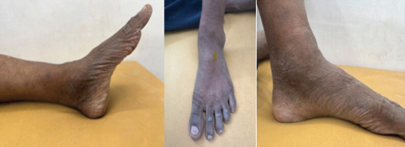
- Diffuse swelling over distal tibia anteromedially
- Local tenderness
- Mild painful restriction of ankle dorsiflexion
- No neurovascular deficit (Fig. 1).
Radiological findings
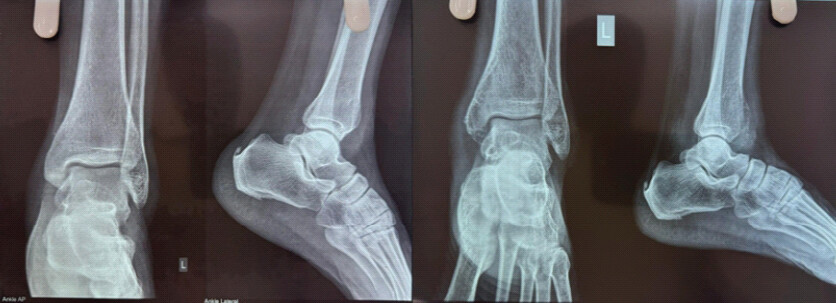
X-ray (Anteroposterior and Lateral)
- Eccentric expansile lytic lesion
- Involving distal tibial epiphysis extending to metaphysis
- Cortical thinning
- No matrix calcification
- No periosteal reaction
Campanacci Grade II lesion (Fig. 2).
Magnetic resonance imaging (MRI) findings
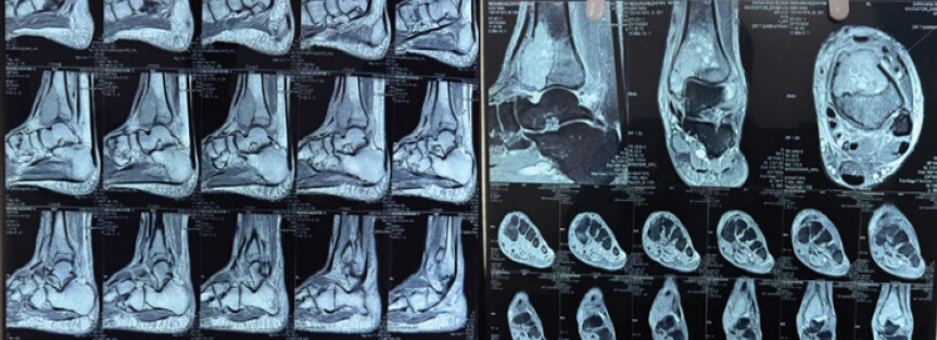
- Subarticular involvement
- Cortical thinning without breach
- No significant soft-tissue component
- No skip lesions (Fig. 3).
Treatment
The patient underwent:
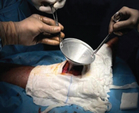
- Extended intralesional curettage
- High-speed burring
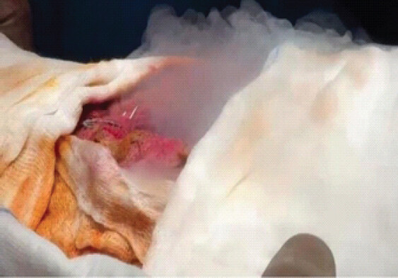
- Chemical cauterization with Liquid Nitrogen
- Cavity filling with polymethylmethacrylate (PMMA) bone cement with prophylactic distal tibia locking compression plating
Intraoperative findings confirmed intact articular cartilage (Figs. 4 and 5).
Histopathology
- Sample Sent: Sample obtained from extended intralesional curettage
- Findings: Predominant osteocartilaginous tissue with marrow adipose tissue, along with a scant amount of lesional tissue showing a giant cell-rich lesion consistent with GCTs.
Post-operative course
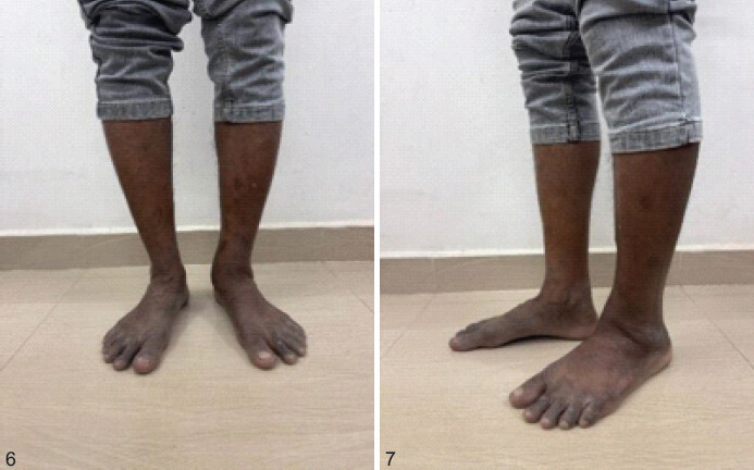
- Non-weight-bearing for 6 weeks
- Gradual weight-bearing with Ankle range of motion (ROM)
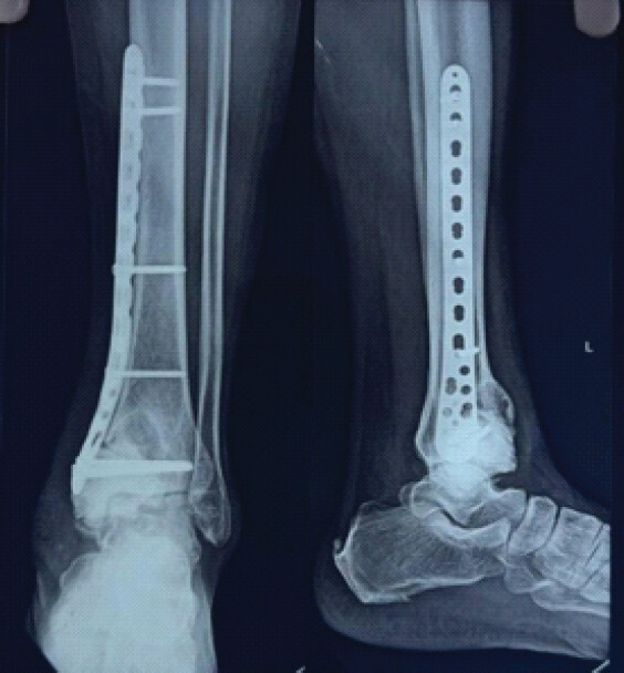
- Full weight-bearing at 8 weeks (Fig. 6 and 7).
Follow-up
At 12 months:
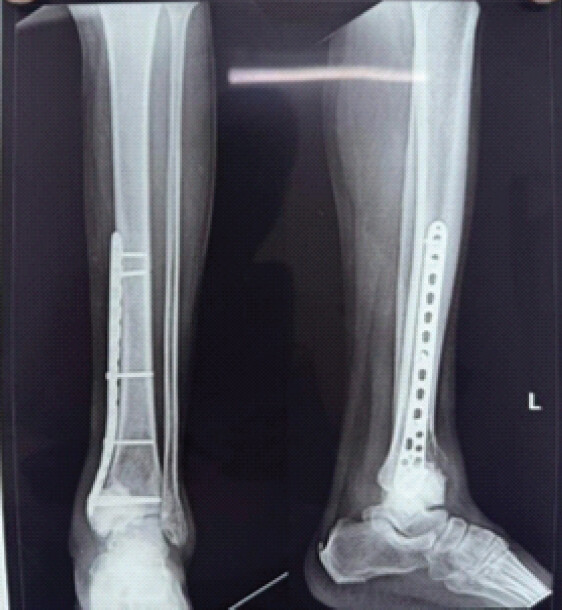
- No radiological recurrence
- Pain-free ambulation
- Ankle ROM: 0–20° dorsiflexion, 0–40° plantarflexion
- Functional outcome was assessed using the musculoskeletal tumor society scoring system, with the patient achieving a score of 28/30 at 12 months
- No metastasis on chest radiograph (Figs. 8 and 9).
Although the follow-up duration is limited to 12 months, most recurrences of GCT occur within the first 2 years. Continued follow-up is ongoing.
Discussion
According to the large series by Campanacci et al. [1], the majority of GCTs occur in patients between 20 and 40 years, with only a small percentage occurring after the age of 50. This atypical age group may contribute to diagnostic delay, as clinicians often initially attribute symptoms to degenerative conditions or minor trauma. In the present case, the patient was initially treated as an ankle sprain for several months before imaging revealed the lesion. Similar delayed presentations in older patients have also been reported by Turcotte [2], who noted that tumors in older individuals may mimic degenerative ankle pathology and lead to late diagnosis. The risk of local recurrence following treatment of GCT varies depending on the surgical technique and use of adjuvants. Klenke et al. [3] identified several factors associated with recurrence and emphasized the importance of adequate local tumor control. Sobti et al. [4] provided an overview of GCT, including its clinical presentation, radiological features, biological behavior, and treatment options.
Distal tibial involvement accounts for only 3–4% of all GCTs according to a study conducted by Errani et al. [5]. The most common locations for GCT include the distal femur, proximal tibia, and distal radius, accounting for the majority of cases. Distal tibial involvement is relatively rare, comprising approximately 3–4% of all GCT cases. Errani et al. [5], in their review of 349 cases of GCTs, reported that distal tibial lesions represented a very small subset compared to other peri-knee locations. Balke et al. [6], in their analysis of 214 cases, reported favorable outcomes with extended curettage and cementation in extremity GCTs. The rarity of this location adds to the diagnostic challenge, especially when symptoms are mild or resemble ankle sprain or arthritis.
Radiographically, GCT typically appears as an eccentric, expansile lytic lesion with cortical thinning and a “soap-bubble” appearance, which is consistent with the findings in the present case. MRI findings in this patient demonstrated subarticular extension without significant soft-tissue involvement, which corresponds to a Campanacci Grade II lesion. Similar imaging characteristics have been described by Chakarun et al. [7], who emphasized that MRI is essential for assessing cortical breach and soft-tissue extension.
GCT is characterized by a complex interaction between neoplastic stromal cells and osteoclast-like giant cells. Werner [8] described the morphological, biological, and histogenetic aspects of GCT. Although GCT is generally considered benign, secondary malignant transformation is a rare complication. Rock et al. [9] reported secondary malignant giant-cell tumors of bone and emphasized the importance of long-term clinical and radiological surveillance.
Intralesional curettage remains the most commonly used treatment modality for GCT, although recurrence rates vary depending on the technique used. Simple curettage alone has been associated with recurrence rates of up to 40–50%, as reported in early studies. Prosser et al. [10] evaluated recurrence following curettage and highlighted the importance of adequate local treatment. Van der Heijden et al. [11] emphasized that treatment should be individualized according to tumor location, extent, cortical integrity, soft-tissue involvement, and the possibility of preserving the adjacent joint.
Occurrence beyond 50 years is rare, reported in <10% of cases. [12] Similar cases of distal tibial GCT in older patients have also been rarely reported. In contrast to typical GCT presentation in younger individuals, this case highlights a diagnostic delay due to atypical age and misleading clinical presentation as an ankle sprain. This emphasizes the need for early imaging in persistent ankle pain even in older patients. Compared to previous reports, our case demonstrated absence of cortical breach despite delayed presentation, allowing joint preservation through extended curettage rather than resection.
Intralesional curettage remains the most commonly used treatment modality for GCT, although recurrence rates vary depending on the technique used. To reduce recurrence, various adjuvants such as high-speed burring, chemical cauterization, cryotherapy, phenol, or PMMA cementation are frequently used. Cryotherapy using liquid nitrogen causes tumor cell necrosis through ice crystal formation and vascular thrombosis. Compared to phenol, it provides deeper penetration and lower recurrence rates. Faur et al. [13] described the use of liquid nitrogen in the treatment of GCT. However, it carries risks such as fracture and soft tissue damage, which were minimized in this case by careful technique and prophylactic plating. In the present case, the patient underwent extended curettage with high-speed burring and liquid nitrogen chemical cauterization followed by PMMA cementation and prophylactic locking compression plating. PMMA cementation has several advantages. In addition to filling the defect and providing immediate structural support, the exothermic polymerization reaction produces a local tumoricidal effect, which may reduce residual tumor cells. Furthermore, cement allows for easier radiographic detection of recurrence due to the clear contrast between cement and bone. Kim et al. [14] discussed the modern interpretation of GCT and its biological behavior.
Functional preservation of the joint is an important goal in the management of GCT, particularly when the lesion is located close to the articular surface. Intralesional curettage with cementation allows preservation of the native joint while maintaining structural stability. In this case, the patient achieved pain-free ambulation and satisfactory ankle ROM at 12-month follow-up, with no evidence of recurrence. Similar functional outcomes were reported by Errani et al. [5], who noted that intralesional procedures combined with adjuvant therapy allow good joint preservation and functional recovery in most patients with extremity GCT. Sumari et al. [15], in a systematic review, reported the global prevalence and recurrence of GCT and emphasized the importance of adequate local control and follow-up.
Distal tibial GCT is rare and may be misdiagnosed as:
- Chronic ankle sprain
- Ankle arthritis
- Subchondral simple cyst
Differential diagnoses include:
- Aneurysmal bone cyst
- Chondroblastoma
- Brown tumor (hyperparathyroidism)
- Metastasis (especially in older patients).
Conclusion
Although rare in patients over 50 years, distal tibial GCT should be considered in persistent ankle pain with lytic lesions on imaging. Early diagnosis prevents joint destruction. Extended curettage with cement augmentation offers excellent functional and oncological outcomes.
This case highlights that persistent ankle pain in older patients should not be overlooked as benign pathology. Early imaging and biopsy are essential. Extended curettage with cryotherapy and cement augmentation allows joint preservation with good early functional outcomes.
Clinical Message
Persistent/Chronic ankle pain in older adults should not be ignored as a simple sprain; radiographic evaluation is much essential to detect rare lesions such as distal tibial giant cell tumor early.
Conflict of Interest:
Source of Support:
Nil
Consent:
The authors confirm that informed consent was obtained from the patient for publication of this article
References
- Campanacci M, Baldini N, Boriani S, Sudanese A.Giant-cell tumor of bone. J Bone Joint Surg Am 1987;69:106-14. Google Scholar | PubMed
- Turcotte RE.Giant cell tumor of bone. Orthop Clin North Am 2006;37:35-51. Google Scholar | PubMed
- Klenke FM, Wenger DE, Inwards CY, Rose PS, Sim FH.Giant cell tumor of bone: Risk factors for recurrence. Clin Orthop Relat Res 2011;469:591-9. Google Scholar | PubMed
- Sobti A, Agrawal P, Agarwala S, Agarwal M.Giant cell tumor of bone - an overview. Arch Bone Jt Surg 2016;4:2-9. Google Scholar | PubMed
- Errani C, Ruggieri P, Asenzio MA, Toscano A, Colangeli M, Rimondi E.Giant cell tumor of the extremity: A review of 349 cases. Cancer Treat Rev 2010;36:1-7. Google Scholar | PubMed
- Balke M, Schremper L, Gebert C, Ahrens H, Streitbuerger A, Koehler G.Giant cell tumor of bone: Treatment and outcome of 214 cases. J Cancer Res Clin Oncol 2008;134:969-78. Google Scholar | PubMed
- Chakarun CJ, Forrester DM, Gottsegen CJ, Patel DB, White EA, Matcuk GR.Giant cell tumor of bone: Review, mimics, and new developments in treatment. Radiographics 2013;33:197-211. Google Scholar | PubMed
- Werner M.Giant cell tumour of bone: Morphological, biological and histogenetical aspects. Int Orthop 2006;30:484-9. Google Scholar | PubMed
- Rock MG, Sim FH, Unni KK, Witrak GA, Frassica FJ, Schray MF.Secondary malignant giant-cell tumor of bone. Clinicopathological assessment of nineteen patients. J Bone Joint Surg Am 1986;68:1073-9. Google Scholar | PubMed
- Prosser GH, Baloch KG, Tillman RM, Carter SR, Grimer RJ.Does curettage without adjuvant therapy provide low recurrence rates in giant cell tumor of bone?. Clin Orthop Relat Res 2005;435:211-8. Google Scholar | PubMed
- Van der Heijden L, Dijkstra PD, Van de Sande MA, Kroep JR, Nout RA, Van Rijswijk CS.The clinical approach toward giant cell tumor of bone. Oncologist 2014;19:550-61. Google Scholar | PubMed
- Mani KC, Ramesh V, Balamurugan S.Giant cell tumor of distal tibia: A rare case report. J Orthop Case Rep 2022;12:45-8. Google Scholar | PubMed
- Faur CI, Anglitoiu B, Chiriac A, Rotaru H.The use of liquid nitrogen in the treatment of giant cell tumor of bone. Appl Sci (Basel) 2020;10:6310. Google Scholar | PubMed
- Kim Y, Nizami S, Goto H, Lee FY.Modern interpretation of giant cell tumor of bone. Clin Orthop Surg 2012;4:107-6. Google Scholar | PubMed
- Sumari SN, Mangi AA, Khandaker S, Islam MT, Rahman MS.Global prevalence and recurrence of giant cell tumor of bone: A systematic review. J Orthop Surg Res 2022;17:346. Google Scholar | PubMed
© 2026 Journal of Orthopaedic Case Reports - Published by Indian Orthopaedic Research Group
About the Authors
-

Dr. Saminathan Thiyagarajan
-

Dr. N. Prabakaran
-
Dr. R. Sivabalan
-

Dr. S. Pavithra
Related Articles in Journal of Orthopaedic Case Reports
 March 1, 2026 Patient-specific Distal Tibia Prosthesis for Reconstruction in a Case of Recurrent Giant Cell Tumor – A Case Report
 March 1, 2026 Functional Outcome of Curettage and Chemical Cauterization with Bone Grafting and Fixation with Neo-Adjuvant Denosumab Therapy in a Case of Giant Cell Tumor of Navicular Bone of a Young Adult – A Case Report
 January 1, 2026 Reconstruction of Recurrent First Metatarsal Giant Cell Tumor with Vascularized Metatarsal Transfer: Long-Term Functional Outcomes
 September 1, 2025 Chondroblastoma of the Right Distal Femur Medial Condyle: A Case Report