


Introduction
Fractures around the pediatric elbow are common; however, T-condylar distal humerus fractures represent one of the rarest injury patterns, accounting for <2% of all pediatric elbow fractures. These injuries occur predominantly in older children and adolescents because of the progressive ossification and maturation of the distal humerus. Unlike conventional supracondylar fractures, T-condylar fractures extend into the articular surface and therefore require precise reduction and stable fixation to restore elbow congruity and function.
The mechanism of injury is typically a direct impact on a flexed elbow, resulting in splitting of the distal humerus into medial and lateral condylar fragments. Failure to recognize the intra-articular extension may result in malunion, stiffness, loss of motion, post-traumatic arthritis, and deformity. Several studies have emphasized that high-quality imaging and anatomical reconstruction are essential to achieving favorable outcomes.
Computed tomography (CT) has become increasingly important in delineating fracture morphology, particularly in adolescents, in whom the fracture pattern often resembles the adult distal humerus fracture. Associated capitellar coronal shear fragments are uncommon and can be difficult to identify on plain radiographs alone. Their presence further complicates treatment and necessitates restoration of radiocapitellar congruity.
Although Kirschner-wire fixation remains the standard treatment for many pediatric distal humerus fractures, complex intra-articular fracture patterns often require more rigid fixation. Screw fixation provides compression across fracture surfaces and may facilitate earlier mobilization. We report a rare case of a CT-confirmed adolescent T-condylar distal humerus fracture associated with a capitellar coronal shear fragment managed successfully using a triangular cannulated cancellous screw construct and Herbert screw fixation through a posterior paratricipital approach.
Case Report
Patient information
A 12-year-old right-hand dominant male presented to the emergency department with severe pain, swelling, and inability to move the right elbow following a self-fall injury. There was no history of previous elbow pathology, and he was otherwise healthy.
Clinical findings
On examination, diffuse swelling and tenderness were present around the elbow. The injury was closed, with no skin compromise. Distal neurovascular examination was normal, and there was no evidence of radial, median, or ulnar nerve injury or vascular compromise.
Diagnostic assessment
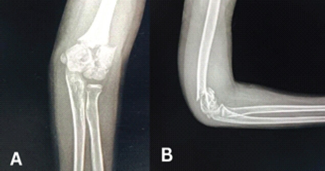
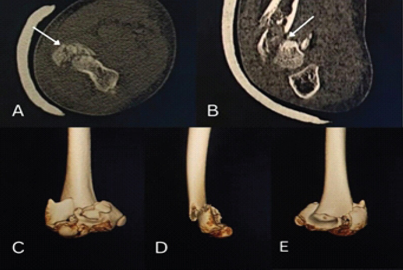
Anteroposterior and lateral radiographs of the elbow demonstrated a displaced intra-articular distal humerus fracture with a T–Y configuration and supracondylar extension. Because of suspicion of a complex intra-articular injury, CT imaging with three-dimensional reconstruction was obtained (Fig. 1).
CT imaging confirmed a T-condylar distal humerus fracture with intercondylar separation and supracondylar extension. Additionally, a displaced capitellar coronal shear fragment was identified, which was not fully appreciated on conventional radiographs (Fig. 2).
Therapeutic intervention
Given the intra-articular displacement and complexity of the fracture pattern, operative treatment was planned. Surgery was performed within 12 h of injury under general anesthesia.
A posterior paratricipital approach was utilized to preserve the extensor mechanism while allowing adequate visualization of the articular surface. Intraoperatively, significant displacement and rotation of the intercondylar fragment were noted.
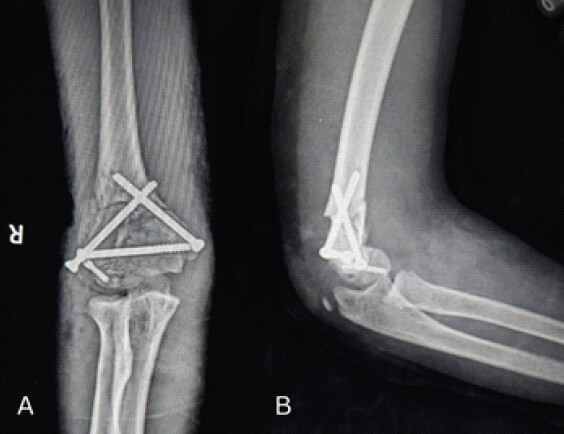
The articular surface was anatomically reduced first. A transverse cannulated cancellous screw was inserted across the condyles to achieve intercondylar compression. The supracondylar component was subsequently stabilized using two obliquely directed cannulated cancellous screws inserted proximally, creating a triangular fixation construct. This configuration provided stable fixation and rotational control of the distal humerus.
The capitellar coronal shear fragment was then reduced and fixed with a Herbert screw, restoring radiocapitellar congruity while minimizing hardware prominence (Fig. 3).
Post-operative management
Post-operative radiographs confirmed satisfactory reduction and fixation. The limb was immobilized in an above-elbow splint. Following wound healing, supervised range-of-motion exercises were initiated.
Outcome and follow-up
The patient was routinely followed up at 1 month, 6 months, and 12 months, respectively. Serial radiographs demonstrated progressive union without loss of reduction or implant failure. Functional scores such as the Mayo Elbow Performance Score (MEPS), Flynn Criteria, and QuickDASH score were calculated at these intervals (Table 1 and Fig. 4).
MEPS, QuickDASH, and Flynn's criteria grading for 1, 6, and 9-month follow-up post-operative status
| Follow-up | MEPS (0–100) | QuickDASH (0–100) | Flynn's criteria |
|---|---|---|---|
| 1 month | 55 | 68.2 | Poor (>15° loss of motion) |
| 6 months | 90 | 9.1 | Good (6–10° loss of motion) |
| 1 year | 100 | 0 | Excellent (≤5° loss of motion and carrying angle) |
MEPS: MAYO Elbow Performance Score
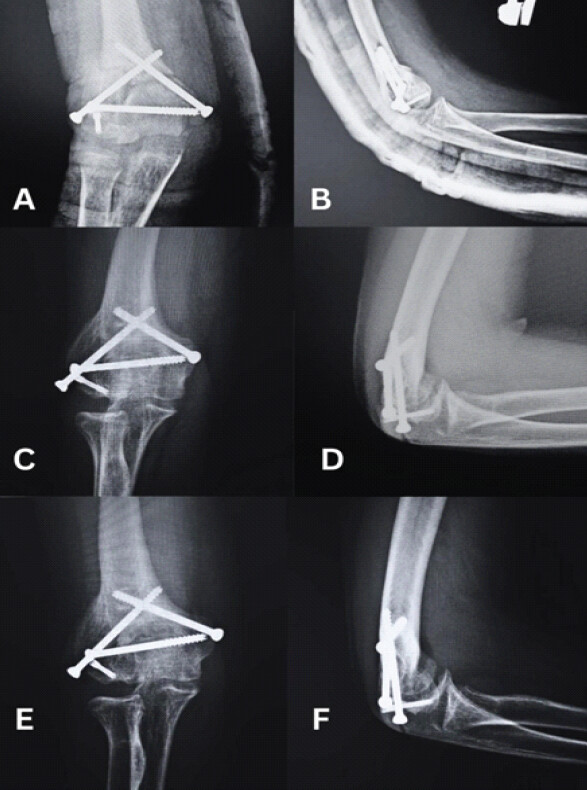
At final follow-up, the fracture had united completely. The patient underwent implant removal after 1 year, following which the patient achieved near-full elbow flexion of 115° and extension deficit of 5° with forearm pronation and supination of approximately 80–90°. No residual deformity, instability, heterotopic ossification, cubitus varus, cubitus valgus, infection, or neurovascular deficit was observed (Fig. 5 and 6).
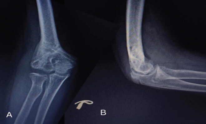
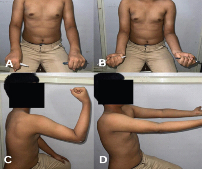
The patient returned to routine daily activities and sports participation without pain or functional limitation.
Discussion
T-condylar distal humerus fractures account for <2% of pediatric elbow fractures and occur mainly in older children because of progressive distal humeral ossification [1, 2]. Popkin et al. and Ducic et al. highlighted their rarity, diagnostic difficulty, and need for anatomical reduction [3, 4].
Our case is unusual because of the associated capitellar coronal shear fragment, a combination rarely reported. CT accurately defined the fracture pattern and guided surgical planning, identifying injuries that may be overlooked on plain radiographs [3, 4]. Uncommon distal humeral fracture patterns are associated with higher complication rates than extension-type injuries, reinforcing the need for careful assessment of atypical elbow trauma [5].
Displaced intra-articular T-condylar fractures generally require open reduction and internal fixation to restore articular congruity and permit early motion, as found in systematic reviews [4, 6]. Although K-wire fixation is suitable for selected fractures, screw fixation offers superior compression and rotational stability, particularly in complex patterns [7, 8, 9].
We used a transverse intercondylar screw with two oblique cannulated screws forming a triangular construct, together with Herbert screw fixation of the capitellum. This provided rigid fixation, maintained reduction, and allowed progressive rehabilitation. Reports describing this construct combined with capitellar fixation in adolescents are extremely limited [10, 11].
The posterior paratricipital approach provided excellent exposure while preserving the extensor mechanism and avoiding olecranon osteotomy-related morbidity [4, 6]. Stable fixation enabled early mobilization, and the patient achieved excellent MEPS, Flynn criteria, and QuickDASH outcomes with complete union.
Our findings reinforce key principles
CT-based assessment, meticulous anatomical reduction, stable fixation, recognition of associated capitellar injuries, and structured rehabilitation are essential for successful management of these rare fractures. This case adds to the limited literature on adolescent T-condylar fractures with capitellar shear fragments. Long-term follow-up studies demonstrated that even when radiographic union is achieved, residual elbow stiffness and carrying angle abnormalities may persist if anatomical reduction is not restored, emphasizing meticulous reconstruction of the articular surface [12].
Conclusion
CT-confirmed adolescent T-condylar distal humerus fractures associated with capitellar coronal shear fragments represent exceptionally rare and complex injuries. Accurate pre-operative characterization using CT imaging is essential for understanding fracture morphology and planning treatment. Stable fixation using a triangular cannulated cancellous screw construct combined with Herbert screw fixation can achieve anatomical reduction, reliable union, and excellent functional outcomes. This technique offers a reproducible option for managing selected adolescent intra-articular distal humerus fractures while facilitating early rehabilitation and restoration of elbow function.
Clinical Message
Rare adolescent T-condylar distal humerus fractures may harbor associated capitellar shear fragments that are easily missed on plain radiographs. Pre-operative CT evaluation and stable anatomical fixation using a triangular cannulated screw construct with Herbert screw fixation can provide excellent articular restoration, reliable union, and near-normal elbow function.
Conflict of Interest:
Source of Support:
Nil
Consent:
The authors confirm that informed consent was obtained from the patient for publication of this article
References
- Vaquero-Picado A, González-Morán G, Moraleda L.Management of supracondylar fractures of the humerus in children. EFORT Open Rev 2018;3:526-40. Google Scholar | PubMed
- Kumar V, Singh A.Fracture supracondylar humerus: A review. J Clin Diagn Res 2016;10:RE01-6. Google Scholar | PubMed
- Popkin CA, Rosenwasser KA, Ellis HB Jr.Pediatric and adolescent t-type distal humerus fractures. J Am Acad Orthop Surg Glob Res Rev 2017;1:e040. Google Scholar | PubMed
- Ducic S, Stojanovic B, Lazovic M, Bukva B, Radlovic V, Bumbasirevic V.T-condylar humerus fracture in children: Treatment options and outcomes. Int Orthop 2020;45:1065-70. Google Scholar | PubMed
- Kuoppala E, Parviainen R, Pokka T, Sirviö M, Serlo W, Sinikumpu JJ.Low incidence of flexion-type supracondylar humerus fractures but high rate of complications. Acta Orthop 2016;87:406-11. Google Scholar | PubMed
- Anari JB, Neuwirth AL, Carducci NM, Donegan DJ, Baldwin KD.Pediatric T-condylar humerus fractures: A systematic review. J Pediatr Orthop 2017;37:36-40. Google Scholar | PubMed
- Dabash S, Gerzina C, Prabhakar G, Thabet AM, Jeon S, Heinrich SD.Screw fixation for supracondylar humerus fractures in children: A report of seventeen cases. Eur J Orthop Surg Traumatol 2018;29:575-81. Google Scholar | PubMed
- Julfiqar A, Pant A, Huda N, Ahmed W.Closed reductions and percutaneus 'K' wire fixation for adolescent intercondylar fractures of the distal humerus. J Clin Diagn Res 2013;7:1666-8. Google Scholar | PubMed
- Wendling-Keim DS, Teschemacher S, Dietz HG, Lehner M.Lateral condyle fracture of the humerus in children: Kirschner wire or screw fixation?. Eur J Pediatr Surg 2021;31:374-9. Google Scholar | PubMed
- Are A, Tornatore I, Theodorakis E.Operative management of a shear fracture of the bilateral capitellum: A case report and review of the literature. Chin J Traumatol 2016;19:231-4. Google Scholar | PubMed
- Aishah NZ, Noor HA.Treatment of capitellum and trochlear fracture using headless screw with concomitant olecranon fracture through posterior approach: A case report. Malays Orthop J 2025;19:119-22. Google Scholar | PubMed
- Sinikumpu JJ, Victorzon S, Pokka T, Lindholm EL, Peljo T, Serlo W.The long-term outcome of childhood supracondylar humeral fractures: A population-based follow up study with a minimum follow up of ten years and normal matched comparisons. Bone Joint J 2016;98-B:1410-7. Google Scholar | PubMed
© 2026 Journal of Orthopaedic Case Reports - Published by Indian Orthopaedic Research Group
About the Authors
-

Dr. Rajath S Shetty
-

Dr. Sachin S Sonune
-

Dr. Dharya H Pimpale
-

Dr. Jaideep Das
-

Dr. Ajit S Rathod
-

Dr. Shabad Sehgal
Related Articles in Journal of Orthopaedic Case Reports
 August 10, 2023 Medial Patellofemoral Ligament Tear Associated with an Intra-articular Axial Spilt Osteochondral Fracture of the Lateral Femoral Condyle: a “Knee Trap-door” Fracture
 June 1, 2026 Standardized Preparation and Multi-Site Application of Leukocyte-Poor Platelet-Rich Plasma in Arthroscopic Anterior Cruciate Ligament Reconstruction: A Technical Note with Early Functional Outcomes
 June 1, 2025 Transphyseal Separation of Distal Humerus Extending to the Lateral Condyle Mimics Isolated Lateral Condyle Fracture in Children: A Case Report
 January 6, 2014 Delayed Case of Congenital Bilateral Trigger Thumb: A Case Report and Review of Literature