


Introduction
Lumbar discal cysts are rare intraspinal, extradural cystic lesions that are frequently misdiagnosed as disk herniations or large annular tears and are characterized by a distinct communication with the corresponding intervertebral disk [1]. They commonly present with radiculopathy and/or low back pain [2,3]. Discal cysts have been reported more frequently in Asian male populations, with a median patient age of approximately 30 years [2, 4].
Various treatment modalities have previously been described for discal cysts, including conservative management, microscopic cyst resection with or without discectomy, microendoscopic cyst resection, and computed tomography (CT)-guided cyst aspiration [2,5,6,7]. In recent years, these lesions have increasingly been managed using minimally invasive endoscopic techniques due to advances in spinal endoscopy.
Several reports have described endoscopic excision of discal cysts performed through percutaneous transforaminal and interlaminar endoscopic approaches [8,9,10,11,12,13,14,15]. However, to the best of our knowledge, reports of excision of lumbar discal cysts using the unilateral biportal endoscopic (UBE) approach are limited.
Case Report
History and clinical examination
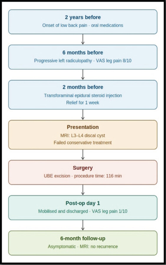
A 27-year-old male presented with low back pain for 2 years and radiating pain to the left lower limb for the last 6 months, with leg pain (visual analog scale [VAS] score 8/10) predominating over low back pain (VAS score 3/10) with a progressive aggravation of symptoms. Symptoms were aggravated by prolonged sitting and forward bending. He denied a history of sensory or bowel and bladder disturbances and had no known medical comorbidities. He had previously been managed conservatively in the form of oral non-steroidal anti-inflammatory and gabapentinoid drugs and had received a transforaminal epidural steroid injection at a different center approximately 2 months before presentation, but experienced only short-term symptomatic relief lasting 1 week (Fig. 1). A neurological examination revealed a positive Lasegue’s sign (passive straight leg raising test) on the left side at 30° and was negative on the right side with intact motor and sensory functions. Deep tendon reflexes were preserved, with mild tenderness over the mid-lumbar spine.
Investigations
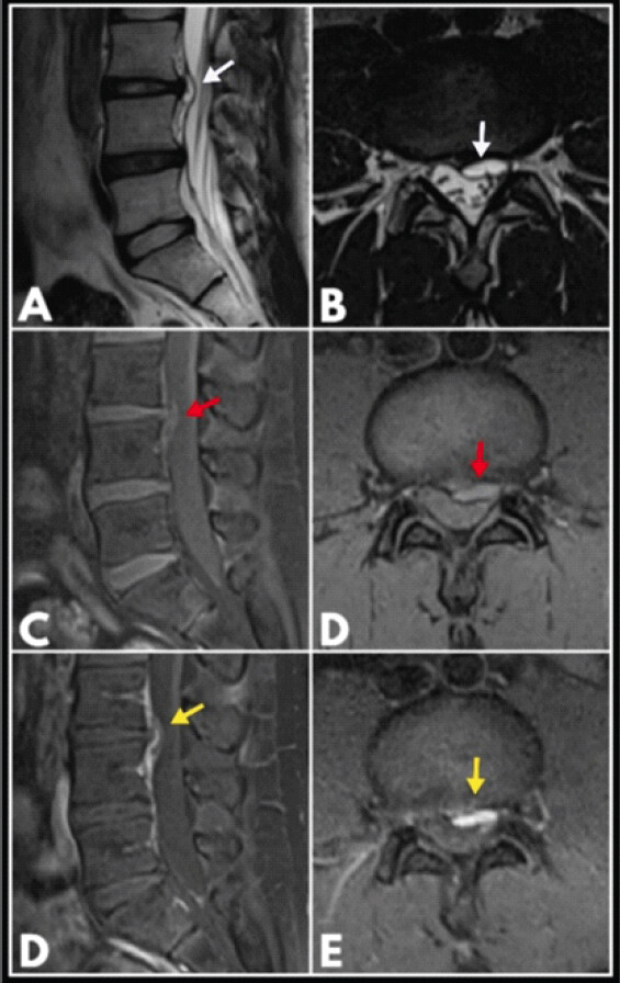
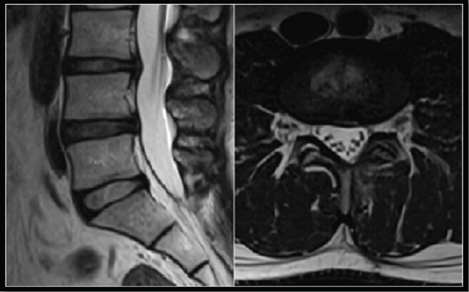
Plain radiographs and magnetic resonance imaging (MRI) – (plain and gadolinium contrast-enhanced) of the lumbar spine demonstrated a large left paracentral discal cyst at the L3–L4 level which was hyperintense on T2 (Fig. 2a and b) and isointense on T1 (Fig. 2c and d) sequences with septations and post-contrast enhancement (Fig. 2e and f), causing predominant compression of the traversing L4 nerve root, with mild indentation of the exiting L3 nerve root. Degenerative disk changes were also noted at L3–L4 and L4–L5 levels with no signs of a herniated nucleus pulposus (HNP). No radiographic instability was identified.
Treatment plan
Given the persistent radicular symptoms and failure of conservative management, including an epidural steroid injection, and imaging-confirmed neural compression, the patient was planned for UBE excision of the discal cyst. The procedure was performed under general anesthesia with the patient positioned in a prone position over a Wilson frame. A left-sided UBE approach to the L3–L4 level was employed using standard portal placement techniques to establish separate viewing and working portals after level confirmation using biplanar fluoroscopy. The viewing portal was marked along the medial pedicular line of L3, aligned with the disk space (approximately 6 mm in length). The working portal (approximately 8 mm in length) was marked along the medial pedicular line of L4, positioned about 3 cm from the viewing portal. The fascial incision for the viewing portal was made in line with the skin incision, whereas that for the working portal was oriented obliquely. This configuration allowed the endoscope and working instruments to converge effectively at the spino-laminar junction of the target level.
Serial dilation was performed at the working portal up to 10 mm to facilitate the establishment of free saline flow. A semi-tubular sheath was then introduced through the working portal to regulate saline outflow and guide instrumentation. Gravity-assisted irrigation was utilized, with 3-L saline bottles positioned approximately 3 feet above the level of the operating table. A 0-° arthroscope was introduced, and a left interlaminar window was created using radiofrequency (RF) ablation and a high-speed burr.
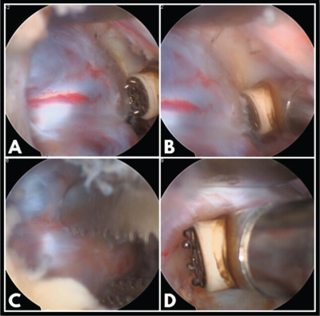
The traversing L4 nerve root was found to be stretched over the cyst and displaced laterally and dorsally (Fig. 3a). Careful adhesiolysis was performed using a fine dissector and nerve hooks to expose the discal cyst. The thecal sac and nerve root were gently retracted medially using an angled retractor. The cyst appeared as a globular, bluish-white lesion with prominent vascularity over its wall and was easily deformable on gentle palpation with a blunt probe. Circumferential dissection was carried out using an RF probe in coagulation mode (Fig. 3b). A biopsy specimen was obtained using pituitary forceps (Fig. 3c), following which cyst debulking was completed. Residual cystic tissue was ablated using the RF probe (Fig. 3d).
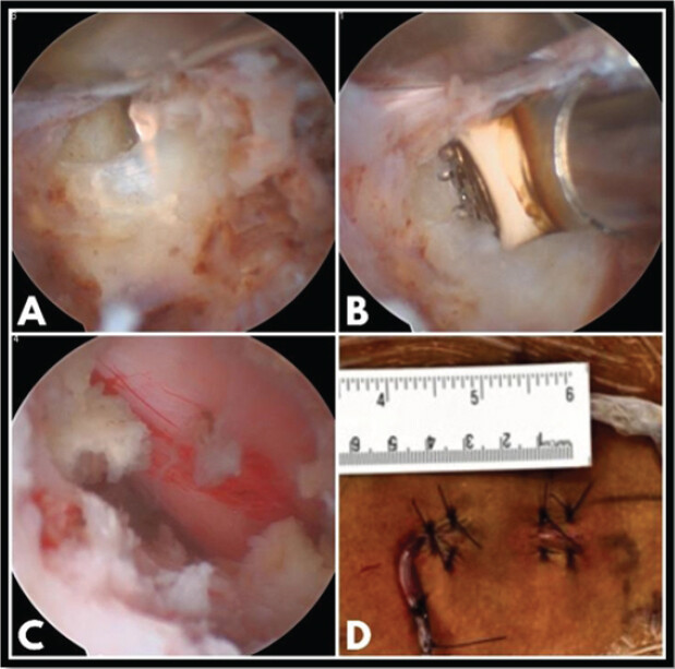
An annular rent was noted intraoperatively (Fig. 4a); however, no HNP fragments were identified. Annuloplasty was performed using the RF probe in coagulation mode (Fig. 4b). At the conclusion of the procedure, the nerve root and thecal sac were free and demonstrated normal pulsatility (Fig. 4c). Wound closure was performed over a drain (Fig. 4d). The duration of the procedure was 116 min. Quantification of intraoperative blood loss is challenging in UBE surgery due to continuous saline irrigation; however, pre-operative and post-operative hemoglobin levels remained comparable, suggesting minimal blood loss. Postoperatively, the patient reported significant improvement in leg pain (VAS 1/10) and low back pain (VAS 2/10). He was mobilized on the same evening and discharged the following morning.
Histopathological examination of the biopsy specimen (three grey-white soft-tissue fragments, aggregate size 1.1 × 0.6 × 0.3 cm) revealed fibrocollagenous and fibroadipose tissue with a cystic space showing denuded lining, areas of mucoid degeneration, hemorrhage, and sparse chronic inflammatory infiltrate, consistent with a fibrotic cyst wall. No granulomatous or malignant features were identified. The patient resumed routine daily activities within 2 weeks of surgery. At the 6-month follow-up, he remained asymptomatic, with sustained clinical improvement and no recurrence of symptoms. Follow-up MRI demonstrated no evidence of recurrent discal cyst. Follow-up MRI demonstrated no evidence of recurrence, with preservation of the posterior soft-tissue structures (Fig. 5).
Discussion
Kazuo Chiba and colleagues first described lumbar discal cysts as intraspinal extradural cysts communicating with the corresponding intervertebral disk and presenting with symptoms similar to lumbar disk herniation [1]. In their series of eight cases, characteristic MRI findings were identified, and surgical excision resulted in immediate relief of radicular symptoms.
The exact pathogenesis of discal cysts remains unclear. Discal cysts may develop during the absorption process of a herniated disk, with macrophages playing a key role, as demonstrated in histological and ultrastructural studies [16]. Other proposed mechanisms include focal annular degeneration with fluid accumulation, organization of an epidural hematoma following disk injury, or an inflammatory reaction to disk material [1, 2, 4]. The absence of epithelial lining on histopathology supports a degenerative rather than synovial origin [1].
Since the initial description of discal cysts, multiple treatment strategies have been reported, ranging from conservative therapy and CT-guided aspiration to open microscopic cyst excision with or without discectomy [2]. Although spontaneous regression and successful conservative management have been described [17], persistent radiculopathy and failure of non-operative treatment often necessitate surgical intervention to achieve rapid symptom relief and early functional recovery. The optimal surgical strategy for discal cysts remains debated. Isolated cyst excision appears appropriate in patients without segmental instability or significant mechanical back pain, whereas fusion procedures may be considered in the presence of instability or advanced degenerative changes [18].
CT-guided aspiration has been described as a minimally invasive treatment option [5, 6, 7]. However, evidence regarding long-term outcomes remains limited; the procedure involves significant radiation exposure, and reliable data on recurrence rates are scarce. With advances in spinal endoscopy, full endoscopic approaches such as transforaminal and interlaminar techniques have increasingly been utilized for excision of lumbar discal cysts, offering the advantages of minimally invasive surgery while providing effective neural decompression [8, 9, 10, 11, 12, 13, 14, 15].
Percutaneous endoscopic surgery for lumbar discal cysts has demonstrated favorable clinical outcomes with high patient satisfaction and minimal complications. Four primary studies involving a total of 26 patients have reported consistent results. Ha et al. reported satisfactory outcomes in 87.5% of eight patients [8]; Chen et al. reported excellent or good outcomes in 88.9% of nine patients [12]; Ding et al. reported satisfactory outcomes in 85.7% of seven patients [15]; and Suo et al. reported successful symptom relief in two cases [11]. Across these studies, mean VAS scores for leg pain improved markedly from pre-operative values of 6.86–8.25 to post-operative values of 1.57–2.25, with similarly significant improvements in Oswestry Disability Index scores. Importantly, no recurrent lesions or serious complications were reported during the respective follow-up periods. Although the current evidence base remains limited in scale, the available literature consistently supports the safety and effectiveness of endoscopic management for lumbar discal cysts.
Interlaminar endoscopic surgery for lumbar discal cysts has also demonstrated favorable clinical outcomes, although the evidence remains limited to small case series [14]. Kim reported successful symptom relief following interlaminar endoscopic decompression using a Ho: YAG laser [13]. These techniques allow targeted decompression with minimal disruption of surrounding structures; however, full endoscopic procedures may be technically demanding due to the limited working channel and restricted instrument maneuverability.
Recently, increasing attention has been directed toward UBE, which combines principles of microscopic surgery with arthroscopic triangulation. This technique allows greater freedom of movement for surgical instruments, as the camera and instruments are introduced through separate viewing and working portals, enabling effective two-handed dissection [19]. Compared with full endoscopy, UBE provides a wider working corridor and improved instrument maneuverability [20], which may be advantageous in cases with dense adhesions, vascular cyst walls, or distorted neural anatomy. Continuous saline irrigation further enhances visualization and facilitates hemostasis. In addition, UBE appears to have a shorter and more manageable learning curve compared with full endoscopic techniques [21]. The infrastructure and equipment costs for UBE are also generally lower than those of dedicated full-endoscopic systems, potentially improving accessibility and wider adoption [22].
To the best of our knowledge, only a single report has described excision of a lumbar discal cyst using the UBE technique. Lin and Pao [23] reported two cases of rare hemorrhagic lumbar discal cysts treated using the UBE approach. Both patients presented with radicular symptoms mimicking lumbar disk herniation. En bloc excision of the cysts was performed, providing excellent visualization and enabling complete removal while preserving surrounding structures. Both patients experienced immediate post-operative symptom relief, with no recurrence noted during follow-up at 12 months. The authors concluded that UBE is a safe and effective minimally invasive alternative for the management of discal cysts, offering advantages such as minimal tissue disruption and favorable clinical outcomes. Available literature suggests that UBE demonstrates clinical outcomes, complication rates, and recovery profiles comparable to those of full-endoscopic techniques in the management of lumbar degenerative pathology [24]. Nevertheless, UBE carries specific risks related to fluid management, including elevated epidural and intracranial pressures, post-operative headache, and the possibility of overlooking small dural tears within a continuous irrigation field [25]. Awareness of these factors and meticulous intraoperative technique are essential to minimize complications.
The literature regarding the surgical management of lumbar discal cysts remains sparse, with most available evidence limited to case reports and small case series. Although UBE offers potential advantages, including direct visualization, precise neural decompression, and limited tissue disruption, there is currently insufficient comparative evidence to establish its superiority over other surgical techniques. In our case, UBE enabled successful excision of the discal cyst with satisfactory clinical and radiological outcomes at 6-month follow-up and rapid post-operative recovery. However, these findings are based on a single patient and are subject to individual patient characteristics and therefore cannot be generalized to the broader population. This report adds to the currently limited evidence regarding the feasibility of UBE for lumbar discal cyst excision rather than establishing its superiority. Further studies involving larger cohorts, comparative analyses, and longer follow-up are warranted to determine recurrence rates, long-term outcomes, and the appropriate role of UBE in the treatment algorithm for this rare pathology.
Patient perspective
The patient reported severe pre-operative pain that significantly affected daily activities. Following surgery, there was marked improvement in symptoms with a smooth recovery. At follow-up, the patient expressed satisfaction with the outcome and return to normal functional status.
Limitations
This report describes a single L3–L4 paracentral discal cyst with 6-month follow-up, which limits the ability to draw definitive conclusions regarding the long-term efficacy, recurrence rates, and durability of symptom relief following UBE. The findings are also subject to individual patient characteristics and clinical circumstances and therefore cannot be generalized to the broader population. In addition, the patient had no significant degenerative changes or spinal instability, representing a single-level, single-pathology experience. Consequently, the favorable outcome observed may not be directly applicable to discal cysts occurring at other spinal levels or to patients with concomitant degenerative disease, spinal instability, multilevel pathology, or other complex anatomical features. Larger studies involving patients with diverse clinical and radiological presentations, longer follow-up, and comparative analyses are required to establish the long-term outcomes, recurrence rates, and broader applicability of UBE and to better define its role in the management of this rare pathology.
Conclusion
UBE may represent a feasible minimally invasive approach for selected lumbar discal cysts, providing direct visualization and effective decompression with favorable short-term clinical outcomes. However, given the rarity of discal cysts and the limited available evidence, including the single-case nature and short follow-up of the present report, these findings should be interpreted cautiously. Larger case series with longer follow-up and comparative studies are required to establish the long-term efficacy, safety, recurrence rates, and relative advantages of UBE over other treatment modalities.
Clinical Message
Lumbar discal cysts are rare causes of lumbar radiculopathy and should be considered in young patients presenting with unilateral radicular symptoms, particularly when imaging demonstrates a ventrolateral extradural cystic lesion adjacent to an intervertebral disk. Accurate diagnosis with MRI and careful correlation with clinical findings are essential. UBE excision provides a minimally invasive approach that allows direct visualization and decompression of the affected nerve root while minimizing tissue disruption. In appropriately selected patients, this technique can provide satisfactory short-term clinical and radiological outcomes.
Conflict of Interest:
Nil
Source of Support:
Nil
Consent:
The authors confirm that informed consent was obtained from the patient for publication of this article
References
- Chiba K, Toyama Y, Matsumoto M, Maruiwa H, Watanabe M, Nishizawa T. Intraspinal cyst communicating with the intervertebral disc in the lumbar spine: Discal cyst. Spine (Phila Pa 1976) 2001;26:2112-8. Google Scholar | PubMed
- Agosti E, De Maria L, Belotti F, Gatti E, Bruzzone M, Amaddeo P. Lumbar discal cysts: A systematic literature review of diagnostic features and treatment strategies. Eur Spine J 2025;34:5539-55. Google Scholar | PubMed
- Kim SY. Radiculopathy caused by discal cyst. Korean J Pain 2014;27:86-9. Google Scholar | PubMed
- Aydin S, Abuzayed B, Yildirim H, Bozkus H, Vural M. Discal cysts of the lumbar spine: Report of five cases and review of the literature. Eur Spine J 2010;19:1621-6. Google Scholar | PubMed
- Dasenbrock HH, Kathuria S, Witham TF, Gokaslan ZL, Bydon A. Successful treatment of a symptomatic L5/S1 discal cyst by percutaneous CT-guided aspiration. Surg Neurol Int 2010;1:41. Google Scholar | PubMed
- Kang H, Liu WC, Lee SH, Paeng SS. Midterm results of percutaneous CT-guided aspiration of symptomatic lumbar discal cysts. AJR Am J Roentgenol 2008;190:W310-4. Google Scholar | PubMed
- Endo Y, Miller TT, Saboeiro GR, Cooke PM. Lumbar discal cyst: Diagnostic discography followed by therapeutic computed tomography-guided aspiration and injection. J Radiol Case Rep 2014;8:35-40. Google Scholar | PubMed
- Ha SW, Ju CI, Kim SW, Lee S, Kim YH, Kim HS. Clinical outcomes of percutaneous endoscopic surgery for lumbar discal cyst. J Korean Neurosurg Soc 2012;51:208-14. Google Scholar | PubMed
- Jha SC, Higashino K, Sakai T, Takata Y, Abe M, Nagamachi A. Percutaneous endoscopic discectomy via transforaminal route for discal cyst. Case Rep Orthop 2015;2015:273151. Google Scholar | PubMed
- Manabe H, Higashino K, Sugiura K. A rare case of a discal cyst following percutaneous endoscopic lumbar discectomy via a transforaminal approach. Int J Spine Surg 2019;13:92-4. Google Scholar | PubMed
- Suo S, Chen Y, Mao X, Chen S, Fu Z. Percutaneous endoscopic surgery for lumbar discal cyst: Two case reports. J Med Cases 2020;11:178-81. Google Scholar | PubMed
- Chen S, Suo S, Li C, Wang Y, Li J, Zhang F. Clinical application of percutaneous transforaminal endoscopic surgery in lumbar discal cyst. World Neurosurg 2020;138:e665-73. Google Scholar | PubMed
- Kim JS, Choi G, Jin SR, Lee SH. Removal of a discal cyst using a percutaneous endoscopic interlaminar approach: A case report. Photomed Laser Surg 2009;27:365-9. Google Scholar | PubMed
- Kasempipatchai P, Kesornsak W, Kuansongtham V. Removal of discal cyst using percutaneous full endoscopic via interlaminar route: A case report. Egypt J Neurosurg 2023;38:48. Google Scholar | PubMed
- Ding S, Zhang G, Gao Y, Hou Z, Shao F. Percutaneous endoscopic resection of lumbar Discal cyst. Pain Physician 2024;27:E873-9. Google Scholar | PubMed
- Kobayashi S, Takeno K, Uchida K, Yayama T, Nakajima H, Miyazaki T. Pathogenesis of the discal cysts communicating with an adjacent herniated disc. Histological and ultrastructual studies of two cases. Joint Bone Spine 2010;77:184-6. Google Scholar | PubMed
- Jadhav N, Sivakumar L, Talibi SS, Momoh P, Rasul F, Hussain RA. Lumbar discal cyst and post-operative discal pseudocyst: A case series. J Surg Case Rep 2022;2022:rjac239. Google Scholar | PubMed
- Huang H, Li G, Deng J, Chen R, Zhou Y. Case Report: Endoscopic cystectomy vs Lumbar interbody fusion for liquid- and gas-filled discal cysts: A case series and literature review.. Front Surg 2025;12:1646605. Google Scholar | PubMed
- Wu PH, Kavishwar RA, Kim HS. A narrative review of current and future of unilateral biportal endoscopic (UBE) transforaminal lumbar interbody fusion. Semin Spine Surg 2024;36:101084. Google Scholar | PubMed
- Dai J, Liu XF, Wang QL, Peng YJ, Zhang QZ, Jiang FX. A new method for establishing operative channels in unilateral biportal endoscopic surgery: Technical notes and preliminary results. J Back Musculoskelet Rehabil 2023;36:367-75. Google Scholar | PubMed
- Liu Y, Li X, Tan H, Hao X, Zhu B, Yang Y. Learning curve of uniportal compared with biportal endoscopic techniques for the treatment of lumbar disc herniation. Orthop Surg 2025;17:513-24. Google Scholar | PubMed
- Yu Z, Ye C, Alhendi MA, Zhang H. Unilateral biportal endoscopy for the treatment of lumbar disc herniation. J Vis Exp 2023;202:e65497. Google Scholar | PubMed
- Lin CH, Pao JL. Minimally invasive en bloc excision of rare hemorrhagic discal cysts using unilateral biportal endoscopic technique: A report of two cases. J Minim Invasive Spine Surg Tech 2020;5:76-80. Google Scholar | PubMed
- Motten TL. Surgical outcomes of unilateral biportal endoscopy versus full endoscopy for lumbar canal stenosis: A meta-analysis. Cureus 2024;16:e76219. Google Scholar | PubMed
- Yang L, Yu T, Jiao J, Hou T, Wang Y, Zhao B. Comprehensive analysis of UBE-related complications: Prevention and management strategies from 4685 patients. Med Sci Monit 2024;30:e944018. Google Scholar | PubMed
© 2026 Journal of Orthopaedic Case Reports - Published by Indian Orthopaedic Research Group
About the Authors
-

Dr. S R Sunkappa
-

Dr. Somashekar Doddabhadregowda
-

Dr. Rajendra Reddy
Related Articles in Journal of Orthopaedic Case Reports
 March 1, 2026 Assessment of Lumbar Foraminal Stenosis: Diagnostic Value and Clinical Correlation: A Systematic Review
 August 1, 2026 Thoracolumbar Fascial Defect in Persistent Low Back Pain After Lumbar Surgery: Ultrasound Findings and Response to Fascial Plane Injection
 June 1, 2026 Influence of Menopause Onset on the Progression of Lumbar Degenerative Disease: A Clinical Correlation Study
 June 1, 2026 Acute Post-operative Lumbar Paraspinal Compartment Syndrome Following Revision Total Joint Arthroplasty: A Case Report