


Introduction
Giant cell tumor of bone (GCTB) is a benign, locally aggressive neoplasm composed of mononuclear stromal cells and numerous osteoclast-like giant cells. It accounts for 4–5% of all primary bone tumors and approximately 20% of benign bone
tumors. Most GCTs arise in the epiphyses of long bones such as the distal femur and proximal tibia. Pelvic involvement is uncommon (1.5–6% of cases), with acetabular lesions particularly rare [1].
Management of pelvic GCTs is challenging due to the complex anatomy, proximity to major vessels and nerves, and the weightbearing function of the hip joint. Extended intralesional curettage with adjuvants such as high-speed burr, phenol, or cryotherapy remains the mainstay of joint-preserving surgery, whereas en bloc resection is reserved for extensive or inaccessible lesions [2]
Denosumab, a RANKL inhibitor, has gained interest as neoadjuvant therapy in large or surgically difficult tumors. It can reduce tumor volume and facilitate joint-preserving surgery, but pre-operative use may increase recurrence risk due to peripheral sclerosis that hampers complete tumor removal. Preoperative selective arterial embolization has also been employed to minimize intraoperative blood loss and facilitate safe tumor resection [3].
We present a rare case of acetabular GCT with protrusio acetabuli, successfully managed with extended curettage, local adjuvants, and structural bone graft reconstruction, highlighting surgical planning, reconstruction strategy, and early functional outcomes.
Case Report
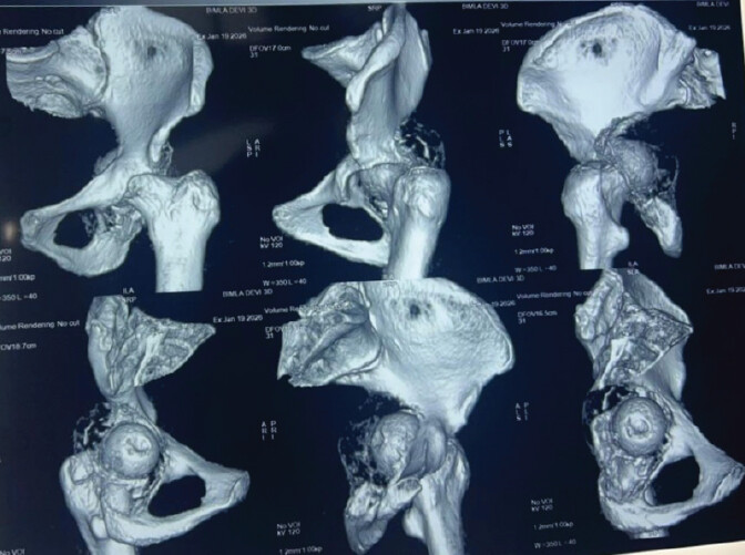
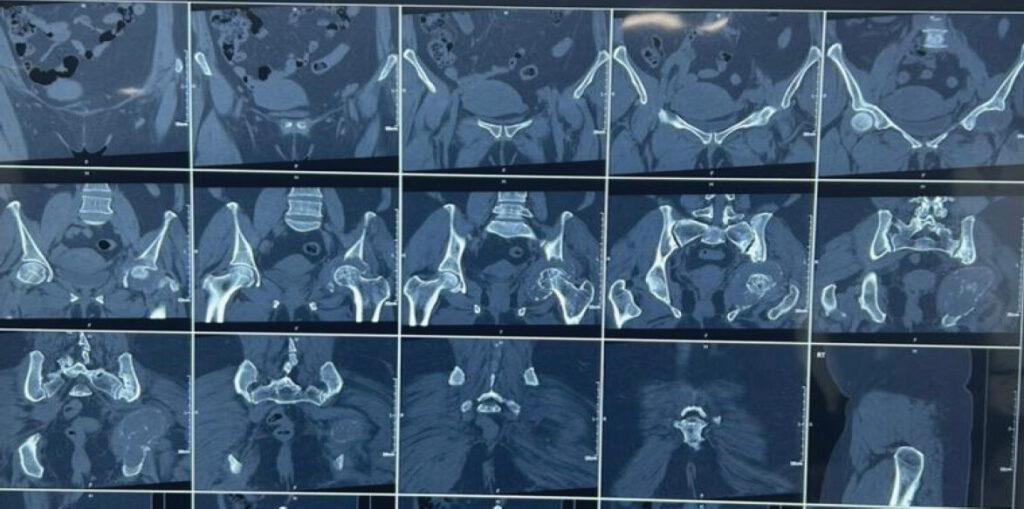
A 50-year-old female presented to the outpatient department of MMCMSR with progressive left hip pain, restricted range of motion, and inability to bear weight for 1 month. She reported a trivial fall at home 1 month prior, which significantly worsened her preexisting mild dull aching hip pain that had been present for 4–5 months but had not previously limited her daily activities. Her medical history included type 2 diabetes mellitus and hypertension, both suboptimally controlled. She denied night pain, fever, weight loss, prior malignancy, tuberculosis, or long-term steroid use. On clinical examination, the patient was hemodynamically stable. Local assessment revealed tenderness over the anterior hip joint, apparent shortening of the left lower limb due to medial migration of the femoral head, and severely restricted and painful movements in all planes. Distal neurovascular status was intact, and sacroiliac stress tests on the left side were positive, suggesting pelvic instability. Plain radiographs of pelvis with both hip in anteroposterior (Fig. 1a) and lateral view of hip with proximal thigh (Fig. 1b and 1c) demonstrated medial displacement of the femoral head consistent with protrusio acetabuli and an expansile, lytic lesion involving the entire acetabulum with cortical thinning and areas of breach . Computed tomography confirmed cortical thinning and breach of the anterior and medial acetabular walls, with involvement of the weight-bearing dome, and aided in surgical planning (Fig. 2). Magnetic resonance imaging showed a T1 hypointense, T2/STIR hyperintense lesion with cystic changes, mild joint effusion, and partial articular cartilage involvement, while also helping exclude differential diagnoses such as aneurysmal bone cyst or chondroblastoma (Fig. 3). Laboratory investigations, including complete blood count, serum electrolytes, and inflammatory markers, were within normal limits, except for mildly elevated erythrocyte sedimentation rate and c-reactive protein, and glycated hemoglobin of 8.1%, indicating suboptimal glycemic control. A CT-guided core needle biopsy revealed numerous multinucleated osteoclast-like giant cells interspersed with mononuclear stromal cells, without cytological atypia or necrosis, consistent with a benign GCTB (Campanacci Grade II–III). Pre-operative management focused on optimizing comorbidities, pain control, and joint alignment. Skin traction was applied to relieve pain and prevent further medial migration of the femoral head. Pre-operative imaging was used to delineate the lesion’s extent, cortical breach, and relationship to neurovascular structures, guiding surgical planning and patient counseling regarding the options of extended curettage versus en bloc resection and the potential need for structural reconstruction.
![Figure 1: [a]Pre-operative anteroposterior radiograph and [b &c ]lateral view of the pelvis with hip showing protrusio acetabuli.](https://jocr.co.in/wp/wp-content/uploads/260FF1-converted-1024x459.jpg)
Pre-operative denosumab was not administered because the lesion was considered amenable to complete extended intralesional curettage with preservation of the native hip joint. Although denosumab can reduce tumor volume and facilitate surgery in selected aggressive or unresectable GCTs, its preoperative use before curettage remains controversial because of the development of peripheral sclerosis, potential difficulty in identifying residual tumor, and concerns regarding increased local recurrence following subsequent curettage. Therefore, in view of the feasibility of thorough curettage and jointpreserving reconstruction, surgery without neoadjuvant denosumab was elected.
The patient underwent surgical management under combined spinal-epidural anesthesia in the lateral decubitus position through a posterior Kocher–Langenbeck approach. The gluteus maximus was split, and the short external rotators were tagged and reflected with careful identification and protection of the sciatic nerve. Following adequate exposure, a cortical window was created and extended intralesional curettage was performed using angled curettes and a high-speed burr to remove all macroscopically visible tumor tissue. Phenol was applied as a local adjuvant, followed by copious saline irrigation. The resulting acetabular defect involved the medial wall and weight-bearing dome and was reconstructed using structural corticocancellous bone grafts to restore the deficient acetabular architecture, with additional cancellous bone graft packed into the residual cavity. Because of the extensive acetabular bone loss, protrusio, and loss of structural support of the native acetabulum, reconstruction was augmented with a customfitted acetabular reinforcement cage. The cage was securely fixed to the remaining viable pelvic bone, providing structural support for the acetabular component. A cementless total hip replacement was subsequently performed, with restoration of the hip center, limb length, and joint stability. Intraoperative fluoroscopy and clinical assessment confirmed satisfactory implant position, hip stability, and restoration of limb length. The wound was irrigated thoroughly and closed in layers over a suction drain.
Postoperatively, the patient received intravenous antibiotics, analgesia, thromboprophylaxis, and strict glycemic control. Early physiotherapy consisting of quadriceps-setting and ankle-pump exercises was initiated. In view of the extensive acetabular reconstruction with cage augmentation and total hip replacement, the patient was maintained on protected non-weight-bearing mobilization with a walker initially. Weight bearing was gradually advanced according to clinical progress and serial radiographic assessment of the reconstructed acetabulum and prosthesis[Fig. 4].
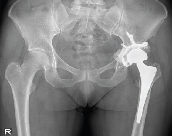
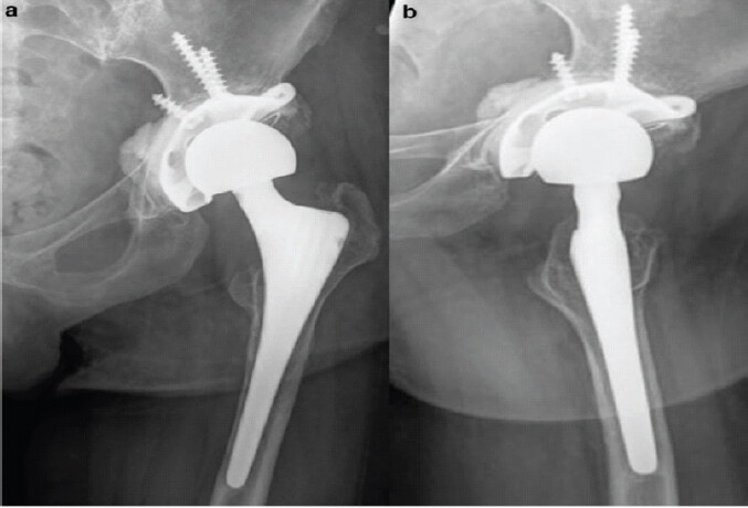
At 6-month follow-up, the patient demonstrated painfree hip movements with restoration of limb length and satisfactory hip stability. Radiographs showed a wellpositioned acetabular cage and total hip prosthesis with incorporation of the structural bone graft and no evidence of implant migration, loosening, or local tumor recurrence [Fig. 5 a and 5b]. The patient achieved a Musculoskeletal Tumor Society score (MSTS) of 25/30 and was independently ambulatory with progressive improvement in function.
Discussion
GCTB is a locally aggressive primary bone tumor that most commonly affects the epiphyseal regions of long bones. Pelvic involvement is uncommon, and acetabular GCTB represents a particularly challenging presentation because of the complex three-dimensional anatomy, proximity to major neurovascular structures, and the critical role of the acetabulum in maintaining hip stability and weight transmission. Patients with acetabular GCTB may present with progressive hip pain and restricted movement, while advanced lesions may produce pathological fracture, protrusio acetabuli, medial migration of the femoral head, and pelvic instability. The present case represents an extensive Campanacci grade II–III lesion involving essentially the entire acetabulum with cortical breach and protrusio acetabuli [4].
The management of pelvic GCTB remains controversial and must be individualized according to tumor extent, cortical integrity, soft-tissue extension, feasibility of complete tumor removal, and the amount of residual bone available for reconstruction. Intralesional curettage with a high-speed burr and local adjuvants remains an important limb- and jointpreserving strategy when adequate tumor clearance can be achieved. However, curettage of extensive acetabular lesions presents a particular challenge because aggressive removal of tumor may leave a large structural defect incapable of supporting the hip joint. Conversely, wide resection can provide improved local control in selected aggressive lesions but may result in substantial loss of pelvic bone and require complex reconstruction [5].
In the present case, the tumor was extensively distributed throughout the acetabulum with cortical thinning and breach of the anterior and medial walls and involvement of the weightbearing dome. Despite the extensive involvement, there was no described extensive involvement of the remaining hemipelvis or major neurovascular structures that would mandate internal hemipelvectomy. Therefore, an intralesional approach was selected to achieve tumor clearance while preserving as much viable pelvic bone as possible. Extended curettage was performed using curettes and a high-speed burr, followed by phenol application and copious irrigation. The use of a highspeed burr allows mechanical removal of residual tumor from the walls of the cavity, while phenol provides an additional local cytotoxic effect. The combination is particularly useful when preservation of the surrounding pelvic architecture is desirable [6].
A major consideration in this case was reconstruction of the extensive acetabular defect following tumor clearance. Simple cancellous bone grafting is generally insufficient when the weight-bearing dome and medial acetabular wall have been substantially compromised. Structural corticocancellous grafts can restore def icient bone and prov ide biological incorporation; however, in a large defect associated with protrusio and loss of the native acetabular architecture, additional mechanical reinforcement is required. In our patient, structural bone grafting was therefore combined with an acetabular reinforcement cage and total hip replacement. The cage provided immediate structural support across the deficient acetabular region and created a stable foundation for implantation of the acetabular component, while total hip replacement restored the hip Center, limb length, joint stability, and functional mobility [7, 8].
Previous reports have described acetabular-preserving procedures using structural grafts and total hip arthroplasty in selected pelvic GCTs with partial acetabular involvement, demonstrating satisfactory graft incorporation and functional outcomes. However, those techniques are most applicable when adequate pelvic columns and acetabular bone remain available for reconstruction. In the present case, the extent of acetabular destruction and protrusio made simple acetabular reconstruction inadequate; hence, cage augmentation was used to provide additional mechanical stability. Custom threedimensional printed hemipelvic implants are another increasingly used option for massive periacetabular defects, particularly when conventional reconstruction cannot obtain reliable fixation. Such implants may be considered when tumor resection leaves inadequate residual bone for standard cage or component fixation [9, 10].
The role of denosumab in GCTB should also be considered carefully. Although denosumab can reduce tumor-associated osteolysis and facilitate surgical downstaging in selected aggressive or unresectable tumors, its use before curettage remains controversial. Systematic reviews and meta-analyses have reported an association between pre-operative denosumab followed by curettage and increased local recurrence, although interpretation is complicated by selection bias because denosumab is preferentially used in more aggressive tumors. One proposed explanation is the formation of a dense peripheral osteosclerotic rim that may make complete identification and removal of residual neoplastic stromal cells more difficult. Therefore, in a surgically accessible lesion such as the present case, where adequate intralesional clearance could be achieved without neoadjuvant therapy, proceeding directly to extended curettage was reasonable [11, 12].
The principal challenge in acetabular GCTB is therefore to balance oncological control with preservation of pelvic stability and restoration of hip function. In our patient, extended intralesional curettage combined with phenol, high-speed burring, str uctural bone grafting, acetabular cage reconstruction, and total hip replacement provided both tumor-directed treatment and immediate mechanical reconstruction. At 6 months, the patient demonstrated restoration of limb length, pain-free hip movement, satisfactory radiographic reconstruction, and an MSTS score exceeding 25/30, with no radiographic evidence of local recurrence. Nevertheless, the 6-month follow-up is relatively short for a locally aggressive tumor such as GCTB. Local recurrence can occur after apparently adequate treatment, particularly in anatomically complex pelvic lesions where complete visualization and clearance are difficult. Continued clinical and radiographic surveillance is therefore essential. Longer followup will also be necessary to evaluate graft incorporation, cage stability, polyethylene or bearing-related complications, prosthetic loosening, infection, and long-term functional outcome.
Conclusion
Giant cell tumor of the acetabulum with extensive bone destruction and protrusio acetabuli is a rare and technically demanding condition. Successful treatment requires careful assessment of tumor extent, residual pelvic bone stock, and the mechanical requirements of the hip. In the present case, extended intralesional curettage with highspeed burring and phenol, followed by structural bone grafting, acetabular cage reconstruction, and total hip replacement, provided satisfactory early oncological and functional results. The acetabular cage was particularly useful in compensating for the extensive loss of the medial wall and weight-bearing acetabular architecture and provided a stable foundation for total hip arthroplasty.
This case demonstrates that cage-augmented acetabular reconstruction with total hip replacement can be an effective option for extensive acetabular GCTB when adequate residual pelv ic bone per mits reconstr uct ion, and inter nal hemipelvectomy is not required. However, treatment should be individualized according to tumor extent and residual bone stock, and long-term surveillance is essential because of the potential for delayed local recurrence and prosthetic complications.
Clinical Message
Extensive acetabular giant cell tumor with cortical destruction and protrusio acetabuli requires meticulous pre-operative imaging and individualized surgical planning. Extended intralesional curettage with high-speed burring and phenol, combined with structural bone grafting, acetabular cage reconstruction, and total hip replacement, can provide effective structural reconstruction and good early functional outcomes in appropriately selected patients. Long-term surveillance is essential because of the risk of local recurrence and prosthetic complications.
Conflict of Interest:
Source of Support:
Nil
Consent:
The authors confirm that informed consent was obtained from the patient for publication of this article
References
- Balke M, Schremper L, Gebert C, Ahrens H, Streitbürger A, Koehler G.Giant cell tumor of bone: Treatment and outcome of 214 cases. J Cancer Res Clin Oncol 2008;134:969-78. Google Scholar | PubMed
- Klenke FM, Wenger DE, Inwards CY, Rose PS, Sim FH.Giant cell tumor of bone: Risk factors for recurrence. Clin Orthop Relat Res 2011;469:591-9. Google Scholar | PubMed
- Tsukamoto S, Tanaka Y, Mavrogenis AF, Tamiya S, Kido A, Kawaguchi M.Is treatment with denosumab associated with local recurrence in patients with giant cell tumor of bone treated with curettage? A systematic review. Clin Orthop Relat Res 2020;478:1076-85. Google Scholar | PubMed
- Palmerini E, Staals EL, Alberghini M, Donati DM.Denosumab in giant cell tumor of bone: Current role and open questions. Cancer Treat Rev 2017;54:113-20. Google Scholar | PubMed
- Thomas D, Henshaw R, Skubitz K, Chawla S, Staddon A, Blay JY.Denosumab in giant-cell tumour of bone: An open-label, phase 2 study. Lancet Oncol 2010;11:275-80. Google Scholar | PubMed
- Zhao Y, Cai Z, Tang X, Du Z, Yang Y, Guo W.Preoperative denosumab may increase the risk of local recurrence of giant-cell tumor of bone treated with curettage: A systematic review and meta-analysis. J Cancer 2021;12:508-17. Google Scholar | PubMed
- Yanagisawa M, Nakano K, Sato T, Nishida Y.Preoperative selective arterial embolization of pelvic giant cell tumour: Clinical effectiveness in reducing intraoperative blood loss. J Bone Joint Surg Br 2003;85:1095-9. Google Scholar | PubMed
- He SH, Xu W, Sun ZW, Liu WB, Liu YJ, Wei HF.Selective arterial embolization for the treatment of sacral and pelvic giant cell tumor: A systematic review. Orthop Surg 2017;9:139-44. Google Scholar | PubMed
- Errani C, Tsukamoto S, Leone G, Hawkins AL.Current concepts for the management of giant cell tumor of bone. J Clin Orthop Trauma 2020;11(Suppl 2)321-7. Google Scholar | PubMed
- Luengo-Alonso G, Mellado-Romero M, Shemesh S, Ramos-Pascua L, Pretell-Mazzini J.Denosumab treatment for giant-cell tumor of bone: A systematic review of the literature. Arch Orthop Trauma Surg 2019;139:1339-49. Google Scholar | PubMed
- Lipplaa A, Dijkstra S, Gelderblom H.Challenges of denosumab in giant cell tumor of bone, and other giant cell-rich tumors of bone. Curr Opin Oncol 2019;31:329-35. Google Scholar | PubMed
- Miles DT, Voskuil RT, Dale W, Mayerson JL, Scharschmidt TJ.Integration of denosumab therapy in the management of giant cell tumors of bone. J Orthop 2020 Mar 28:22:38-47.doi: 10.1016/j.jor.2020.03.020.Erratum in: J Orthop. 020 Dec 14;23:275.doi: 10.1016/j.jor.2020.12.003. PMID: 32280167; PMCID: PMC7136643. Google Scholar | PubMed | CrossRef
© 2026 Journal of Orthopaedic Case Reports - Published by Indian Orthopaedic Research Group
About the Authors
-

Dr. Abhishek Singh
-

Dr. Ayushi Lakhanpal
-

Dr. Abhijit Mahajan
-

Dr. Abhijeet Sandhu
-

Dr. Akhilesh Saini
-

Dr. Shubham Gupta
Related Articles in Journal of Orthopaedic Case Reports
 September 1, 2026 Unnoticed Giant Cell Tumor of the Left Distal Tibia in a 52-Year-Old Male: A Rare Case Report
 April 1, 2026 A Novel Approach to Proximal Humerus Giant Cell Tumor: Cement Spacer and Screw Interdigitation for Optimal Stability
 March 1, 2026 Functional Outcome of Curettage and Chemical Cauterization with Bone Grafting and Fixation with Neo-Adjuvant Denosumab Therapy in a Case of Giant Cell Tumor of Navicular Bone of a Young Adult – A Case Report
 January 1, 2026 Reconstruction of Recurrent First Metatarsal Giant Cell Tumor with Vascularized Metatarsal Transfer: Long-Term Functional Outcomes