


Introduction
Osteogenesis imperfecta (OI) is a rare clinical disorder, occurring at a rate of between 1/10,000 and 1/25,000 worldwide [1]. Previously, OI was thought to be caused solely by dominant mutations in the genes encoding Type I collagen (COL1A1 and COL1A2); however, with the discovery of other rare disease-causing genes, OI is now considered a “type I collagen-associated” disorder. The skeletal phenotype of OI patients is characterized by reduced bone density, increased bone fragility, recurrent fractures, and progressive skeletal deformities; however, because the pathogenic mechanisms vary among OI subtypes, clinical features are also highly diverse [2]. Furthermore, in cases with mild symptoms, such as sillence Type I, it is not uncommon for diagnosis to be delayed and confirmed only after multiple fractures have occurred.
Cubitus varus (CV) is the most frequent complication of treatment for supracondylar humeral fractures in children. It is a 3D deformity consisting of varus angulation in the coronal plane, internal rotation in the axial plane, and extension in the sagittal plane. Although CV has been conventionally described as a cosmetic deformity with little functional significance, there is growing awareness of long-term complications, including chronic pain, ulnar nerve palsy [3, 4], tardy posterolateral rotatory instability [5, 6], and an increased risk of lateral condyle and other secondary fractures [7, 8].
Although CV can indeed occur following a supracondylar humeral fracture in OI, there have been no reports of severe varus deformity exceeding 40°. Here, we report a case of very severe CV and discuss the challenges of treating fractures around the elbow in OI.
Case Report
The patient is a male who initially suffered a supracondylar fracture of the right humerus at 2 years and 6 months of age. He underwent cross-pinning at a hospital on the day of the injury, but the Kirschner wire subsequently came out shortly thereafter, resulting in mild varus deformity of the elbow. After that, the condition was monitored without surgery, but 6 months after the initial fracture, the patient fell and suffered a recurrent supracondylar fracture of the right elbow, which was treated conservatively with a cast at hospital B. Subsequently, at 12, 14, and 16 months after the initial fracture, the patient repeatedly sustained fractures of the lateral condyle of the right humerus due to falls, and pinning and cast treatments were repeatedly performed. Due to the recurrence of frequent fractures, the patient underwent evaluation at the pediatric department of A hospital, where a diagnosis of OI was made (COL1A2 c693+1G>A heterozygous splice donor variant). The patient was then referred to our facility for management of severe CV and OI.
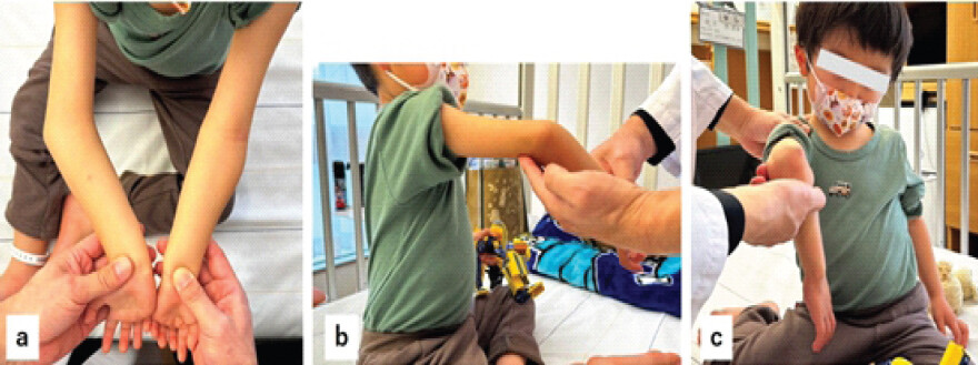
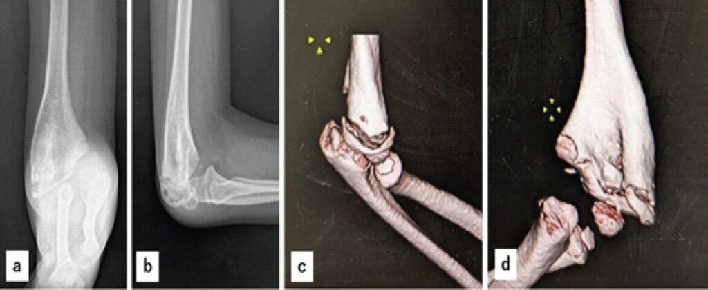
When he first visited our facility at the age of four, his height was 90 cm, and the body weight was 11.6 Kg. He had a severe CV of approximately 45° in the right elbow, and approximately 20° of extension deformity and some internal rotation deformity were observed (Fig. 1). However, the deformity was so severe that accurate three-dimensional assessment was difficult. The patient had blue sclera and no apparent family history of OI. As shown in Fig. 2, X-rays and computed tomography scans revealed severe varus deformity of the distal humerus and a fracture line at the lateral condyle. The dominant hand of the patient is right and suffered significant limitations in activities of daily living. Furthermore, due to the poor alignment of the right elbow, the patient had fallen into a vicious cycle where fractures recurred immediately upon falling and bracing with the hand; therefore, a plan was established to correct the deformity via osteotomy. Rather than forcibly correcting rotational or extension deformities, the plan prioritized achieving stability by increasing the contact surface at the osteotomy site. Therefore, a stepped cut osteotomy in the sagittal plane and 45° valgus correction in the coronal plane were planned (Fig. 3a), followed by plate fixation. It was decided that if, after union was achieved, there were insufficient corrections such as rotational alignment, the case would be re-evaluated in detail before performing additional precise osteotomies and internal fixation; this was fully explained to the patient’s parents before the surgery.
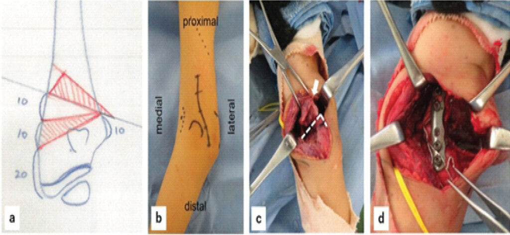
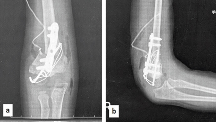
The first surgery was performed under general anesthesia in the prone position at 4 years and 10 months old. An incision was made as shown in Fig. 3b, Fig. 3 and the distal humerus was exposed from the posterior approach. After performing the osteotomy as planned (Fig. 3c), the bone was fixed with a plate from the posterolateral side; and due to concerns about stability, additional fixation was provided using Kirschner wires and soft wires (Fig. 3d and 4). However, due to poor bone quality, the Kirschner wires gradually loosened over time, causing slight displacement at the osteotomy site; nevertheless, bone union was achieved by extending the duration of external fixation. Six months postoperatively, mild varus deformity persisted, and there were concerns about re-fracture due to falls, so we decided to perform a second corrective osteotomy.
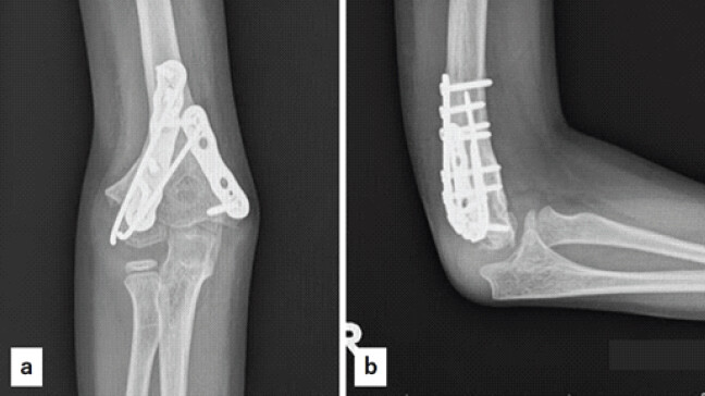
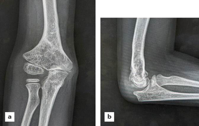
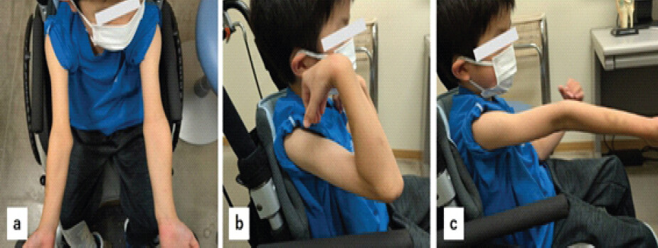
The second surgery was also performed in the prone position using the same incision at 5 years and 8 months old. Since the bone thickness at the planned osteotomy site had increased following the initial surgery, it was determined that a standard wedge osteotomy would provide sufficient contact between the osteotomy surfaces. A wedge-shaped osteotomy was performed to achieve 20° of valgus and 10° of flexion, and the bone was firmly fixed from the posterior aspect using two plates (Fig. 5). Sufficient bone union was achieved 6 months after the second surgery, so the internal fixation hardware was removed. At the final follow-up at age seven, the carrying angle is 15°, and the good range of motion for elbow flexion and extension is maintained (Figs. 6 and 7), thanks to the improved alignment; no re-fractures have occurred following falls.
Discussion
Cases like the present one, involving severe CV exceeding 40° in patients with OI, are extremely rare even in existing reports. CV involves a complex combination of varus, internal rotation, and extension deformities. When the deformity is extremely severe, it becomes difficult to accurately assess the three-dimensional deformity even with plain radiographs or CT scans. Various osteotomy techniques for CV have been reported, and none is generally considered superior to the others [9, 10]. In the current case, due to the patient’s small stature and small bones, combined with the severe deformity, we determined that attempting to achieve perfect 3D anatomical correction in a single surgery would risk reducing the contact area of the osteotomy surfaces, thereby compromising fixation stability. Therefore, given the bone fragility associated with OI, we prioritized “achieving contact and stability at the osteotomy sites” over “perfect correction of rotation and extension” during the initial surgery, limiting the procedure to simple valgus correction in the coronal plane. Furthermore, Takagi et al. have reported that good outcomes can be achieved with valgus correction alone in CV cases under 10 years of age [11]; our judgment is therefore considered reasonable from this perspective as well. Thus, for complex elbow deformities involving underlying bone fragility, a strategy that allows for staged correction – rather than aiming for “perfection in a single surgery” – is more realistic.
The bones of OI patients are extremely fragile, presenting challenges that differ from those of typical fracture treatments and deformity correction surgery. During the initial surgery, we reinforced the fixation with K-wires in addition to plates and screws; however, due to poor bone quality, wire laxity and displacement occurred. This strongly suggests that in OI patients, even when fixation is believed to be “secure,” there is always a risk of post-operative loss of correction. For the second surgery, partly because the bone had thickened following the initial procedure, we opted for robust fixation using two plates. In cases involving OI, careful management is required, including preparing more backup fixation methods than usual and setting a longer period for post-operative external fixation.
In the treatment of severe deformities in patients with OI, the success of treatment depends not only on medical expertise but also on prior communication with the family. Given the fragility of the bones and the complexity of the deformity, the risk of post-operative displacement or insufficient correction is higher than usual. In the current case, it was crucial that we explained to the parents before the initial surgery that “there is a significant possibility that additional surgery will be necessary” and obtained their understanding. Ultimately, after two surgeries, we achieved good alignment and range of motion, breaking the vicious cycle of re-fractures. Precisely because this was a case with a high degree of uncertainty, incorporating a “stepwise approach” into the treatment plan from the pre-operative stage and sharing it with the patient is key to enhancing both the final treatment satisfaction and safety.
Conclusion
Fractures around the elbow in OI require special attention, as they often result in severe deformities during the healing process, unlike typical fractures. We reported a case of extremely severe CV in a patient with OI that was successfully managed through a two-stage surgical approach. In cases with profound bone fragility and complex multi-planar deformities, prioritizing osteotomy site stability over immediate anatomical perfection is a valid and realistic strategy. To minimize the risk of post-operative loss of correction, robust internal fixation and an extended period of external support are essential. Furthermore, establishing a shared understanding with the family regarding the potential necessity of staged procedures is crucial for ensuring treatment safety and satisfaction in such challenging clinical scenarios.
Clinical Message
Fractures associated with osteogenesis imperfecta can result in severe malunion that would not normally occur.
For extremely severe deformities, it may be beneficial to perform corrective surgery in multiple stages.
Conflict of Interest:
Source of Support:
Nil
Consent:
The authors confirm that informed consent was obtained from the patient for publication of this article
References
- Dahlin DC..Bone Tumors: General Aspects and Data on 6221 Cases. 3rd ed. Springfield: Thomas; 1978. Google Scholar | PubMed
- Meller I, Bickels J, Kollender Y, Ovadia D, Oren R, Mozes M.Malignant bone and soft tissue tumors of the shoulder girdle. A retrospective analysis of 30 operated cases. Acta Orthop Scand 1997;68:374-80. Google Scholar | PubMed
- Kassab M, Dumaine V, Babinet A, Ouaknine M, Tomeno B, Anract P.Twenty nine shoulder reconstructions after resection of the proximal humerus for neoplasm with mean 7-year follow-up. Rev Chir Orthop Reparatrice Appar Mot 2005;91:15-23. Google Scholar | PubMed
- Delepine G, Goutallier D.La résection dans le traitement des ostéosarcomes des membres. L'amputation conserve-t-elle des indications? Rev Chir Orthop Reparatrice Appar Mot 1985;71:451-61. Google Scholar | PubMed
- Simon MA, Aschliman MA, Thomas N, Mankin HJ.Limb-salvage treatment versus amputation for osteosarcoma of the distal end of the femur. J Bone Joint Surg Am 1986;68:1331-7. Google Scholar | PubMed
- Rosen G, Murphy ML, Huvos AG, Gutierrez M, Marcove RC.Chemotherapy, en bloc resection, and prosthetic bone replacement in the treatment of osteogenic sarcoma. Cancer 1976;37:1-11. Google Scholar | PubMed
- Petterson H, Hamlin DJ, Manusco A, Scott KN.Magnetic resonance imaging of the musculoskeletal system. Acta Radiol Diag 1985;26:225-34. Google Scholar | PubMed
- O'Connor MI, Sim FH, Chao EY.Limb salvage for neoplasms of the shoulder girdle. Intermediate reconstructive and functional results. J Bone Joint Surg Am 1996;78:1872-88. Google Scholar | PubMed
- Gebhart M, McGuire MH, Mankin HJ.Resection and allograft arthrodesis for malignant bone tumors of the extremity Enneking WF..Limb Salvage in Musculoskeletal Oncology New York: Churchill Livingstone; 1987;567-82. Google Scholar | PubMed
- Peabody TD, Finn HA, Simon MA.Allograft arthrodesis of the shoulder after extra-articular resection of malignant tumors of the proximal humerus Brown KL..Complications of Limb Salvage: Prevention, Management and Outcome. Montreal: International Symposium of Limb Salvage; 1991; 589-92. Google Scholar | PubMed
© 2026 Journal of Orthopaedic Case Reports - Published by Indian Orthopaedic Research Group
About the Authors
-

Dr. Masanori Wako
-

Dr. Tetsuhiro Hagino
-

Dr. Jiro Ichikawa
-

Dr. Tetsuo Hagino
-

Dr. Hirotaka Haro
Related Articles in Journal of Orthopaedic Case Reports
 May 1, 2026 Oblique Lateral Closing-Wedge Osteotomy for Cubitus Varus in Skeletally Immature Children: A Case Series
 December 1, 2024 Effects of Bisphosphonates on Osteotomy Healing in Osteogenesis Imperfecta: A Case Series
 July 1, 2026 Step-cut and Butterfly Femoral Osteotomies Following Intraoperative Fracture Propagation during Distraction Osteogenesis: Report of Two Cases
 June 1, 2026 Oblique Close Wedge versus Modified Step Cut Osteotomy for Cubitus Varus Deformity in Children: A Prospective Non-Randomized Study